
Ophthalmology and Vision Care
OPEN ACCESS | Volume 6 - Issue 1 - 2026
ISSN No: 2836-2853 | Journal DOI: 10.61148/2836-2853/OVC


Dr. Omar Faroque1*, Dr. Md. Abid Akbar2, Dr. Abir Bin Sajj3, Mohammad Shamsal Islam4
1Consultant, Department of Glaucoma, Vision Eye Institute & Hospital, Dhaka, Bangladesh.
2Consultant, Department of Oculoplastic, Vision Eye Institute & Hospital, Dhaka, Bangladesh.
3Department of Cornea, Anterior Segment & Refractive Surgery, Vision Eye Institute & Hospital, Dhaka, Bangladesh.
4Epidemiologist, Ispahani Islamia Eye Institute & Hospital, Dhaka, Bangladesh.
*Corresponding author: Dr. Omar Faroque, Consultant, Department of Glaucoma, Vision Eye Institute & Hospital, Dhaka, Bangladesh, email: faroque76omar@gmail.com.
Received: November 08, 2026 | Accepted: November 18, 2026 | Published: November 23, 2026
Citation: Faroque O, Md. Abid Akbar, Abir B Sajj, Mohammad S Islam, (2026). “Clinical Profile, Management Outcomes, and Prognosis of Patients with Lens-Induced Glaucoma: A Hospital-Based Observational Study” Ophthalmology and Vision Care, 6(1); DOI: 10.61148/2836-2853/OVC/071.
Copyright: © 2026 Omar Faroque. This is an open access article distributed under the Creative Commons Attribution License, which permits unrestricted use, distribution, and reproduction in any medium, provided the original work is properly cited.
Background:
Lens-induced glaucoma (LIG) is a severe type of secondary glaucoma caused by an advanced, unoperated cataract. The clinical profile typically involves elderly patients presenting with acute, unilateral eye pain, redness, and profound vision loss, while the visual prognosis is heavily dependent on how quickly the intraocular pressure (IOP) is managed and the cataract is surgically removed.
Objective:
The study aims to find the clinical profile and prognosis of Lens induced glaucoma patient attending in a tertiary eye hospital.
Methods:
A descriptive cross-sectional study was conducted, and a total of 63 patients of LIG from January 2026 to March 2026 attending the outpatient department were included in this study. After taking informed consent, detail history, detailed examination, IOP by Applanation tonometry, Gonioscopy, examination of posterior segment, patients diagnosed as lens induced glaucoma admitted and managed with medical and surgical treatment. Data analyzed with descriptive statistics and chi square test.
Results:
In our study about 64.7% of the patients were males and 35.3% of the females. The mean age of the patients was 33.35±13.64 years (Range: 18–56 years). About 40.6% of the patients had 6/6 visual acuity in the right eye followed by 6.2% (6/9), 15.6% (6/12), 3.1% (6/18), 3.1% (6/24), 9.4% (6/36), 6.2% (6/60), 3.1% (3/60), 9.4% (FC) and 3.1% of the patients had NPL. About 43.8% of the patients had 6/6 visual acuity in the left eye followed by 9.4% (6/9), 9.4% (6/12), 6.2% (6/18), 9.4% (6/24), 3.1% (6/36), 6.2% (6/60), 3.1% (6/36), 6.2% (6/60), 3.1% (FC) and 3.1% of the patients had HM. About 4.2% of the patients had 1-5 mmHg of IOP in the right eye, followed by 41.6% (6-10 mmHg), 45.9% (11-15 mmHg), and 8.3% of the patients had 16-20 mmHg of the Intraocular Pressure. The average mean Intraocular Pressure of the patients was 11.75±3.082 mmHg (Range: 3–18 mmHg). About 8.3% of the patients had 1-5 mmHg of IOP in the left eye, followed by 33.4% (6-10 mmHg), 37.5% (11-15 mmHg), and 20.8% of the patients had 16-20 mmHg of the Intraocular Pressure. The average mean Intraocular Pressure of the patients was 13.04±6.531 mmHg (Range: 3–36 mmHg). Majority of the patients underwent SICS + PCIOL (65.5%).
Conclusion:
Lens induced glaucoma is still a challenge for complicated of cataract that may prevented by early treatment of cataract. Clinical profile show that SICS is still gold standard that effectively controls IOP and gives better visual outcome.
Lens induced glaucoma, Phacomorphic, Phacolytic, SICS, PCIOL, Bangladesh
Lens-induced glaucoma (LIG) is an important and potentially preventable cause of secondary glaucoma, which may present as either secondary angle-closure or open-angle glaucoma. It develops as a consequence of structural, biochemical, or positional abnormalities of the crystalline lens that interfere with normal aqueous humour outflow. The condition is commonly associated with mature or hypermature senile cataracts and includes several clinical subtypes, particularly phacomorphic and phacolytic glaucoma.¹,² LIG remains an important public health concern, particularly in developing countries, where socioeconomic barriers, limited access to ophthalmic services, inadequate awareness, and delayed cataract surgery contribute to late presentation and avoidable visual impairment.²,³
The condition predominantly affects older individuals, particularly those aged 60–70 years, with some studies reporting a female preponderance. In a prospective observational study involving 165 patients, the mean age at presentation was 63.8 years, with a female-to-male ratio of 1.4:1.³ The clinical presentation varies according to the underlying mechanism but commonly includes sudden or subacute onset of severe unilateral ocular pain, redness, headache, blurred vision, and associated systemic symptoms such as nausea and vomiting. Patients may experience a rapid deterioration of their remaining vision, sometimes presenting with only hand-movement perception. Slit-lamp examination frequently reveals circumcorneal congestion, corneal edema, microcystic epithelial changes, anterior chamber cells and flare, and a sluggish or mid-dilated pupil. The presence of an intumescent or hypermature cataract is an important diagnostic finding.¹,²
Patients with LIG frequently present with markedly elevated baseline intraocular pressure (IOP), sometimes exceeding 40 mmHg. The degree of IOP elevation depends on the underlying mechanism, severity of the disease, and duration of presentation.²,⁴ Effective management requires prompt medical stabilization followed by definitive surgical intervention. Initial treatment generally involves topical aqueous suppressants, including beta-blockers and carbonic anhydrase inhibitors, together with systemic carbonic anhydrase inhibitors and hyperosmotic agents when indicated. These measures aim to reduce IOP, control associated inflammation, and improve corneal clarity before surgery. Definitive management involves removal of the cataractous lens through manual small-incision cataract surgery (MSICS/SICS) or phacoemulsification. MSICS is particularly valuable in resource-limited settings because it provides an effective and relatively accessible surgical approach for managing advanced cataracts.¹,²,³
The visual and IOP outcomes following surgery are substantially influenced by the severity of the disease at presentation, duration of elevated IOP, pre-existing optic nerve damage, and the presence of peripheral anterior synechiae (PAS). Early diagnosis and timely cataract extraction generally result in significant visual improvement and satisfactory postoperative IOP control, with many patients not requiring long-term antiglaucoma medication. In the prospective study by the investigators, 75.6% of patients achieved a postoperative best-corrected visual acuity of 6/18 or better, while only 7.9% required additional glaucoma management.³ Conversely, delayed presentation has been associated with poorer visual outcomes, particularly when prolonged IOP elevation results in irreversible glaucomatous optic neuropathy, optic atrophy, or permanent angle damage.³,⁴
Despite advances in cataract surgery and glaucoma management, LIG continues to be encountered in ophthalmic clinical practice, particularly in resource-constrained healthcare settings. Evidence regarding clinical characteristics, factors associated with delayed presentation, and prognostic indicators remains important for improving patient outcomes5-8. Therefore, the present study aims to describe the clinical presentation of patients with LIG, identify factors associated with poor visual and IOP outcomes, and evaluate postoperative recovery following surgical intervention. The findings may contribute to improved clinical decision-making, appropriate surgical timing, and the development of effective management strategies to reduce preventable blindness.
Methods:
Study design:
The study was a descriptive cross-sectional study and Department of Glaucoma, Vision Eye Institute & Hospital, Dhaka, Bangladesh. All Glaucoma patients aged above 18 years attending the Glaucoma department was included. A total of 95 patients were included in this study.
Patient Selection
Clinical and Ocular Examination
Data Analysis and Interpretation:
Descriptive analysis was used both continues and categorical analysis. Continuous variables were summarized using means, standard deviations, medians, and ranges. Categorical variables were summarized using frequencies, proportions, and percentages. Graphs and charts were also used to present the data. Bivariate analysis was used to establish the statistical significance of associated between the dependent variable and the various independent variables. A simple linear regression was used to establish the effect of the independent variables on the dependent continuous variable. Correlations were done for relationship between depended variables and continue variables. The factors were presented with their respective p-values and those with p-values less than 0.05 were considered significant. Multivariate analysis was done on variables which had p-values less than 0.05 at the bivariate analysis level by use of the multiple linear regression model.
Results:
Sociodemographic Characteristics and Clinical Profile of the Patients
In our study, the majority of the patients were male, accounting for 64.7% of the study population, while females constituted 35.3%. The higher proportion of male patients may reflect differences in healthcare-seeking behaviour, disease distribution, referral patterns, or other demographic and socioeconomic factors. The age distribution revealed that the patients belonged predominantly to the young and middle-aged adult population. The mean age of the study participants was 33.35 ± 13.64 years, with an age range of 18–56 years. The relatively wide standard deviation indicated considerable variation in the age of the patients. The relatively young mean age of the participants also emphasizes the possible socioeconomic implications of ocular diseases requiring surgical intervention, particularly among individuals in their productive years.
About 40.6% of the patients had normal visual acuity of 6/6, representing the largest proportion of the study population. Among the remaining patients, 6.2% had visual acuity of 6/9, while 15.6% had 6/12 visual acuity. Furthermore, 3.1% of the patients had visual acuity of 6/18, and another 3.1% had 6/24.
About 9.4% of the patients had 6/36 VA, whereas 6.2% had 6/60 vision. More severe visual impairment was observed in a proportion of patients, with 3.1% having visual acuity of 3/60. Additionally, 9.4% of the patients presented with finger counting (FC) vision, indicating substantially reduced visual function. A further 3.1% of the patients had no perception of light (NPL), representing the most severe level of visual impairment recorded in the right eye.
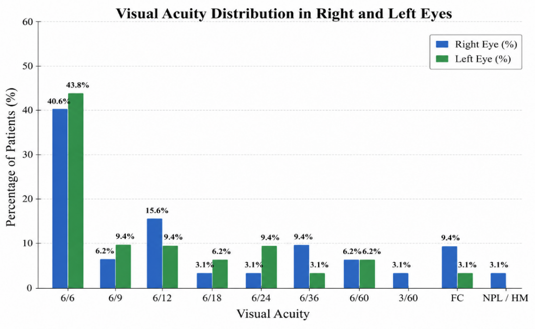
About 43.8% of the patients had normal visual acuity of 6/6, representing the largest category. Among the remaining participants, 9.4% had visual acuity of 6/9, while another 9.4% had 6/12 vision. Furthermore, 6.2% of the patients had visual acuity of 6/18, and 9.4% had 6/24.
About 3.1% had 6/36 vision, while 6.2% had 6/60 visual acuity. The reported distribution also included 3.1% of patients with finger counting (FC) vision and 3.1% with hand movement (HM) vision. The relatively high proportion of patients with 6/6 visual acuity in both eyes suggests that a substantial number of participants retained good central visual function. However, the presence of patients with markedly reduced visual acuity, including finger counting and hand movement vision, indicates that significant visual impairment was also present in some cases. The differences in visual acuity between the right and left eyes further emphasize the importance of evaluating each eye independently during clinical assessment.
About 4.2% of the patients had IOP values ranging from 1–5 mmHg. The largest proportion, accounting for 45.9%, had IOP measurements between 11–15 mmHg. Meanwhile, 41.6% of the patients had IOP values between 6–10 mmHg, and 8.3% had IOP measurements ranging from 16–20 mmHg.
The mean intraocular pressure in the right eye was 11.75 ± 3.082 mmHg, with a minimum value of 3 mmHg and a maximum value of 18 mmHg. The findings indicated that the majority of patients had IOP measurements within the 6–15 mmHg range. In particular, the concentration of patients within the 11–15 mmHg category suggested that this was the predominant IOP range observed in the right eye.
The relatively low mean IOP observed in the right eye may have clinical significance, particularly in relation to the underlying ocular condition and its associated pathophysiological changes. However, IOP measurements should always be interpreted in conjunction with other clinical findings, including anterior chamber depth, lens status, optic nerve appearance, corneal condition, and other relevant ophthalmological parameters. A single IOP measurement may not adequately represent the patient's overall pressure profile.
The distribution of intraocular pressure in the left eye demonstrated some differences compared with the right eye. Approximately 8.3% of the patients had IOP measurements ranging from 1–5 mmHg, while 33.4% had values between 6–10 mmHg. The largest proportion, accounting for 37.5%, had IOP measurements between 11–15 mmHg. Additionally, 20.8% of the patients had IOP values ranging from 16–20 mmHg.
The mean IOP in the left eye was 13.04 ± 6.531 mmHg, with a reported range of 3–36 mmHg. The mean pressure in the left eye was therefore higher than that observed in the right eye. Furthermore, the relatively greater standard deviation indicated considerable variability in IOP measurements among the patients. Although most patients had IOP values between 6 and 15 mmHg, a notable proportion demonstrated measurements within the 16–20 mmHg category.
The comparison between the two eyes revealed that the mean IOP was relatively higher in the left eye (13.04 ± 6.531 mmHg) than in the right eye (11.75 ± 3.082 mmHg). However, the majority of patients in both eyes demonstrated IOP measurements within the 6–15 mmHg range. These findings suggest some degree of inter-eye variation in intraocular pressure among the study participants.
About surgical management, small-incision cataract surgery (SICS) with posterior chamber intraocular lens implantation (PCIOL) was the most frequently performed surgical procedure, accounting for 65.5% of the patients. This finding demonstrated that SICS combined with PCIOL implantation was the predominant surgical approach adopted in the study population.
The preference for SICS with PCIOL implantation may be related to its established role in cataract management, relatively cost-effective nature, suitability for different cataract presentations, and potential for satisfactory postoperative visual rehabilitation. In resource-limited healthcare settings, SICS is particularly relevant because it can provide favourable visual outcomes while reducing dependence on expensive surgical equipment.
The predominance of SICS with PCIOL implantation in the present study reflects its important role in the surgical management of the included patients. Nevertheless, the remaining 34.5% underwent other surgical procedures, although the specific distribution of these procedures was not provided. Further categorization of the alternative surgical techniques would help provide a more comprehensive understanding of the surgical management patterns.
Discussion:
In the present study, the majority of the participants (64.7%) are males, whereas females accounted for 35.3%. This male predominance is associated with differences in healthcare-seeking behaviour, accessibility to ophthalmic services, socioeconomic conditions, referral patterns, and gender-related disparities in healthcare utilization. In developing countries, financial limitations and unequal access to specialized eye care services may influence the demographic distribution of patients seeking surgical treatment. However, this finding should be interpreted cautiously, as the higher proportion of male participants does not necessarily indicate a greater prevalence of the underlying ocular condition among males.
The age distribution revealed that the mean age of the study participants was 33.35 ± 13.64 years, ranging from 18 to 56 years. This indicates that the study population predominantly comprised young and middle-aged adults. The relatively high standard deviation reflects considerable age variation among the participants. The occurrence of significant ocular morbidity during the productive years of life may have substantial socioeconomic consequences, including reduced working capacity, loss of productivity, increased financial dependency, and deterioration in quality of life. These findings highlight the importance of early diagnosis, timely intervention, and appropriate postoperative follow-up to minimize the long-term functional and economic burden associated with ocular diseases.
Assessment of preoperative visual acuity demonstrated considerable variation in visual function between the two eyes. In the right eye, 40.6% of patients had a visual acuity of 6/6, representing the largest proportion of the study population. Other recorded visual acuity levels included 6/9 in 6.2%, 6/12 in 15.6%, 6/18 in 3.1%, and 6/24 in 3.1% of patients. Furthermore, 9.4% had 6/36 vision, while 6.2% had 6/60 vision. Severe visual impairment was also observed, with 3.1% of patients having 3/60 vision, 9.4% having finger counting (FC) vision, and 3.1% having no perception of light (NPL). These findings indicate that, although a considerable proportion of patients maintained good central visual acuity, a subset presented with substantial visual impairment.
A similar pattern was observed in the left eye, where 43.8% of patients had a visual acuity of 6/6, followed by 6/9 and 6/12 in 9.4% each. Additionally, 6.2% had 6/18 vision, 9.4% had 6/24, and 3.1% had 6/36 visual acuity. More severe visual impairment was identified in some patients, including 6.2% with 6/60 vision, 3.1% with finger counting (FC), and 3.1% with hand movement (HM) vision. The relatively high proportion of patients with 6/6 visual acuity in both eyes suggests that many participants retained satisfactory central visual function. Nevertheless, the presence of markedly reduced visual acuity in some patients demonstrates the heterogeneous nature of the clinical presentation.
The observed differences in visual acuity between the right and left eyes emphasize the importance of evaluating each eye independently during ophthalmological assessment. The presence of severe visual impairment in a proportion of patients also highlights the need for timely clinical evaluation and appropriate management to prevent further deterioration of visual function. Moreover, visual acuity alone cannot provide a comprehensive assessment of ocular health or disease severity and should therefore be interpreted alongside other relevant clinical and ophthalmological findings.
Regarding intraocular pressure (IOP), the mean IOP in the right eye was 11.75 ± 3.082 mmHg, with a range of 3–18 mmHg. The majority of patients (45.9%) had IOP measurements between 11 and 15 mmHg, followed by 41.6% with measurements between 6 and 10 mmHg. A smaller proportion had IOP values between 16 and 20 mmHg (8.3%), while 4.2% had measurements between 1 and 5 mmHg. These findings suggest that most patients had relatively low-to-moderate IOP measurements in the right eye at the time of assessment.
In the left eye, the mean IOP was 13.04 ± 6.531 mmHg, with a wider range of 3–36 mmHg. Approximately 37.5% of patients had IOP values between 11 and 15 mmHg, while 33.4% had measurements between 6 and 10 mmHg. Additionally, 20.8% had IOP values between 16 and 20 mmHg, and 8.3% had measurements between 1 and 5 mmHg. Compared with the right eye, the left eye demonstrated a higher mean IOP and greater variability in pressure measurements, suggesting differences in the distribution of IOP between the two eyes.
The inter-eye comparison revealed a higher mean IOP in the left eye (13.04 ± 6.531 mmHg) than in the right eye (11.75 ± 3.082 mmHg). This variation may be related to differences in individual ocular characteristics, underlying pathology, disease severity, or previous treatment. However, in the absence of appropriate statistical testing, the observed difference cannot be considered statistically significant. Importantly, relatively low mean IOP values do not necessarily indicate the absence of clinically significant ocular disease. IOP measurements may be influenced by the underlying pathology, disease stage, previous medical or surgical treatment, and timing of examination. Therefore, IOP assessment should be integrated with other clinical parameters, including anterior chamber depth, lens status, corneal condition, optic nerve appearance, and relevant diagnostic investigations. Repeated IOP measurements may also be beneficial in obtaining a more reliable assessment of pressure variation.
Regarding surgical management, small-incision cataract surgery (SICS) with posterior chamber intraocular lens implantation (PCIOL) was the most frequently performed procedure, accounting for 65.5% of the patients. This finding indicates that SICS with PCIOL implantation was the predominant surgical approach in the present study. Its frequent utilization may be attributed to its established role in cataract management, cost-effectiveness, relatively less demanding technological requirements, and suitability for managing different cataract presentations.
In resource-constrained healthcare settings, particularly in developing countries such as Bangladesh, SICS represents an important surgical option because it offers the potential for satisfactory visual rehabilitation without requiring some of the expensive equipment associated with alternative cataract surgical techniques. Its relatively affordable nature and applicability in settings with limited infrastructure make it a practical approach for expanding access to cataract surgery. Furthermore, posterior chamber intraocular lens implantation following cataract extraction helps restore the optical function of the eye, improves postoperative visual rehabilitation, and reduces dependence on high-powered aphakic spectacles.
Nevertheless, 34.5% of patients underwent alternative surgical procedures, indicating that surgical management was not uniform across the study population. The selection of different surgical approaches may have been influenced by individual clinical presentations, cataract characteristics, associated ocular conditions, and surgeon preference. A more detailed classification of these alternative procedures would help clarify the factors influencing surgical decision-making and provide a better understanding of the overall management pattern.
Overall, the findings of the present study demonstrate considerable variation in the demographic and clinical characteristics of the participants, particularly in visual acuity and intraocular pressure between the two eyes. The predominance of SICS with PCIOL implantation reflects the important role of this surgical approach in the management of the included patients, particularly in resource-limited settings. These findings emphasize the importance of comprehensive preoperative evaluation, individualized surgical planning, appropriate selection of surgical techniques, and regular postoperative monitoring to optimize visual rehabilitation and achieve favourable clinical outcomes.
Conclusion:
Lens induced glaucoma is still a challenge for complicated of cataract that may prevented by early treatment of cataract. Clinical profile show that SICS is still gold standard that effectively controls IOP and gives better visual outcome.
Acknowledgement:
The authors express their sincere gratitude to the Department of Glaucoma, Dhaka, Bangladesh, for their continuous academic support during the preparation of this manuscript. The authors are grateful to all cases for providing written informed consent for publication of their anonymized clinical information and retinal images for scientific and educational purposes.
Author Contributions:
Dr. Omar Faroque conceived the study, managed the cases, interpreted the clinical findings, and prepared the initial manuscript. Dr. Md. Abid Akbar, and Dr. Abir Bin Sajj, contributed to patient management, interpretation of data, and critical revision of the manuscript. Mohammad Shamsal Islam contributed to the study design, scientific writing, interpretation of the findings, editorial revision, and overall coordination of manuscript development. Dr. Omar Faroque and Mohammad Shamsal Islam contributed to data organization, literature review, methodological support, and manuscript revision. All authors critically reviewed the manuscript, approved the final version, and agree to be accountable for all aspects of the work.
Funding:
The authors received no external funding for this study.
Conflict of Interest:
The authors declare that they have no competing interests.
Data availability Statement:
All data generated or analysed during this study are included in this published article. Additional information may be obtained from the corresponding author upon reasonable request.



Open Access By Aditum Open Access Journals id licensed under Creative Commons Attribution 4.0 International License. Based On a Work at aditum.org


