Ophthalmology and Vision Care
OPEN ACCESS | Volume 6 - Issue 1 - 2026
ISSN No: 2836-2853 | Journal DOI: 10.61148/2836-2853/OVC


P D Gupta
Founder Director Iladevi Cataract and IOL Research Centre, Ahmadabad, India
*Corresponding Author: P D Gupta, Founder Director Iladevi Cataract and IOL Research Centre, Ahmadabad, India.
Received: April 15, 2021
Accepted: April 20, 2021
Published: April 26, 2021
Citation: P D Gupta. (2021) “Eye Infections: A Review”, Ophthalmology and Vision Care, 1(2); DOI: http;//doi.org/04.2021/1.1007.
Copyright: © 2021 P D Gupta. This is an open access article distributed under the Creative Commons Attribution License, which permits unrestricted use, distribution, and reproduction in any medium, provided the original work is properly Cited.
The Eyes are the windows to the world. It works like a camera; light has to pass through the lens to form the image. To perform this primary and most important function, there should be complete transparency, Infections in the front layers brings about opacity in the light path and hence, it is important to keep eyes infection free. In this series of articles, we are describing various pathogens infecting the tissues of the eye, their effects on vision, manifestations, remedies, etc. The present paper deals with protections provided by the Nature to the eye, pathogens (viruses including recently described SARS-CoV2, various types of bacteria) infecting the eyelids and conjunctiva.
Introduction:
The eye, on human body is constantly exposed to the atmospheric component and that is why they are vulnerable and can easily be damaged due to the ill effects of bio- pollutants present in the environment [1,2]. Though the nature has provided double protection to the eye, viz. eyelids and oily layer on the cornea, still sometimes certain microbes can cross these two hurdles and infect eyes. [3]. The superficial tissue infections will usually clear up in 7 to 14 days without treatment and without any long-term consequences. However, in some cases, viral conjunctivitis can take 2 to 3 weeks or more to clear up. A doctor can prescribe antiviral medication to treat more serious forms of conjunctivitis.
The eye is important sense organs which connect the external world to the brain and due to infections, that are not only interfere with the vision but create other problems too. The eye infection can spread to the brain (meningitis) and spinal cord [4], or blood clots can form and spread from the veins around the eye to involve a large vein at the base of the brain (the cavernous sinus) and result in a serious disorder called cavernous sinus thrombosis [5].
The eye as an organ composed of many tissues all put together fits in the eye orbit created by the facial bones. Externally eyes are covered by lower and upper eyelids both upper and the lower eyelids are decorated by eyelashes and on the upper eyelid at the margins of orbit eyebrows are provided [6]. The eyelids, eyelashes and eyebrows are the part of skin and all tissue are vulnerable to infections; they can be infected with viruses, bacteria or fungus, the other infections are rare. In this review we will deal with tissue wise.
Natural protection to the eye:
Since eye is an important sense organ and always exposed to the environment, nature has provided multi-fold protection e.g., eyebrows, eyelashes, eyelids and the protective layer of oil from the external atmosphere. Eyelashes keep foreign pollutants particles away from the eye by acting as a physical barrier and by causing the person to blink reflexively at the slightest sensation. As far as eye lids are concerned the upper and lower eyelids are thin flaps of skin and muscle that can cover the eye, and they reflexively blink to form a mechanical barrier that protects the eye from foreign objects, dust wind, and very bright light [7,8].The conjunctiva provides cellular defense and protects the sensitive tissues underneath it from pollution, where on the moist back surface of the eyelid, the conjunctiva coils around to cover the visible surface of the eyeball and up to the edge of the cornea [9].The eyelids help to spread tears uniformly over the surface of the eye when it got blinked. Small glands at the edge of the upper and lower eyelids secrete an oily substance that contributes to the tear film and keeps tears from evaporating. Tears contain three components, e.g., water, mucous, and oil. The tear glands are responsible for the production of water layer. Mucous glands in the conjunctiva produce mucus, and oil [lipid] glands in the eyelid margin produce oil. The mucus and oil mix with the watery portion of the tears to create a more protective tear film [10]. Tears constantly cleanse the surface of the eye and keep the cornea transparent. The cornea gets oxygen and nutrients through tears by keeping the surface moist. In the absence of moisture, the cornea becomes dry and opaque; in turn can easily injured and infected. Tears also trap and sweep away small pollutant particles that enter into the eye. Moreover, tears are rich in antibodies that help prevent infection. The eyelids and tears shield the eye while undesirable light rays entering the eye [11].
Though human eyes are well protected, still some times pathogenic organism by pass the all protective hurdles and infect the eye tissues. Below we describe the pathogeneses of the eye tissues.
1. Eyelids:
The oil glands on the eyelids can also become infected and blocked. A stye (hordeolum) is an infection of a gland in the eyelid. The most common type of stye infects the tear glands that are at the base of the eyelashes. Styes also sometimes occur inside the eyelid due to infected oil glands. Styes usually begin as red, itchy, painful, swollen lumps. In most cases, the infection only affects a single tear or oil gland and requires no treatment [12]. Warm compresses can help with the pain.
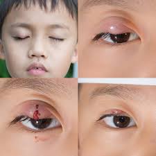
Various types of Styes on the upper eyelid Orbital cellulitis: Orbital cellulitis is an infection deep in the tissue of the eyelid infection affecting the tissue within the orbit and around and behind the eye.. It can spread quickly and is often extremely painful. Even a tiny cut can introduce enough bacteria to trigger orbital cellulitis. Usually, computed tomography or magnetic resonance imaging of the orbits is done [13]. Both orbital cellulitis and preseptal cellulitis are more common among children. Preseptal cellulitis is far more common than orbital cellulitis. However, orbital cellulitis is more dangerous. Orbital cellulitis usually is caused by spread of an infection to the orbit from the sinuses around the nose (nasal sinuses) but can also be spread from infection of the teeth or bloodstream.
Ocular herpes: Ocular herpes is a herpes infection in and around the eyes. Though anyone can develop ocular herpes, it is most common in children. Ocular herpes can look a lot like pink eye but does not always produce distinct lesions [14]. To diagnose herpes, a doctor will need to take an eye culture to check for the presence of the virus. Though the virus remains in the body and there is no cure, antiviral medications can manage the symptoms.
2. Conjunctiva:
The conjunctiva is a loose connective tissue that covers the surface of the eyeball (bulbar conjunctiva) and reflects back upon itself to form the inner layer of the eyelid (palpebral conjunctiva). The conjunctiva of the eye provides protection and lubrication of the eye by the production of mucus and tears. It prevents microbial entrance into the eye and plays a role in immune surveillance. It lines the inside of the eyelids and provides a covering to the sclera.
3. Conjunctivitis:
By far, the number one type of eye infection is conjunctivitis, more commonly known as pink eye. Pink eye infections enter through a part of the eye called the conjunctiva, the thin mucous membrane that that lines the inside of the eyelids and covers the sclera. The conjunctiva of the eye provides protection and lubrication of the eye by the production of mucus and tears. It prevents microbial entrance into the eye and plays a role in immune surveillance. Antibiotic treatment is usually effective in treating this disease. However conjunctivitis spreads easily, so it is important to take some precautions if you have this eye infection, such as washing your hands often, not touching your eyes, and ensuring that you don't share a washcloth or a towel with another person [15].
It can be caused by a bacteria or virus, although sometimes you might get it from an allergic reaction or irritants. It's common to get pinkeye when you have a cold. It's an infection of your conjunctiva and usually gives your eyes a pink tint. It's characterized by burning, itchy eyes, as well as a thick yellow discharge and redness of the inner eyelid.
Contagious: Viral Conjunctivitis Infection of the eye caused by a a number of different viruses, Sometimes can result in large outbreaks depending on the virus.
Not contagious: Allergic Conjunctivitis The result of the body’s reaction to allergens, such as pollen from trees, plants, grasses, and weeds; dust mites; molds; dander from pets; medicines; or cosmetics Occurs more frequently among people with other allergic conditions, such as hay fever, asthma, and eczema. It can occur seasonally, when allergens such as pollen counts, are high or can also occur year-round due to indoor allergens, such as dust mites and animal dander. Conjunctivitis caused by irritation from a foreign body in the eye or contact with smoke, dust, fumes, or chemicals can occur when contact lenses are worn longer than recommended or not cleaned properly.
3.1. Viral conjunctivitis:
Viral conjunctivitis is highly contagious and the most common cause of infectious conjunctiva. It is caused by a virus such as the common cold or herpes simplex virus. This infection is more common in adults than in children. Around 65–90% of cases are caused by adenovirus Eye discharge associated with viral pink eye typically is clear and watery but may include a white or light-yellow mucus component.
Patients can generally be advised that viral conjunctivitis is self-limiting and, as there are no specific treatments, for comfort they can use cold compresses, artificial tears or topical antihistamines. [16,17] Antibiotics are not needed, are costly and may increase antibiotic resistance. If there is evidence of herpes simplex or zoster virus then antivirals should be prescribed. When viral conjunctivitis is severe or the patient experiences symptoms after its resolution, the patient should be referred to an ophthalmologist. This is to consider topical steroids and to exclude an immune ‘post-viral’ keratitis.
Severe acute respiratory syndrome coronavirus (SARS-CoV2) can be transmitted through the conjunctiva. However, it is currently unclear whether conjunctival epithelial cells express. Nevertheless, for the first time, Clemens Lange and Julian Wolf [18] showed the presence of receptor ACE2 in the cornea of human eye. Recently it is found that daily wearers of eyeglasses may be less susceptible to COVID-19 [19,20]. Once the eyes are infected, they become storehouse for transmission of virus to the other tissues and/or organs or body fluids, through tears and/or by touching eyes with hands and fingers. On analysing tears of some Covid-10 patient occasionally, coronavirus was detected in the samples. Given the presence of viral particles, it is, therefore, possible to transmit COVID-19. Nevertheless, since sample size is small either way conclusions cannot be drawn. Therefore, additional studies are needed for conclusive results.
3.2. Bacterial conjunctivitis:
Bacterial Conjunctivitis Infection of the eye caused by certain bacteria, such as Staphylococcus aureus, Streptococcus pneumoniae, Haemophilus influenzae, Moraxella catarrhalis, or, less commonly, Chlamydia trachomatis and Neisseria gonorrhoeae [22]
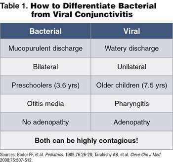
Bacterial conjunctivitis is more common in children, therefore can be spread easily, Children with conjunctivitis without fever or behavioural changes can usually continue going to school especially with certain bacteria and in certain, climatic conditions December through Apr il. It is more common in kids than adults,
Gonococcal conjunctivitis Conjunctivitis caused by Neisseria gonorrhoeae is uncommon but should be considered in neonates and sexually active young adults. If suspected, the practitioner should take conjunctival swabs for Gram staining and special culture for Neisseria species.
Chlamydial conjunctivitis Most cases of chlamydial conjunctivitis are unilateral and have concurrent genital infection. Symptoms usually include conjunctival hyperemia, mucopurulent discharge and lymphoid follicle formation.
3.3. Allergic conjunctivitis:
Allergic conjunctivitis occurs when the outer part of the eye becomes swollen or irritated in a reaction to pollen, dander, mould, or other substances that trigger allergies such as, chemical scents such as household detergents or perfumes [24]. Some people may also experience allergic conjunctivitis in reaction to certain medications or substances dropped into the eyes, such as contact lens solution or medicated eye drops.
Allergy-causing substances release a chemical called histamine into the eyes, which causes the blood vessels widened in the outer layer, so it becomes swollen. The eyes may quickly become red, itchy, and watery. Other symptoms of allergic conjunctivitis include:
Allergies affect people of all ages, though they are more common in children and young adults [25]. If one has allergies and lives in locations with high pollen counts, you are more susceptible to allergic conjunctivitis.


Open Access By Aditum Open Access Journals id licensed under Creative Commons Attribution 4.0 International License. Based On a Work at aditum.org