
International Surgery Case Reports
OPEN ACCESS | Volume 8 - Issue 2 - 2026
ISSN No: 2836-2845 | Journal DOI: 10.61148/2836-2845/ISCR


Dr. Sofija Ivan Popevska
Cardiac surgery, University Clinical Hospital Rebro, Kispaticeva 12, 10000 Zagreb, Croatia.
Corresponding author: Sofija Popevska MD, Clinic for Cardiac surgery, University Hospital Rebro Kispaticeva 12, 10000 Zagreb Croatia, popevskasofija@hotmail.com.
Received: November 05, 2026 | Accepted: November 16, 2026 | Published: November 19, 2026
Citation: Sofija I Popevska. (2026) “The Treatment Of Multiple Heart Valve Disease And The Test Of Time: Replacing A Tricuspid Biological Valve In Presence Of Normally Functioning Mechanical Mitral Prosthesis”, International Surgery Case Reports, 8(3); DOI: 10.61148/2836-2845/ISCR/121.
Copyright: © 2026. Sofija Ivan Popevska. This is an open access article distributed under the Creative Commons Attribution License, which permits unrestricted use, distribution, and reproduction in any medium, provided the original work is properly cited.
The patients who require tricuspid valve reoperation are usually high-risk surgical candidates with high early and late mortality. According to the latest recommendations, there is a lack of data on mixed and multiple valve diseases which does not allow for evidence-based recommendations for surgical management of mixed or multiple valve disease (1,7). However this requires early treatment of heart valve disease in presence of symptoms, especially in aortic regurgitation
A 65-year-old woman, with multiple valve disease from rheumatic origin, had normally functioning Starr-Edwards (S-E) caged-disk mechanical mitral valve model 6520 implanted in the year 1975. Also a tricuspid valve was been replaced with a biological prosthesis in the year 1992. Through median sternotomy, under total-bypass using peripheral femoral and superior cava vein cannulation, tricuspid bioprosthesis was replaced with a biological Hancock II Medtronic mitral valve prosthesis 33 mm. She had right-heart-failure symptoms resistant to medical therapy, normally functioning mechanical mitral, a severe tricuspid bioprosthetic deterioration with estimated area of 0.6-0.8 cm2, calcificated leaflets and suspected thrombus formations. Aortic valve was sclerotic with regurgitation grade +2. Giant right atrium was noted of 101cm² size. Following TVR with biological prosthesis size 33mm the patient was extubated on the day 1 after the operation. On the 6th postoperative day, she was referred to the ward. On the 11th day after the operation haemodialysis was indicated because of oligoanuria. She was transferred on the day 15 after the operation, to the cardiology ward for further medical recovery. Trans-thoracic echocardiography before discharged showed normal left ventricle with EF 55%, moderate to severe aortic valve regurgitation, with dilatated right ventricle, with moderate reduced systolic function. A normally functioning tricuspid bioprosthesis with mean diastolic gradient of 3 mmHg and no signs of regurgitation was noted. Unfortunatly, she died on the 90th day after the operation because of multi-organ-system failure. Last control echocardiography showed volume and pressure overloaded, dilatated, dysfunctional right ventricle, with akinetic free wall and dyskinetic interventriclar septum. Right atrial dilatation with dilatated inferior caval vein and coronary sinus size 45 mm was present. Systolic pressures in right ventricle and pulmonary artery could not be measured but PvAcct was 60 ms in context of severe pulmonary hypertension. Conclusion: The right side of the heart provides its own special set of anatomical and functional considerations, often necessitating the use of a bioprosthetic valve or valved conduit, all of which have a variable but limited longevity. The main cause of mortality in tricuspid valve surgery is reoperation. The choice of the prosthesis remains a subject of debate. Earlier AVR for th. of AR 1+ may be important for preventing pulmonary hypertension and improving survival after operation.
Heart Valve Disease
Background
Tricuspid valve reoperation is not a common operation and, in most series, is associated with high intra-operative and post-operative mortality, despite advances in perioperative care. The patients who require tricuspid valve reoperation are usually high risk surgical candidates with high early and late mortality. According to the latest recommendations, there is a lack of data on mixed and multiple valve diseases which does not allow for evidence-based recommendations for surgical management of mixed or multiple valve disease [1,7]. Tricuspid valve surgery is very high-risk since can result with in-hospital mortality in second stage operation, where biological prosthesis is degenerative and requires reoperation. Tricuspid valve replacement threatment with valve-invalve surgery has been currently done in high volume centers and still it may result with valve thrombosis and dysfunction.
On-x heart valves prosthesis may be used in this case and can reduce RV load especially in dilated right ventricles and RV failure in presence of mechanical functional prosthesis since requires lower INR ratio of 2 or less in absence of Atrial fib. in setting of AR for AVR.
Materials and Methods: 65-year-old woman, with multiple valve disease from rheumatic origin, had normally functioning Starr-Edwards (S-E) caged-disk mechanical mitral valve model 6520 implanted in the year 1975. The tricuspid valve has been replaced with a biological prosthesis in the year 1992. In the last 3 years, she presented several times with right-heart-failure symptoms. Prior to the operation, she had right-heart-failure symptoms resistant to medical therapy, normally functioning mechanical mitral valve with mean peak gradient of 4 mmHg and area 1.5-1.6 cm2, a severe tricuspid bioprosthetic deterioration with estimated area of 0.6-0.8 cm2, calcificated leaflets and suspected thrombus formations. Aortic valve was sclerotic with regurgitation grade +2 (PISA 0.6 cm). Giant right atrium was noted of 101cm2 size. Estimated EuroSCORE2 was 15%. Through median sternotomy, under total-bypass using peripheral femoral and superior caval vein cannulation, tricuspid bioprosthesis was replaced with a biological Hancock II Medtronic mitral valve prosthesis 33 mm. Exploration of the mechanical mitral valve showed a normal functioning prosthesis, with small surface area.
Results: Patient was extubated on the day 1 after the operation. On the 6th postoperative day, she was referred to the ward. On the 11th day after the operation haemodialisis was indicated because of oligoanuria. She was transferred on the day 15 after the operation, to the cardiology ward for further medical recovery. Trans-thoracic echocardiography before discharged showed normal left ventricle with EF 55%, moderate to severe aortic valve regurgitation, with dilatated right ventricle, with moderate reduced systolic function. A normally functioning tricuspid valve bioprosthesis with mean diastolic gradient of 3 mmHg and no signs of regurgitation was noted. Unfortunately, she died on the 90th day after the operation because of multi-organ-system failure. Last control echocardiography showed volume and pressure overloaded, dilatated, dysfunctional right ventricle, with akinetic free wall and dyskinetic interventricular septum. Right atrial dilatation with dilatated inferior cava vein and coronary sinus size 45 mm was present. Systolic pressures in right ventricle and pulmonary artery could not be measured but PvAcct was 60 msec in context of severe pulmonary hypertension.
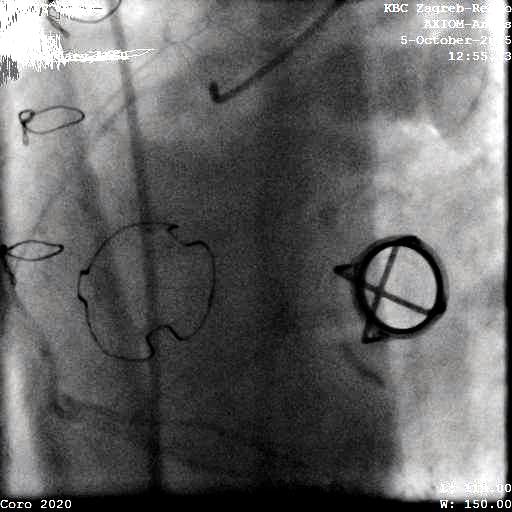
Figure: Pre-operative cardiac catheterization showing tricuspid biological prosthetic valve and caged-disc mechanical mitral prosthesis
Conclusion: The right side of the heart provides its own special set of anatomical and functional considerations, often necessitating the use of a bioprosthetic valve or valved conduit, all of which have a variable but limited longevity [2]. The main cause of mortality in tricuspid valve surgery is reoperation. The choice of the prosthesis remains a subject of debate [3]. In the long run survival with mechanical prostheses could be superior, given the high rate of bioprosthetic degeneration after 7 to 10 years, and is accelerated in younger patients. However untreated AR grade 1+ can be factor for early in-hospital mortality, due to RV failure. This can be overlooked by AVR with mechanical prosthesis that are simply designed to last forever. Do we tested the time with the SE valve showing no need for reoperation especially in small ventricles and rheumatic mitral valve disease. In rheumatic heart valve disease it should be recognized the importance of aortic regurgitation especially in presence of symptoms. Early treatment with mechanical prosthesis may be indicated in presence of previous mechanical heart valves in place and no need of worry after operating the TV for stenosis. The most relevant finding in this case presented is that AR1+ needs to be carefully treated surgically if present, especially in HF patients. Increased Lv load always results with RV failure due to type 2 pulmonary hypertension.
Acknowledgements: This case-report was presented on the 4th EuroValve Congress, in Brussels Belgium, on March 10-11th 2016.
Financial Disclosure: None Declared.
Funding/Support: None Declared.



Open Access By Aditum Open Access Journals id licensed under Creative Commons Attribution 4.0 International License. Based On a Work at aditum.org


