International Surgery Case Reports
OPEN ACCESS | Volume 8 - Issue 1 - 2026
ISSN No: 2836-2845 | Journal DOI: 10.61148/2836-2845/ISCR


Doui Doumgba Antoine1*, Ngboko Mirotiga Pétula Annicette1, Tapande Yakossa Elemance Eva2
1Service de Chirurgie générale. CHU de l’Amitié Sino Centrafricaine de Bangui.
2Servie d’Anatomie Pathologique, Laboratoire National de Biologie Clinique et de Santé Publique (LNBCSP). Bangui.
*Corresponding author: Professor Antoine DOUI DOUMGBA, Ngola centre KM 11 Avenue du 15 mars, BP: 2184 Bangui.
Received: November 25, 2025 | Accepted: December 22, 2025 | Published: January 03, 2026
Citation: Doui D Antoine, Pétula Annicette MN, Elemance Eva TY. (2026) “Adult Colonic Intussusception. Study about Two Cases Prolapsed by the Anus Operated at the Chu of the Sino-Central African Friendship of Bangui.”, International Surgery Case Reports, 8(1); DOI: 10.61148/2836-2845/ISCR/112.
Copyright: © 2026. Doui Doumgba Antoine. This is an open access article distributed under the Creative Commons Attribution License, which permits unrestricted use, distribution, and reproduction in any medium, provided the original work is properly cited.
Introduction : prolapse of the intussusception tube through the anus is a rare complication of intussusception, even in infants. The objective of this study was to report the diagnostic and therapeutic difficulties of 2 cases of colonic intussusception prolapsed through the anus in adults.
Observations : in a series of 674 acute intestinal obstruction operated on over three years, we recorded 7 cases of intussusception in adults (1.03%) among which 2 cases of colonic intussusception were prolapsed through the anus either 28.57%. Both patients presented with diffuse abdominal pain, a prolapsed intestinal mass in the anus suggesting a rectal prolapse. Apart from the preoperative paraclinical assessment, no morphological exploration had been carried out. Both patients had undergone a laparotomy during which the diagnosis of colocolic intussusception was made intraoperatively. The attempt to manually reduce the invagination tube was unsuccessful. All invaginated colonic portions were necrotic. The procedures performed included colonic resection followed by colostomy. The restoration of digestive continuity was made one to two months later. The postoperative course was simple and the histopathological study of the surgical specimens revealed chronic inflammation of the colonic mucosa.
Conclusion : intussusception is a rare condition in adults. Practitioners should consider the protracted form of intussusception when suspecting rectal prolapse in an adult patient.
Introduction : le prolapsus du boudin d’invagination par l’anus est une complication rare de l’invagination intestinale, même chez le nourrisson. L’objectif de cette étude était de rapporter les difficultés diagnostiques et thérapeutiques de deux cas d’invagination colo colique prolabée par l’anus chez l’adulte.
Observations : sur une série de 674 cas d’occlussions intestinales aiguës opérées en trois ans, nous avions enregistré 7 cas d’invagination chez l’adulte (1,03%) parmi lesquels 2 cas d’invagination colocolique étaient prolabés par l’anus soit 28,57%. Les 2 patients avaient présenté des douleurs abdominales diffuses, une masse intestinale prolabée à l’anus faisant évoquer un prolapsus rectal. En dehors du bilan paraclinique préopératoire, aucune exploration morphologique n’avait été réalisée.
Les 2 patients avaient bénéficié d’une laparotomie au cours de laquelle le diagnostic d’invagination colo-colique était fait en per opératoire. La tentative de réduction manuelle du boudin d’invagination était infructueuse.
Toutes les portions coliques invaginées étaient nécrosées. Les gestes réalisées avaient comporté une résection colique suivie d’une colostomie. Le rétablissement de la continuité digestive avait été fait un à deux mois plus
tard. Les suites opératoires étaient simples et l’étude histopathologique des pièces opératoires avait révélé une inflammation chronique de la muqueuse colique.
Conclusion : l’invagination intestinale est une pathologie rare chez l’adulte. Les praticiens doivent penser à sa forme prolabée lorsqu’ils soupçonnent un prolapsus rectal chez un patient adulte.
Mots clés : invagination, colon, prolapsus, adulte.
intussusception, colon, prolapse, adult
INTRODUCTION
Acute intussusception is a rare condition in adults, accounting for 1 to 2% of intestinal obstructions [1]. Prolapse of the intussusception loop through the anus is a rare complication, even in infants [2]. However, in coloanal varieties, the intussusception may protrude through the anus, where a rectal examination can differentiate it from rectal prolapse. Colorectal intussusception protruding through the anus in adults is unusual. A few cases have been reported in the global literature, most of which were secondary to a tumor, particularly a lipoma, carcinoma, or villous adenoma [3]. It often poses a differential diagnosis problem with a large polyp or rectal prolapse [4]. Treatment is always surgical in adults and often does not allow for reduction by hyperpressure under radiological control [5]. This study reports the observation of two cases of colo-colic intussusception prolapsed through the anus in adults, with their diagnostic and therapeutic difficulties.
OBSERVATIONS
Observation 1: Ms. X... Lucie, aged 44, was transferred from the Gynecology-Obstetrics department on 11/11/2019 for diffuse abdominal pain that had been developing for 4 days, accompanied by an abdominal mass and swelling around the anal margin. She had no specific medical history. She was in her fourth trimester, fourth pregnancy. On examination, her general condition was good, her blood pressure was 120/80 mmHg, and her pulse was 88 beats per minute. The patient did not have a fever. The abdomen was asymmetrical, flat, and on palpation, diffuse pain was noted without abdominal guarding. A firm mass was palpated in the left iliac fossa. At the anal margin, there was a dark red tumor prolapsed into the anus, approximately 15 cm in size, which was oozing bloody mucus. Attempts to reduce the mass were unsuccessful.
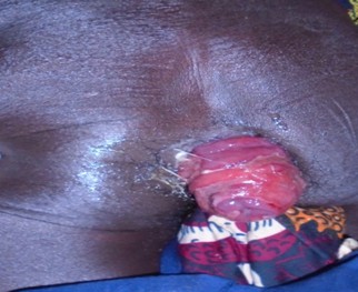
Figure 1 : view of a total intussusception prolapsed through the anus
Two days later, there was a cessation of bowel movements and gas. The paraclinical assessment included an abdominal X-ray without preparation, which showed hydro-aerial levels higher than wide with peripheral topography, a hemoglobin level of 11g/dl, hyperleukocytosis at 13,000 white blood cells/mm³, and creatinine at 10mg/dl. An exploratory laparotomy was indicated and performed on November 15, 2019. The procedure was performed under general anesthesia and tracheal intubation. The incision was midline above and below the umbilicus. Surgical exploration revealed total ileocecal intussusception. Desinvagination maneuvers were difficult and impossible.
The entire transverse colon and left colon were necrotic, prompting a subtotal colectomy. The rectal stump was closed and a right ileostomy was performed.
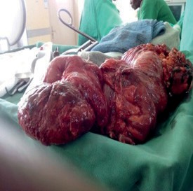
Figure 2 : Surgical specimen from subtotal colectomy for ileocecal intussusception prolapsed into the anus.
The surgical specimen examined in the pathology laboratory revealed chronic inflammation of the mucosa, with no other abnormalities. The postoperative course was uneventful and the patient was followed up on an outpatient basis from the sixth postoperative day. Three months later, she underwent a second operation to restore digestive continuity, and an ileo-rectal anastomosis was performed. One month after the operation, complications arose in the form of an eventration. Six (6) months later, we repaired the eventration by inserting a Trulene prosthesis. The postoperative course was uneventful after a 12-month follow-up.
Observation 2: Mr. Y..., 49 years old, farmer, with no particular medical history, was admitted to our department on 12/07/2022 for diffuse, intermittent abdominal pain, rectal bleeding, and the presence of a pink mass prolapsing from the anus. The interview did not reveal any constipation or gas retention. Clinical examination revealed that he was in good general health, with a temperature of 37°C, blood pressure of 100/60 mmHg, and a pulse rate of 76 beats per minute. He was in good nutritional condition (BMI of 18). The abdomen was flat, supple, depressible, with no palpable mass but with hypogastric tenderness. However, examination of the anal margin revealed a purplish mass approximately 20 cm in size, prolapsed from the anus, oozing bloody mucus, and irreducible. A diagnosis of rectal prolapse was made, and abdominal rectopexy was indicated. The results of the preoperative assessment were within normal limits: creatinine level at 10.7 mg/L, blood urea at 0.11 g/L, blood glucose at 0.70 g/L, hemoglobin level at 10.7 g/dL. The patient was blood type O Rh positive. On 12/10/2022, the surgical procedure was performed under general anesthesia with tracheal intubation. After a midline laparotomy above and below the umbilicus, the exploration revealed a colo-colic intussusception involving the transverse and sigmoid colon segments.
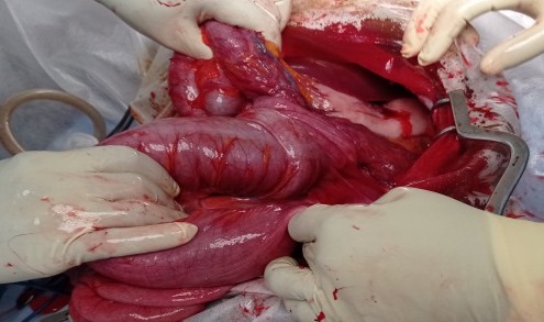
Figure 3 : Surgical view of the colonic intussusception
Total disintussusception was performed. However, the sigmoid colon was completely ischemic, leading us to perform a resection. The procedure ended with a Hartmann colostomy, and the surgical specimen was sent to the pathology laboratory for histological examination. The postoperative course was uneventful. The patient was discharged from the hospital on the seventh postoperative day. Six months after surgery, he underwent a second operation to restore digestive continuity via a colo-colonic anastomosis. The postoperative course was uneventful.
DISCUSSION
Intussusception in adults remains a rare condition. It accounts for only 1% of causes of mechanical obstructions and 1% of hospital admissions [4]. One of the rare complications of intussusception is prolapse of the loop through the anus. It accounts for one-third of intussusception cases in adults in our practice. Cases of total intussusception with prolapse of the loop through the anus have been reported in infants, but remain extremely rare in adults [6,7]. These cases are most often secondary to an underlying condition such as a lipoma, carcinoma, or villous adenoma [3]. The symptoms of intussusception in adults can be overlooked. They are often misleading, and the diagnosis suggests a large anal polyp or rectal prolapse [8,9]. In our study, both patients presented with diffuse abdominal pain and a prolapsed intestinal mass at the anus, suggesting rectal prolapse. The first patient had an occlusive syndrome and a palpable abdominal mass corresponding to the intussusception loop. This is why it is essential to perform a complete and careful physical examination of the abdomen, combined with a rectal examination in these patients and to emphasize the importance of examining the perineum, as highlighted by Nouri et al [6], so as not to confuse prolapsed tissue with rectal prolapse. The presence of a space between the anal canal and the rectum on the one hand and the prolapsed intestine on the other, large enough to accommodate a finger, confirms the diagnosis of intussusception with prolapsed intestine at the anus. In contrast, in cases of anorectal prolapse, the finger stops 1 or 2 cm from the anal margin. Abdominal ultrasound and computed tomography (CT) scans can be used to make the diagnosis and identify the site of the intussusception. Neither patient had undergone an abdominal CT scan because the abdominal mass was visible at the anus and mistaken for rectal prolapse, which did not require morphological examination. Teyha et al [10] did not perform an abdominal ultrasound or abdominal CT scan in their reported case because the intestinal mass was clearly visible at the anus. Teyha et al [10] did not perform an abdominal ultrasound or abdominal CT scan in their reported case because the intestinal mass had clearly protruded beyond the anus and did not warrant complex investigations. In both patients, the diagnosis of prolapsed colo-colic intussusception at the anus was made intraoperatively. Dennis et al [11] believe that it is likely that few general surgeons will see more than one or two such patients during their career, because most cases will not be diagnosed before surgery. The advent of laparoscopy now provides a reliable means of diagnosis and sometimes treatment of small bowel intussusception [12]. Treatment of intussusception in adults is essentially surgical, because a organic cause is found in 70% to 90% of cases [1]. Our patients underwent conventional surgery with colonic resection. The delayed restoration of digestive continuity was due to ischemia. In fact, in colo-colonic or ileo-colonic intussusceptions, due to the frequency of cancer as the causal lesion, primary resection (right or left colectomy) is recommended by most authors in order to limit the risk of metastatic spread [13]. The pathological examination revealed chronic inflammation of the colonic mucosa, which was also reported by Teyha et al. [10]. Generally, postoperative outcomes are straightforward, as in our two patients, but due to laparotomy, the most common postoperative complication is postoperative eventration.
CONCLUSION
Intussusception is a rare condition in adults. The form with the intussusception loop prolapsing into the anus remains extremely rare. The diagnosis is often confused with that of rectal prolapse. Surgeons should always keep it in mind as a differential diagnosis when rectal prolapse is suspected in an adult patient, hence the importance of a complete and thorough physical examination.
Conflict of interest: the authors declare no conflict of interest.


Open Access By Aditum Open Access Journals id licensed under Creative Commons Attribution 4.0 International License. Based On a Work at aditum.org