
Immunity Inflammation and Infection Diseases
OPEN ACCESS | Volume 2 - Issue 1 - 2025
ISSN No: - | Journal DOI: 10.61148/IIID


Hamidreza Mehdizadeh¹, Abbas Samim², Mehdi Raei³, Ali Ghazvini², Emad Dadgar¹, Mohammad Javad Behzadnia⁴*
¹Student Research Committee, Baqiyatallah University of Medical Sciences, Tehran,.
²Chemical Injuries Research Center, Systems Biology and Poisoning Institute, Baqiyatallah University of Medical Sciences, Tehran.
³Artificial Intelligence in Health Research Center, Biomedicine Technologies Institute, Baqiyatallah University of Medical Sciences, Tehran.
⁴Trauma Research Center, Baqiyatallah University of Medical Sciences, Tehran.
*Corresponding author: Mohammad Javad Behzadnia, Trauma Research Center, Baqiyatallah University of Medical Sciences, Tehran.
Received: November 09, 2026 | Accepted: November 16, 2026 | Published: November 20, 2026
Citation: Mehdizadeh H, Samim A, Raei M, Ghazvini A, Dadgar E, Mohammad J Behzadnia. (2026) “Pleural Effusion and Clinical Outcomes in COVID-19 Patients Presenting to the Emergency Department: Lessons Learned for Future Pandemics.” Immunity, Inflammation and infection Disease, 2(1); DOI: 10.61148/IIID/004
Copyright: © 2026 Mohammad Javad Behzadnia. This is an open access article distributed under the Creative Commons Attribution License, which permits unrestricted use, distribution, and reproduction in any medium, provided the original work is properly cited.
Background
Pleural effusion is an uncommon radiologic finding in patients with COVID-19, and its association with clinical outcomes is not yet fully understood. This study evaluated the relationship between pleural effusion and mortality in patients with COVID-19.
Methods
We conducted a retrospective cohort study of patients with COVID-19 admitted to Baqiyatallah Hospital in Tehran between 2021 and 2022. Patients with and without pleural effusion were compared regarding demographic characteristics, comorbidities, laboratory findings, echocardiographic parameters, treatment, length of hospital stay, and mortality at one, three, and six months. Associations were evaluated using appropriate comparative tests and multivariable logistic regression.
Results
A total of 122 patients were included, comprising 61 with pleural effusion and 61 without. Three-month mortality was higher among patients with pleural effusion compared to those without (86.9 vs. 13.1%, P = 0.027). Similarly, six-month mortality was elevated in the pleural effusion group (82.0 vs. 18.0%, P = 0.003). Patients with pleural effusion exhibited higher D-dimer levels at admission, as well as increased ferritin, D-dimer, C-reactive protein, and erythrocyte sedimentation rate levels at discharge. Multivariable analysis identified age and ferritin level as independent predictors of six-month mortality.
Conclusions
Among hospitalized patients with COVID-19, pleural effusion was associated with higher mortality and elevated markers of systemic inflammation. Due to the retrospective study design and potential residual confounding, these findings should be interpreted as associations rather than evidence of causation. Larger prospective studies are needed to determine whether pleural effusion provides independent prognostic information.
COVID-19, pleural effusion, mortality, prognosis, retrospective cohort study.
Since its emergence in late 2019, COVID-19 has caused substantial global morbidity and mortality [1] .The World Health Organization declared the outbreak a Public Health Emergency of International Concern on January 30, 2020 [2]. SARS-CoV-2 can cause a broad spectrum of respiratory disease, ranging from mild upper-respiratory-tract symptoms to severe pneumonia and acute respiratory distress syndrome [1, 3]. This infection manifests with a broad spectrum of systemic symptoms, including fever, cough, hemoptysis, gastrointestinal disturbances, myalgia, weakness, body aches, chest pain, and shortness of breath. COVID-19 patients with pleural effusion often present with symptoms such as high fever, shortness of breath, and severe coughing [4, 5]. Diagnosis of COVID-19 is based on medical history, clinical symptoms, and imaging findings, with chest CT scans playing a crucial role in early detection [6-8]. The most common diagnostic findings on chest CT scans include bilateral and peripheral ground-glass opacities [9]. Indeed, most patients with COVID-19 pneumonia exhibit typical imaging features such as ground-glass opacities, vascular enlargement, pneumatoceles, consolidation, or bronchiectasis [10]. Pulmonary involvement is partly related to the expression and distribution of angiotensin-converting enzyme 2, the cellular receptor used by SARS-CoV-2 for entry [11]. Identifying factors that exacerbate the clinical course of patients is crucial for their effective treatment. Pleural effusion, lymphadenopathy, and calcification are observed less frequently in COVID-19 cases [12, 13]. Pleural effusion can arise from various causes, including viral pleuritis, chronic heart failure, cancer, and rheumatic diseases, as well as other underlying conditions such as congestive heart disease, cirrhosis, kidney failure, and viral or bacterial infections [13-15]. The initial step in the differential diagnosis or in determining the pathogenesis of pleural fluid is to classify it as exudative or transudative. Transudates result from increased hydrostatic pressure (e.g., heart failure), decreased oncotic pressure (e.g., hypoproteinemia), increased negative pressure in the pleural space (e.g., atelectasis), or the movement of ascitic fluid through the diaphragm (e.g., hepatic hydrothorax) [16, 17]. Conversely, exudates are caused by increased capillary permeability or lymphatic obstruction, often due to proliferative or inflammatory processes [18]. Therefore, we aimed to evaluate the association between pleural effusion and short- and intermediate-term mortality among hospitalized patients with COVID-19 and to examine whether the characteristics of pleural fluid, echocardiographic findings, and inflammatory markers were associated with outcomes.
Materials and Methods:
In this retrospective cohort study, among patients with confirmed COVID-19 by PCR who underwent chest CT scans or X-rays, 61 individuals with evidence of pleural effusion and 61 individuals without pleural effusion were randomly selected. Additional tests and evaluations—including pleural fluid thoracentesis, characterization of pleural fluid (transudative or exudative), echocardiography findings (ejection fraction), and assessment of pericardial effusion—were performed and recorded for these patients. Furthermore, various data were collected and compared between the two groups, including demographic information (age and gender), body mass index, underlying conditions (diabetes, hypertension, kidney disease, heart disease, liver disease, cancer, smoking history), clinical symptoms (shortness of breath, fever, chills, body pain, chest pain, nausea and vomiting, diarrhea, hemoptysis, headache, and oxygen saturation upon admission), and the number of days from symptom onset to hospital admission. Inflammatory serum markers (ferritin, D-dimer, LDH, CRP, fibrinogen), medications administered during hospitalization (corticosteroids, remdesivir, antibiotics, tocilizumab, famotidine, melatonin, vitamin C, vitamin D, diphenhydramine, bromhexine), as well as one-month, three-month, and six-month prognoses and length of hospital stay were also evaluated and compared. Data analysis was performed using independent t-tests, chi-square or Fisher’s exact tests, and logistic regression to control for potential confounding factors. All analyses were conducted using SPSS statistical software version 22.
Results:
The mean age and standard deviation of patients with pleural effusion were significantly higher than those of patients without pleural effusion. The mean age of patients with pleural effusion was approximately 11 years greater than that of patients without pleural effusion (61/16 ± 20/65 years vs. 72/15 ± 05/54 years ) , P < 0.001 (independent t-test).
Table 1: Demographic information of COVID-19 patients based on the presence or absence of pleural effusion.
|
Demographic Information |
Pleural effusion |
P* |
||||
|
YES |
No |
|||||
|
Number |
Percentage |
Number |
Percentage |
|||
|
Sex |
Male |
38 |
62.3 |
38 |
62.3 |
0.999 |
|
Female |
23 |
37.7 |
23 |
37.7 |
||
|
Underlying diseases |
Diabetes |
23 |
37.7 |
18 |
29.5 |
0.338 |
|
Hypertension |
30 |
49.2 |
21 |
34.4 |
0.099 |
|
|
Chronic Kidney disease |
16 |
26.2 |
3 |
4.9 |
0.001 |
|
|
Heart disease |
23 |
37.7 |
9 |
14.8 |
0.004 |
|
|
Cancer |
12 |
19.7 |
2 |
3.3 |
0.005 |
|
|
History of smoking |
9 |
14.8 |
3 |
4.9 |
0.068 |
|
|
Hyperlipidemia |
4 |
6.6 |
2 |
3.3 |
0.402 |
|
|
Stroke |
3 |
4.9 |
3 |
4.9 |
0.999 |
|
|
Asthma |
4 |
6.6 |
- |
- |
0.042 |
|
* Chi-squared test
Demographic data indicate a significantly higher prevalence of kidney disease (26.9% vs. 4.9%, P = 0.001), heart disease (37.7% vs. 14.8%, P = 0.004), cancer (19.7% vs. 3.3%, P = 0.005), and asthma (6.6% vs. 0%, P = 0.042) among patients with pleural effusion compared to those without (Table 1).
Among the clinical symptoms, the prevalence of body aches in patients with pleural effusion was significantly lower than in those without pleural effusion (0.23% vs. 54.1%, P < 0.001).
The oxygen saturation upon admission in COVID-19 patients with pleural effusion was 89.2 ± 7.7%, compared to 89.95 ± 8.6% in those without pleural effusion (P = 0.57), indicating no significant difference. Additionally, the average number of days from symptom onset to hospital admission was 10.2 ± 8.5 days for patients with pleural effusion and 8.6 ± 4.2 days for those without (P = 0.2), which was also not statistically significant. However, the average length of hospital stay was significantly longer in patients with pleural effusion, at 14.9 ± 12.4 days, compared to 6.4 ± 4.2 days in those without pleural effusion (P < 0.001).
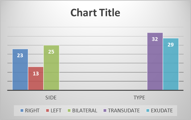
Chart 1- Distribution of Pleural Effusion Characteristics in Two Groups
Among 61 COVID-19 patients with pleural effusion, 23 (37.7%) had right-sided effusion, 13 (21.3%) had left-sided effusion, and 25 (41%) had bilateral pleural effusion (Chart 1). Additionally, 32 patients (52.5%) had transudative pleural effusion, while 29 (47.5%) had exudative effusion. Among the transudative cases, 8 patients showed no evidence of congestive heart failure (CHF) and had an ejection fraction (EF) above 50%. In this study, no significant relationship was found between the Pleural-fluid classification and the prognosis or outcomes of the patients (Chart 1). Furthermore, among the 61 COVID-19 patients with pleural effusion, 4 (6.6%) had positive smear and culture results, all identified as gram-positive cocci, specifically Staphylococcus aureus.
Table 2 - Prognostic assessment of patients based on the nature of pleural fluid
|
Outcome |
Pleural-fluid classification |
P* |
|||||
|
transudate |
exudate |
||||||
|
Number |
Percentage (%) |
Number |
Percentage (%) |
||||
|
1 month follow-up |
Alive |
27 |
84.4 |
24 |
82.8 |
0.865 |
|
|
Dead |
5 |
15.6 |
5 |
17.2 |
|||
|
3 month follow-up |
Alive |
23 |
71.9 |
20 |
69 |
0.8 |
|
|
Dead |
9 |
28.1 |
9 |
31 |
|||
|
6 month follow-up |
Alive |
18 |
56.3 |
17 |
58.6 |
0.85 |
|
|
Dead |
14 |
43.8 |
12 |
41.4 |
|||
|
Echocardiography findings |
Mean |
Standard deviation |
Median |
Max |
Min |
|
EF |
41.43 |
12.96 |
45 |
55 |
10 |
|
PAP |
37.42 |
10.88 |
35 |
70 |
22 |
Table 3: Echocardiographic findings in hospitalized COVID-19 patients with pleural effusion
Among patients with pleural effusion, 51 (83.6%) had no pericardial effusion, 6 (9.8%) had mild pericardial effusion, and only 4 (6.6%) had moderate to severe pericardial effusion. There was no correlation between echocardiographic findings—including Ejection fraction (EF) and Pulmonary Artery Pressure (PAP)—and the prognosis of COVID-19 patients with pleural effusion (Tables 3 and 4). Similarly, no correlation was observed between the final outcome and the presence of pericardial effusion (Table 5).
Table 4 - Examination of the relationship between echocardiographic findings, including PAP and EF, and the outcome
|
Outcome |
Echocardiography findings |
|||||||||
|
EF |
PAP |
|||||||||
|
Number |
Mean |
Standard deviation |
Number |
Mean |
Standard deviation |
|||||
|
1 month follow-up |
Alive |
51 |
41.8 |
13.0 |
47 |
37.72 |
11.0 |
|||
|
Dead |
10 |
39.5 |
13.0 |
8 |
35.63 |
10.4 |
||||
|
P Value |
0.61 |
0.61 |
||||||||
|
3 month follow-up |
Alive |
43 |
41.33 |
13.83 |
39 |
38.79 |
11.02 |
|||
|
Dead |
18 |
41.67 |
10.98 |
16 |
34.06 |
10.0 |
||||
|
P Value |
0.92 |
0.14 |
||||||||
|
6 month follow-up |
Alive |
35 |
42.2 |
13.1 |
32 |
37.9 |
10.8 |
|||
|
Dead |
26 |
40.3 |
12.9 |
23 |
36.7 |
11.1 |
||||
|
P Value |
0.59 |
0.69 |
||||||||
Table 5 - The relationship between pericardial effusion and the outcome
|
Outcome |
Pericardial Effusion |
P* |
||||||
|
No effusion |
Mild |
Moderate to Severe |
||||||
|
Number |
Percentage (%) |
Number |
Percentage (%) |
Number |
Percentage (%) |
|||
|
1 month follow-up |
Alive |
42 |
82.4 |
5 |
83.3 |
4 |
100 |
0.83 |
|
Dead |
9 |
17.6 |
1 |
16.7 |
0 |
0 |
||
|
3 month follow-up |
Alive |
37 |
72.5 |
5 |
83.3 |
1 |
25 |
0.10 |
|
Dead |
14 |
27.5 |
1 |
16.7 |
3 |
75 |
||
|
6 month follow-up |
Alive |
30 |
58.8 |
4 |
66.7 |
1 |
25 |
0.45 |
|
Dead |
21 |
41.2 |
2 |
33.3 |
3 |
75 |
||
Based on the results of this study, among the inflammatory blood tests conducted upon patient admission, those with pleural effusion exhibited significantly higher levels of D-dimer (3.93 ± 3.79 vs. 3.05 ± 1.73 units, P = 0.004) compared to patients without pleural effusion.
Among the blood tests conducted at the time of discharge, patients with pleural effusion exhibited significantly higher levels of ferritin (1285.81 ± 678.19 vs. 661.68 ± 814.6, P = 0.02), D-dimer (4.62 ± 3.25 vs. 2.29 ± 1.76, P = 0.003), C-reactive protein (CRP) (32.25 ± 23.95 vs. 10.36 ± 10.48, P < 0.001), and erythrocyte sedimentation rate (ESR) (44.69 ± 35.67 vs. 24 ± 22.44, P = 0.01) compared to patients without pleural effusion.
In the comparison of inflammatory markers at admission and discharge among patients with pleural effusion, no significant differences were observed. However, among patients without pleural effusion, significant changes were noted: the mean ESR decreased from 44.17 ± 26.05 to 22.44 ± 24 (P = 0.001); the mean CRP decreased from 37.77 ± 28.7 to 10.48 ± 10.26 (P < 0.001); the mean fibrinogen decreased from 521.93 ± 135.8 to 357.5 ± 112.7 (P = 0.008); and the mean platelet count increased from 200.16 ± 74.3 to 240.76 ± 87.2 (P = 0.003). These changes were statistically significant (Table 6).
Table 6 – comparative laboratory results in the presence or absence of pleural effusion
|
Blood test results |
Plural effusion |
P* |
||||
|
Yes |
NO |
|||||
|
Mean |
Standard deviation |
Mean |
Standard deviation |
|||
|
WBC |
Admission |
8.998 |
6.50 |
6.989 |
3.57 |
0.036 |
|
Discharge |
9.4372 |
4.85 |
9.4935 |
3.25 |
0.946 |
|
|
lymphocytes |
Admission |
15.566 |
10.58 |
17.680 |
9.62 |
0.25 |
|
Discharge |
14.254 |
10.47 |
12.940 |
8.47 |
0.496 |
|
|
Neutrophils |
Admission |
76.303 |
14.80 |
76.672 |
10.88 |
0.876 |
|
Discharge |
75.247 |
14.66 |
80.630 |
9.99 |
0.036 |
|
|
Creatinine |
Admission |
2.174 |
2.09 |
1.080 |
0.52 |
0.001> |
|
Discharge |
2.009 |
1.93 |
1.191 |
0.62 |
0.009 |
|
|
AST |
Admission |
53.733 |
72.7 |
89.525 |
306.5 |
0.38 |
|
Discharge |
40.56 |
29.92 |
40.97 |
23.32 |
0.952 |
|
|
ALT |
Admission |
57.80 |
125.1 |
94.03 |
357.3 |
0.46 |
|
Discharge |
36.33 |
22.32 |
72.15 |
55.99 |
0.003 |
|
*Independent T-test
According to Table 6, among admission tests, patients with pleural effusion had significantly higher white blood cell counts (WBC) (8.99 ± 6.5 vs. 6.98 ± 3.57; P = 0.03) and creatinine levels (2.17 ± 2.09 vs. 1.08 ± 0.52; P < 0.001) compared to patients without pleural effusion. Additionally, at discharge, patients with pleural effusion showed significant differences in neutrophil percentages (75.24 ± 14.66 vs. 80.63 ± 9.99; P = 0.03), creatinine levels (2.009± 1.93 vs. 1.19 ± 0.62; P = 0.009), and alanine aminotransferase (ALT) levels (36.33 ± 22.32 vs. 72.15 ± 55.99; P = 0.003) compared to patients without pleural effusion.
Table 7: Patients' outcome based on the presence or absence of pleural effusion
|
Outcome |
pleural effusion |
P* |
||||
|
yes |
No |
|||||
|
Number |
Percentage (%) |
Number |
Percentage (%) |
|||
|
1 month follow-up |
Alive |
51 |
83.6 |
54 |
88.5 |
0.433 |
|
Dead |
10 |
16.4 |
7 |
11.5 |
||
|
3 month Follow- up |
Alive |
43 |
70.5 |
53 |
86.9 |
0.027 |
|
Dead |
18 |
29.5 |
8 |
13.1 |
||
|
6 month Follow- up |
Alive |
35 |
57.4 |
50 |
82.0 |
0.003 |
|
Dead |
26 |
42.6 |
11 |
18.0 |
||
Three-month mortality was higher among patients with pleural effusion than among those without pleural effusion: 18/61 (29.5%) versus 8/61 (13.1%), respectively; P = 0.027.
Accordingly; Six-month mortality was 26/61 (42.6%) among patients with pleural effusion and 11/61 (18.0%) among those without pleural effusion; P = 0.003. (Table 7)
The relationship between the 6-month outcome and the qualitative variables studied, as well as prescribed medications, was examined in all patients, regardless of the presence of pleural effusion. Among the qualitative variables analyzed, a significant association was found between cancer, the presence of pleural effusion, antibiotic use, and the 6-month outcome.
Table 8: Relationship of other Variables and Outcomes of Patients
|
Qualitative Variable |
Outcome |
P
|
||||
|
Alive |
Dead |
|||||
|
Number |
Percentage (%) |
Number |
Percentage (%) |
|||
|
Gender |
Male |
53 |
7.69 |
23 |
3.30 |
00.1 |
|
Female |
32 |
6.69 |
14 |
4.30 |
||
|
Diabetes |
27 |
9.65 |
14 |
1.34 |
51.0 |
|
|
Hypertension |
33 |
7.64 |
18 |
3.35 |
32.0 |
|
|
Kidney Disease |
10 |
6.52 |
9 |
4.47 |
07.0 |
|
|
Heart Disease |
19 |
4.59 |
13 |
6.40 |
17.0 |
|
|
Cancer |
5 |
7.35 |
9 |
3.64 |
003.0 |
|
|
Smoking History |
8 |
7.66 |
4 |
3.33 |
00.1 |
|
|
Hyperlipidemia |
5 |
3.83 |
1 |
7.16 |
66.0 |
|
|
Stroke |
5 |
3.83 |
1 |
7.16 |
66.0 |
|
|
Asthma |
4 |
100 |
0 |
0 |
31.0 |
|
|
Dyspnea |
50 |
7.66 |
25 |
3.33 |
42.0 |
|
|
Fever |
29 |
4.64 |
16 |
6.35 |
41.0 |
|
|
Chills |
29 |
9.65 |
15 |
1.34 |
54.0 |
|
|
Body Pain |
34 |
3.72 |
13 |
7.27 |
68.0 |
|
|
Cough |
58 |
4.74 |
20 |
6.25 |
15.0 |
|
|
Chest Pain |
8 |
80 |
2 |
20 |
51.0 |
|
|
Nausea &Vomiting |
15 |
2.68 |
7 |
8.31 |
00.1 |
|
|
Diarrhea |
8 |
5.61 |
5 |
5.38 |
53.0 |
|
|
Headache |
14 |
70 |
6 |
30 |
00.1 |
|
|
Pleural Effusion |
35 |
4.57 |
26 |
6.42 |
003.0 |
|
|
Corticosteroids |
76 |
71 |
31 |
29 |
55.0 |
|
|
Remdesivir |
15 |
9.78 |
4 |
1.21 |
42.0 |
|
|
Antibiotics |
43 |
7.59 |
29 |
3.40 |
005.0 |
|
|
Actemra(Tocilizumab) |
13 |
4.68 |
6 |
6.31 |
00.1 |
|
To investigate the simultaneous effects of significant variables such as patient age, initial saturation, ferritin, CRP, cancer, presence of pleural effusion, and antibiotic use on the 6-month follow-up, backward logistic regression was employed. The results indicated that only two variables—age and ferritin levels—were significantly associated with mortality. Specifically, for age, the odds ratio was 1.1 (95% confidence interval: 1.02–1.18), indicating that with each additional year of age, the odds of death increase by 10%. For ferritin, the odds ratio was 1.002 (95% confidence interval: 1.00–1.003), meaning that for every 100-unit increase in ferritin, the odds of death increase by approximately 20%.
Discussion
In this single-center retrospective cohort study of hospitalized patients with laboratory-confirmed COVID-19, the presence of pleural effusion was associated with a substantially higher risk of mortality during follow-up. Although one-month mortality did not differ significantly between groups, mortality was significantly higher among patients with pleural effusion at three months (29.5% vs. 13.1%) and six months (42.6% vs. 18.0%). Pleural effusion was also associated with a longer hospital stay and a more pronounced inflammatory and coagulation profile, including higher D-dimer levels at admission and higher ferritin, D-dimer, C-reactive protein, and erythrocyte sedimentation rate levels at discharge. Taken together, these findings suggest that pleural effusion may serve as a clinically important marker of disease burden and adverse prognosis in hospitalized patients with COVID-19.
Pleural effusion is not among the characteristic radiologic manifestations of COVID-19 [17]. Typical thoracic imaging findings include bilateral peripheral ground-glass opacities, consolidation, and other features of viral pneumonitis, whereas pleural effusion is reported less frequently.[10] Its presence should therefore prompt careful clinical assessment rather than being regarded as an incidental imaging finding. Previous observational studies and reviews have similarly described pleural effusion as an uncommon complication of COVID-19 that occurs more often in patients with severe disease, critical illness, or multisystem involvement [19-21]. The present findings are consistent with this literature and extend it by demonstrating that the association with adverse outcomes persisted beyond the acute hospitalization period, particularly at three and six months.
Several mechanisms may explain the observed relationship between pleural effusion and mortality. First, pleural effusion may reflect more extensive pulmonary inflammation and increased vascular permeability associated with severe SARS-CoV-2 infection. Second, it may indicate systemic endothelial dysfunction, capillary leak, and dysregulated inflammatory responses. The higher D-dimer levels observed at admission and the persistently elevated ferritin, D-dimer, CRP, and ESR levels at discharge support the possibility that pleural effusion occurs in the context of sustained inflammation and coagulation activation. These processes are biologically plausible contributors to respiratory deterioration, thromboembolic complications, multiorgan dysfunction, and delayed recovery. However, the present study cannot establish whether pleural effusion directly contributes to these outcomes or simply reflects the severity of the underlying disease.
The clinical characteristics of the pleural-effusion group further support the interpretation that pleural effusion may identify a medically vulnerable population. Patients with pleural effusion were older and had higher prevalences of chronic kidney disease, heart disease, and cancer than patients without pleural effusion. These comorbidities are independently associated with poor outcomes in COVID-19 and may also contribute directly to pleural-fluid formation through heart failure, renal dysfunction, hypoalbuminemia, malignancy, or impaired fluid clearance [22]. Accordingly, pleural effusion should not automatically be attributed to SARS-CoV-2-related pleuritis. The finding may represent a heterogeneous clinical phenotype resulting from several overlapping mechanisms, including viral inflammation, cardiac dysfunction, renal impairment, secondary infection, or pre-existing disease [16]. This is particularly relevant because more than half of the effusions in the present cohort were classified as transudative, while the remainder were exudative.
The lack of a significant association between pleural-fluid classification and mortality is noteworthy. Outcomes were similar among patients with transudative and exudative effusions at one, three, and six months. This finding may indicate that the presence and clinical context of pleural effusion are more important prognostically than the transudative or exudative designation alone. Nevertheless, the analysis should be interpreted cautiously. The sample size was small, the number of outcome events within each subgroup was limited, and the classification of pleural fluid may have been influenced by incomplete sampling or varying clinical indications for thoracentesis. In addition, the cohort included patients with important comorbidities that could produce transudative effusions independently of COVID-19. Future studies should incorporate standardized pleural-fluid analysis, serum and pleural albumin and protein measurements, microbiological testing, cytology, and systematic evaluation for cardiac, renal, hepatic, and malignant causes.
The echocardiographic findings provide additional context. The mean ejection fraction in patients with pleural effusion was reduced, while pulmonary artery pressure was variably elevated; however, neither parameter was significantly associated with mortality. Similarly, the presence or severity of pericardial effusion was not significantly related to outcome. These negative findings should not be interpreted as evidence that cardiac dysfunction is irrelevant. Rather, the study may have lacked sufficient power to detect clinically meaningful associations, and single measurements of ejection fraction or pulmonary artery pressure may not adequately capture dynamic cardiac stress during acute illness. Moreover, pleural effusion can arise from mechanisms that are not reflected by ejection fraction alone, including diastolic dysfunction, right-sided heart failure, pulmonary vascular disease, fluid overload, renal dysfunction, and systemic inflammation.
The inflammatory-marker findings deserve particular attention. Patients with pleural effusion had higher D-dimer levels at admission, suggesting early activation of coagulation and fibrinolysis. At discharge, the pleural-effusion group continued to demonstrate higher ferritin, D-dimer, CRP, and ESR levels than the comparison group. In contrast, patients without pleural effusion showed significant reductions in ESR, CRP, and fibrinogen, together with an increase in platelet count between admission and discharge. This pattern may suggest a slower resolution of systemic inflammation and coagulation abnormalities among patients with pleural effusion. Importantly, these biomarkers may be markers of disease severity rather than independent causal factors. Their prognostic value should therefore be examined alongside age, comorbidity burden, oxygen requirement, radiologic severity, renal function, treatment exposure, and intensive-care utilization.
In the multivariable analysis, age and ferritin level remained significantly associated with six-month mortality, whereas pleural effusion did not retain an independent association after adjustment. This result is important and should be emphasized. The unadjusted association between pleural effusion and mortality may be partly explained by differences in age, comorbidities, and systemic inflammatory burden between the groups. Therefore, pleural effusion should be regarded as a potential risk marker or clinical warning sign, not as an established independent predictor of death. The finding also illustrates the distinction between prognostic association and prognostic independence: a radiologic feature may identify patients at high risk while adding limited predictive information after more fundamental determinants of outcome have been considered.
From a clinical perspective, the detection of pleural effusion in a patient with COVID-19 should encourage a structured reassessment of disease severity and alternative diagnoses. Clinicians should evaluate fluid volume and laterality, oxygenation, hemodynamic status, renal and cardiac function, inflammatory and coagulation markers, medication exposure, and the possibility of bacterial superinfection or malignancy. Pleural effusion alone should not dictate treatment, and the present data do not support routine thoracentesis or a specific intervention solely to improve survival. Instead, it may be useful as a trigger for closer monitoring, more frequent reassessment, and early identification of potentially reversible contributors such as volume overload, heart failure, pulmonary embolism, bacterial infection, or renal deterioration.
This study has several strengths. It included a comparison group of patients with COVID-19 without pleural effusion, assessed outcomes beyond the initial hospitalization, and examined clinical, laboratory, echocardiographic, and pleural-fluid characteristics. The six-month follow-up is particularly valuable because it suggests that the prognostic implications of pleural effusion may extend beyond the acute phase of infection. However, the limitations are substantial. The study was retrospective and conducted at a single tertiary-care center, which limits generalizability. The sample size was modest, and only 122 patients were included; consequently, subgroup analyses and multivariable models may have been vulnerable to imprecision and overfitting. The groups differed in age and several major comorbidities, raising the possibility of residual confounding. The timing, volume, and radiologic definition of pleural effusion were not described in sufficient detail to determine whether effusion preceded clinical deterioration or developed as a consequence of it. Information regarding disease severity, oxygen and ventilatory support, intensive-care admission, vaccination status, viral variant, and other potentially important treatment-related factors may also have been incomplete. Finally, the study was not designed to determine whether pleural effusion independently improves risk prediction beyond established clinical and laboratory variables.
Future research should use prospective, multicenter designs with standardized imaging criteria and serial assessment of pleural-fluid volume and characteristics. Studies should distinguish new-onset effusion from pre-existing effusion and should systematically evaluate competing causes, including heart failure, renal disease, hypoalbuminemia, pulmonary embolism, bacterial infection, and malignancy. Larger cohorts should use time-to-event analyses and clinically relevant adjustment sets that include age, comorbidities, baseline oxygenation, radiologic severity, renal function, inflammatory markers, and treatment exposure. It will also be important to determine whether adding pleural effusion to established prognostic models improves discrimination, calibration, or clinical decision-making. Such work could establish whether pleural effusion is merely a marker of severe disease or a useful independent component of risk stratification.
Conclusion
Among hospitalized patients with COVID-19, pleural effusion was associated with longer hospitalization, persistent inflammatory and coagulation abnormalities, and significantly higher three- and six-month mortality. These findings support considering pleural effusion as a clinically meaningful warning sign that should prompt comprehensive reassessment for severe pulmonary disease, systemic inflammation, cardiac or renal dysfunction, thromboembolic disease, and secondary infection. Nevertheless, because the association was attenuated after multivariable adjustment and because of the retrospective, single-center design, pleural effusion cannot yet be considered an independent causal determinant or standalone prognostic tool. Its prognostic significance should be confirmed in larger prospective multicenter studies using standardized imaging, pleural-fluid characterization, and rigorous adjustment for comorbidity and disease severity.
Acknowledgment
The authors would like to thank the Clinical Research Development Unit of Baqiyatallah Hospital for all their support and guidance during the carrying out of this study. This study received ethical approval from Baqiyatallah University of Medical Sciences, under the approval number IR.BMSU.BAQ.REC.1402.005
Conflict of Interest
The authors declare no conflict of interest.

Open Access By Aditum Open Access Journals id licensed under Creative Commons Attribution 4.0 International License. Based On a Work at aditum.org


