Gastroenterology and Hepatology Research
OPEN ACCESS | Volume 7 - Issue 1 - 2026
ISSN No: 2836-2888 | Journal DOI: 10.61148/2836-2888/GHR


Hilary Denis Solomons
MB BCh, M Med Hematology, Pathology, University of the Witwatersrand, South Africa
Corresponding author: Hilary Denis Solomons, MB BCh, M Med Hematology, Pathology, University of the Witwatersrand, South Africa
Received date: January 21, 2021
Accepted date: February 20, 2021
published date: March 05, 2021
Citation: Hilary D Solomons. “Pathology of Rectal Biopsy in Hirschsprung’s Disease”. J Gastroenterology and Hepatology Research, 2(2); DOI: http;//doi.org/03.2020/1.1006.
Copyright: © 2020 Hilary Denis Solomons. This is an open access article distributed under the Creative Commons Attribution License, which permits unrestricted use, distribution, and reproduction in any medium, provided the original work is properly Cited.
The aim of this study was to review our experience in applying acetylcholinesterase histochemistry for diagnosing colonic dysganglionoses in children
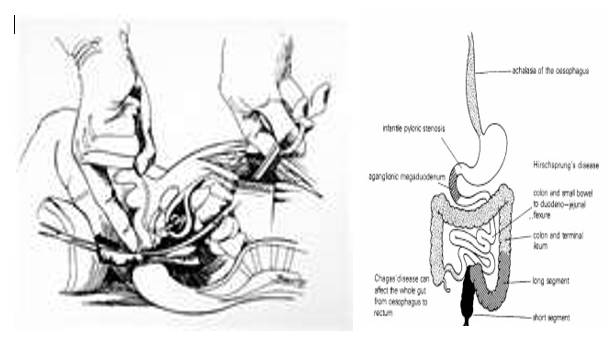
What is Hirschsprung's Disease?
Hirschsprung's disease is characterized by aganglionosis of the enteric neural plexuses and failure of migration of fetal neuroblasts from the cephalic neural crest down the alimentary tract is the most likely embryonal basis for this condition.
The rectum is most commonly involved and classification depends on the length of the ganglionic segment:
- Short segment disease involves rectum and sigmoid.
- In long segment disease aganglionosis extends more proximally than the sigmoid colon.
- The whole colon may be involved and, rarely the entire intestinal tract is aganglionic.
Use of rectal suction mucosal biopsies has now replaced full thickness biopsies.
The diagnosis depends predominantly on clinical presentation, barium studies and rectal biopsy.
Absence of ganglion cells, often accompanied by thickened nerve fascicles, is required for histological diagnosis.
The acetylcholinesterase reaction has substantially facilitated evaluation of suction biopsies and the technique can be carried out fairly rapidly.
Ischemic enterocolitis is a serious complication of untreated Hirschsprung's disease and carries significant morbidity and mortality.
Histopathology of Hirschsprung’s Disease

Experience of acetylcholinesterase histochemistry application in the diagnosis of chronic constipation in children.Medicina (Kaunas). 2007;43(5):376-84.
The aim of this study was to review our experience in applying acetylcholinesterase histochemistry for diagnosing colonic dysganglionoses in children. PATIENTS AND
Methods: We analyzed acetylcholinesterase histochemistry results of rectal biopsy specimens obtained from 85 children. The indications for biopsy were suspicion of Hirschsprung's disease in neonates and infants (Group 1; n=21) and older children (Group 2; n=17); megarectum (Group 3; n=44); and colostomy (Group 4; n=3). Specimens were taken at 5 and 10 cm using endoscopic forceps or excised with scissors at 2.5 cm above the dentate line. Acetylcholinesterase activity was evaluated using Karnovsky-Roots method.
Results: The diagnosis of Hirschsprung's disease was confirmed in 17 children of the first group and in 3 of the second group. In the third group, 2 children were diagnosed with ultrashort-segment Hirschsprung's disease and 3 children with intestinal neuronal dysplasia. In one case, acetylcholinesterase reaction was false positive. Hirschsprung's disease was diagnosed in 2 children with colostomies; in one case acetylcholinesterase activity caused false-positive results. Colonic dysganglionoses were diagnosed in 78% of infants and in 14% of children over 1 year of age. The diagnostic specificity of acetylcholinesterase in Hirschsprung's disease was 92%.
Conclusions: 1) The analysis of acetylcholinesterase activity in children's rectal biopsy specimens is a reliable method for diagnosing Hirschsprung's disease, especially in infants; 2) This method of examination is irreplaceable in diagnosing ultrashort-segment Hirschsprung's disease and remains the only method to confirm the diagnosis of this disease; 3) Acetylcholinesterase histochemistry is not sufficiently informative in diagnosing intestinal neuronal dysplasia type B, because authors applying other neurohistochemical investigation methods have reported higher incidence of this disease.
Calretinin immunohistochemistry: a simple and efficient tool to diagnose Hirschsprung disease

Calretinin immunohistochemistry: a simple and efficient tool to diagnose Hirschsprung disease
(a): Rectal suction biopsy for suspicion of Hirschsprung disease (HD) showing a total absence of staining after calretinin immunohistochemistry either in submucosae, muscularis mucosae or lamina propria. Insert shows an unstained abnormally large nerve fiber. Original magnification 400 and 600 (insert). This pattern diagnosis HD. (b, c and d): Calretinin immunohistochemistry on a rectal suction biopsy for suspicion of HD shown in (b) and (c), positive staining easily detectable at low magnification in submucosae (in b, 250), in muscularis mucosae (in c, 400) or sometimes extended to the lamina propria (in d, 400). This pattern excludes HD. (e): Calretinin immunohistochemistry on a rectal suction biopsy for suspicion of HD showing positivity in Schwann cells and nuclear expression by ganglion cells (insert) in nerve plexus ( 1000). This pattern excludes HD. (f): Calretinin immunohistochemistry on a rectal suction biopsy for suspicion of HD showing a similar pattern than in Figure 1a but with a slight positivity in large nerve fibers (insert).
Biopsy Procedure and Area of Hirschsprung’s Disease


Open Access By Aditum Open Access Journals id licensed under Creative Commons Attribution 4.0 International License. Based On a Work at aditum.org