
Pharmacy and Drug Innovations
OPEN ACCESS | Volume 5 - Issue 1 - 2026
ISSN No: 2994-7022 | Journal DOI: 10.61148/2994-7022/PDI


Short Title: Ferroptosis and Huntington’s disease
Rohan Bansal1, Sanchi Jain1, Neeraj Poni1 and Shamsher Singh1*
Neuropharmacology Division, Department of Pharmacology, ISF College of Pharmacy, Ghal Kalan, Moga, Punjab, India.
*Corresponding author: Prof. (Dr.) Shamsher Singh, Neuropharmacology Division, Department of Pharmacology, ISF College of
Pharmacy, Moga, Punjab, India, 142001.
Received: September 10, 2026 | Accepted: September 22, 2026 | Published: September 25, 2026
Citation: Bansal R, Jain S, PoniN and Singh S. (2026) “Targeting Ferroptosis in Huntington’s disease: Therapeutic Potential of the Nrf2/HO-1/GPX4 Pathway’’ Pharmacy and Drug Innovations. 6(1); DOI: 10.61148/2836-2802/JPCHI /086
Copyright: © 2026. Shamsher Singh. This is an open access article distributed under the Creative Commons Attribution License, which permits unrestricted use, distribution, and reproduction in any medium, provided the original work is properly cited.
Huntington’s disease (HD) is a hereditary neurodegenerative condition marked by gradual impairment in motor control, cognitive deterioration, and widespread neuron death, especially in the striatum and cerebral cortex. Recent studies highlights ferroptosis- a form of regulated cell death reliant on iron, fueled by lipid peroxidation and buildup of reactive oxygen species- as a major factor in HD pathology. Mutant huntingtin (mHTT) disrupts iron metabolism, boosts labile iron levels, intensifies oxidative damage, and weakens antioxidant systems, rendering neurons more vulnerable to ferroptosis. The Nrf2/HO-1/GPX4 pathway acts as a central regulator of ferroptosis, serving as a primary antioxidant signaling network that preserves redox equilibrium and clears lipid peroxides. Upon activation, Nrf2 shifts to the nucleus and drives expression of protective genes such as HO-1 and GPX4. HO-1 regulates heme breakdown and alleviates oxidative stress, whereas GPX4 functions as an essential lipid repair enzyme that prevents ferroptotic damage to membranes. Impaired function or diminished activation of this axis in HD undermines antioxidant defenses, hastens lipid peroxidation, and drives ferroptotic neuron death. Elucidating the interplay between ferroptosis and mHTT-driven metabolic disruptions, along with impact of Nrf2/HO-1/GPX4 modulation on neuronal viability, could reveal new therapeutic strategies. Strategies targeting iron excess, boosting Nrf2-driven transcription, stabilizing GPX4 function, or using ferroptosis inhibitors offer promising approaches to slow neurodegeneration and alter disease progression. Thus, the ferroptosis pathway and Nrf2/HO-1/GPX4 axis emerge as key molecular hubs connecting oxidative stress to neuronal susceptibility in HD, with strong potential as therapeutic targets.
Huntington’s disease, Ferroptosis, Oxidative stress, Nrf2, HO-1, GPX4
Huntington’s disease is an inherited, autosomal dominant neurological disorder caused by mutation in huntingtin’s gene. In mutant huntingtin’s gene there is an extended trinucleotide CAG repetition cause mutation in huntingtin protein with an elongated polyglutamine stretch. These aberrant proteins undergo misfolding and aggregate into insoluble protein assemblies, contributing to cellular dysfunction such as gene regulation, mitochondrial bioenergetics, axonal transport and synaptic dysfunction (Kuwar & Singh, 2025). The medium spiny neurons in striatum are vulnerable to metabolic stress and degeneration is particularly noticeable. In the progression of huntington’s pathology abnormalities in redox signaling and iron handling plays a significant role, despite the fact that toxicity caused by mutant huntingtin has long been thought to be the primary cause of the disease (Niu et al; 2018).
According to extensive pathological and laboratory study, excessive iron is recurrent feature of huntington’s disease with prominent deposition detected in striatum and cortex. Mutant huntingtin protein’s contribute to buildup of free, reactive iron occurs in neurons which disrupts the multiple important processes of neuronal iron regulation, including transferrin receptor uptake, divalent metal transporter-1 (DMT1) levels, and ferritin turnover (Niu et al; 2024). The production of dangerous hydroxyl radicals through Fenton reaction is speeding up by elevated levels of ferrous iron (Fe2+) cause more oxidative stress and make neuronal membrane more susceptible to lipid damage. The condition required for ferroptosis, a controlled, iron-dependent, lipid peroxidation- mediated kind of cell death that is becoming more widely acknowledged as pertinent in the context of neurodegenerative (Rosas et al; 2012).
According to the recent studies, in huntington’s disease there is widespread accumulation of oxidized membrane lipids and increased activity of lipid-modifying enzymes, and displays a number of important molecular indicators of ferroptosis, including decreased glutathione (GSH) levels, decreased glutathione peroxidase 4 (GPX4) expression or function (Jimenez-Jimenez et al; 2025). Increment in reactive oxygen species and impairs cellular antioxidant defenses by mitochondrial damage brought on by mutant huntingtin protein. Together, these elements imply that ferroptosis is an important process of cell death that result in the loss of specific neurons in Huntington’s disease (Simmons et al; 2007).
After the discovery of degenerative mechanism caused by iron and lipid degradation, iron chelators and ferroptosis- blocking therapies for huntington’s disease have gained attention (Butterfield et al; 2012). In experimental model of HD, to enhance the behavioral and pathological parameters, lower brain iron level and lessen oxidative damage, drugs like deferiprone and deferoxamine have been demonstrated. However, the new ferroptosis inhibitors and antioxidants substances which promote GPX4 function such as liproxstatin-1, ferrostatin-1 coenzyme Q10 are showing promise as strategies to combat ferroptotic stress and preserve neuronal integrity (Fan et al; 2021, Liu et al; 2022).
To connect mutant huntingtin toxicity with abnormalities in iron homeostasis, oxidative balance and ferroptosis in the pathogenic model of huntington’s disease is need to be update. This revisement offers prospects for novel therapeutics aimed at altering iron metabolism and regulating ferroptotic cell death, as well as deep understanding of disease mechanism. In the current evidence on ferroptosis in HD pathophysiology, this study evaluates the therapeutic prospects of addressing iron overload and ferroptosis for successful treatment of HD (Muller & Leavitt, 2014).
Ferroptosis is a regulated, iron-dependent type of cell death initiated by excessive lipid peroxidation, which causes oxidative injury to cellular membranes and ultimately result in cell destruction (Awasthi et al; 2025).
The concept of ferroptosis evolved over many years, the discovery of prevention of particular cell death by the eliminating cysteine or preventing the synthesis of glutathione is reported in research performed from 1950s to the 1970s. Suggesting that oxidative stress and lipid peroxidation were causing cell damage in a manner distinct from conventional apoptosis, antioxidants like vitamin E might prevent this cell death (Hirschhorn & Stockwell, 2019). The fenton process links iron metabolism to oxidative cellular damage. Early investigations in the 1980s discovered oxytosis, a type of oxidative glutamate- induced cell death, which was later linked to ferroptosis. Dolma et al; 2003 found Erastin as a tiny chemical that preferentially triggers non- apoptotic cell death in RAS – mutated tumor cells, a mechanism that can be prevented by iron chelators and lipid peroxidation inhibitors. Latest studies shows that for preventing lethal lipid peroxidation glutathione peroxidation (GPX4) is essential, by inhibiting GPX4 cause a certain type of controlled cell death (Yang & Stockwell, 2016). Dixon and his colleagues formally called this process ferroptosis in 2012, emphasizing its dependence on iron involved in redox processes and the accumulation of oxidized polyunsaturated lipids. This type of cell death differs from necrosis, autophagy and apoptosis in both appearance and processes (Dixon et al; 2012). In the field of cancer treatment, neurological illnesses, blood flow restoration damage, metabolic disorders, aging and tissue balance maintenance Ferroptosis gained a lot of attention since from its discovery. Ferroptosis is a crucial connection between metabolism, redox processes and regulated cellular function as demonstrated by the progression from early discoveries on oxidative stress to comprehensive molecular understanding (Li et al; 2020). Medium spiny neurons (MSNs) are thought to be more susceptible to oxidative stress and ferroptosis than striatal interneurons because they have a lesser antioxidant defense capacity. Several ideas have been presented to explain this selective vulnerability, including decreased glutathione availability, increased metabolic load, increased mitochondrial dysfunction, excess glutamate- mediated excitotoxicity, and increased iron accumulation in MSNs. In contrast, striatal interneurons have more robust calcium- buffering and antioxidant systems, which may give resistance to oxidative damage and lipid peroxidation (Bergonzoni et al; 2021).
Ferroptosis is caused by a series of interrelated molecular events, including glutathione (GSH) depletion, decreased activity of glutathione peroxidase 4 (GPX4), and iron- dependent lipid peroxidation. Disrupting cysteine absorption via the system Xc- transporter reduces intracellular cysteine availability and impairs GSH production (Yan et al; 2021). GSH is an important cofactor for GPX4, and its depletion impairs GPX4 function, limiting the conversion of harmful lipid hydroperoxides into non- toxic lipid alcohols. Polyunsaturated phospholipids undergo oxidative alteration to generate lipid peroxides, which is further enhanced by ferrous ion (Fe2+) and the formation of reactive oxygen species via Fenton chemistry (Chen et al; 2020).
Iron- driven lipid peroxidation is the oxidative breakdown of polyunsaturated fatty acids (PUFAs) in membrane phospholipids, which leads to ferroptosis. Redox- active ferrous iron (Fe2+) promotes the generation of highly reactive lipid radicals and reactive oxygen species (ROS) via fenton and Haber- Weiss reactions (Sun et al; 2025). Furthermore, non- heme iron- containing enzyme known as lipoxygenase (LOX) play a crucial role in the start of lipid peroxidation by accelerating the oxidation of PUFAs in membrane phospholipids. These radicals set off a chain reaction that transforms membrane PUFAs into lipid hydroperoxides (PLOOH). When the concentration of these oxidized lipids exceeds the neutralizing capacity of glutathione peroxidase 4 (GPX4), membrane integrity is compromised (Yang & Stockwell, 2016). Finally, rupture of the plasma membrane causes ferroptotic cell death. Therefore, iron- induced lipid peroxidation is a critical pathway coupling iron overload, oxidative damage, and membrane breakdown during ferroptosis (Rochette et al; 2022).
To convert phospholipid hydroperoxides (PL- OOH) and other lipid hydroperoxides (L-OH) into their innocuous alcohol counterpart (L-OH) the selenium-containing enzyme glutathione peroxidase 4 (GPX4) uses reduced glutathione (GSH). This helps to maintain the stability of the cell membrane by halting the chain reaction of lipid peroxidation (Weaver & Skouta, 2022).
Loss of GPX4 function is caused by number of important experimental and clinical factors. When GPX4’s expression is reduced or eliminated through genetic deletion or knockdown GPX4’s enzymatic function is eliminated. The alteration in enzyme’s active-site selenocysteine by small molecules ( like the covalent inhibitor RSL3) prevent catalysis and causes pharmacological inactivation and upstream disruption of GSH synthesis or supply (like erastin’s inhibition of system Xc- or cysteine depletion) deprives GPX4 of its necessary cofactor, functionally mimicking GPX4 loss (Conrad & Friedmann, 2015). The each disruption produced same molecular end point: an inadequate conversion of L-OOH to L-OH and gradual accumulation of lipid hydroperoxides.
The biochemical and cellular repercussions of losing GPX4 are consistent and well-documented. Lipid peroxidation in membranes is accelerated when iron driven processes are undergo by lipid peroxides accumulate in the presence of iron, finally causing membrane damage or rupture. This action starts ferroptosis cell death pathway (Cao & Dixon, 2016). The morphological and biochemical aspects of ferroptosis owing to GPX4 depletion differ from apoptosis or necrosis: cells have smaller mitochondria with tighter membrane and fewer cristae and they perish without activating caspases. Acute GPX4 loss in animals models result in fast death and organ failure, including kidney failure, underscoring the vital and indispensable protective role of GPX4 in living things (Friedmann Angeli et al; 2014).
Cells have backup antioxidant mechanism that impact how vulnerable they are losing GPX4, one example is FSP1- CoQ10 pathway. Which helps reduce lipid oxidation independently of GPX4. The reduction in lipid degradation is due to another process in which tetrahydrobiopterin (BH4) is produced. When GPX4 malfunctioning mechanism is compromised then cells are most vulnerable to undergo ferroptosis. Therefore, even though GPX4 plays a crucial role in avoiding ferroptosis, how sensitive a cell is depends on various antioxidant mechanism acting together (Liu et al; 2022).
Promising strategies for both promoting ferroptosis (helpful in resistant cancers) and preventing it (to protect against acute organ damage and neurodegeneration) by targeting GPX4 and its upstream regulators, such as glutathione biosynthesis, cystine import through Xc- and selenium metabolism, since loss of GPX4 alone can cause ferroptosis. Effective therapeutic manipulation of ferroptosis will likely require combination treatments or techniques customized to specific settings is due to the availability of GPX4 independent antioxidant defences. This complexity underlines the necessity for precision in targeting ferroptosis pathway (Ma et al; 2022).
Glutathione is responsible for the maintenance of the cell’s redox balance and defence against oxidative stress. In ferroptosis, a controlled iron-dependent kind of cell death that differs from apoptosis, GSH depletion is an early and crucial step. Dangerous phospholipid hydroperoxides is detoxified by glutathione peroxidase 4 (GPX4) that buildup in membrane rich in polyunsaturated fatty acids, depends on GSH as its essential reducing agent. Normally, preventing spreading from oxidative damage is due to GPX4 transforms these lipids peroxides into innocuous lipid alcohol. GPX action is hindered and causing lipid peroxides to build up when GSH levels drops below a particular protective threshold. Ferroptotic cell death is due to the cell membrane are harmed by this oxidative stress (Yang & Stockwell, 2016).
Glutathione depletion happens through multiple linked processes, including reduced cysteine uptake via the system Xc- transporter, decreased synthesis due to limited cysteine supply, excessive use during high oxidative stress and problems recycling oxidized glutathione (GSSG) back to its reduced form (GSH). This effectively decreases GSH levels by inhibiting this transporter effectively and is frequently employed in research to induce ferroptosis because cysteine intake through system Xc- controls the critical first stage of GSH synthesis. The consumption of GSH and its depletion significantly enhanced by elevated reactive oxygen species (ROS) especially in tissues with high metabolic rates (Ursini & Maiorino, 2020).
When glutathione (GSH) levels are low, cells transition from a stable antioxidant state to one where lipid peroxidation spreads is uncontrollable. Ferroptosis is a caspase- independent form of controlled cell death that lacks traditional apoptotic hallmarks including caspase activation and DNA fragmentation. It differs from apoptosis in that it has specific morphological alterations such as reduced mitochondrial size, increased membrane density, and weakened or absent cristae. Due to its crucial place in biochemistry, low intracellular GSH acts as a pivotal decision point, deciding if cells withstand oxidative stress or undergo ferroptosis (Sun et al; 2018).
There is substantial therapeutic potential in comprehending the role that glutathione (GSH) deficiency plays in ferroptosis. The main strategies to prevent ferroptosis are using iron chelators, antioxidant that trap radicals and stabilizing GPX4 with medication. These methods are being studied for the treatment of inflammation, ischemia-reperfusion injury and neurodegenerative illness. In treatment of cancer GSH is reducing intentionally to induce ferroptosis, particularly in cases when cancer cells are resistant to apoptosis. Thus addressing glutathione metabolism provides a significant technique to either supress or accelerates ferroptosis depending on the illness situation (Li et al; 2022).
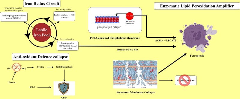
Figure 1: Molecular Mechanisms and key Biochemical events governing Ferroptosis.
To detect ferroptosis experimentally, a thorough multifaceted method that incorporates biochemical markers, lipid peroxidation assessment, iron level measurement in depth ultrastructural investigation and functional rescue testing is needed. In case of Huntington’s disease (HD) these techniques becoming more important for comprehending ferroptosis as a possible cause of neurodegeneration (Stockwell & Jiang, 2020). On a molecular level, ferroptosis is principally characterized by the loss or inactivation of glutathione peroxidase-4 (GPX4), the selenoenzyme responsible for reducing lipid hydroperoxides and protecting polyunsaturated phospholipids from oxidative damage. By using the techniques like spectrophotometry, fluorometry, HPLC and mass spectrometry to determine the GSH to GSSG ratio are commonly used to measure GPX4 and enzymatic activity assays that monitor the reduction of lipid hydroperoxides to lipid alcohols, western blotting or ELISA to detect protein level (Tang et al; 2021). Reduced expression of the SLC7A11/xCT transporter, which regulates cysteine absorption necessary for GSH synthesis, is another characteristic of ferroptosis; this is frequent measured by qrt-pCR, immunoblotting or particular transport activity tests. Increased susceptibility to lipid peroxidation and ferroptotic sensitivity are predicted by elevated levels of ACL4, an enzyme that combines arachidonic and adrenic acids into phosphatidylethanolamine. Additionally, changes in iron regulatory proteins, such as enhanced transferrin receptor (TFRC) and decreased ferritin subunit (FTH1/FTL) or genes involved in ferritin breakdown (ferritinophagy) are analyzed to identify disturbed iron metabolism that enhances ferroptosis (Stockwell, 2022).
A significant biochemical signature of ferroptosis is the excessive peroxidation of lipid and identifying this to validating ferroptosis experimentally. The oxidation-sensitive dye BODIPY-C11 (581/591), which changes its fluorescence from red to green in proportion to the amount of lipid reactive oxygen species (ROS), can be used in flow cytometry or fluorescence microscopy to assess reactive lipid species. Additionally, technique like the TBARS assay, ELISA or immunohistochemistry are frequently used to quantify secondary products of lipid peroxidation including malondialdehyde (MDA) and 4- hydroxynonenal (4-HNE) (Nghiem et al; 2025). For more extensive investigation, targeted lipidomics utilizing LC- MS/MS can offer exact profile of oxidized phospholipids (PUFA-PE-OOH), regarded as the most accurate current technique for detecting lipid peroxidation. Ferroptosis relies on iron; therefore assessing iron balance is essential. Ferrozine- based colorimetric assays; electron paramagnetic resonance, fluorescent iron indicators and calcein-AM quenching are used to quantify labile Fe2+ pools. Meanwhile, total iron concentration and its distribution can be measured via procedures such as inductively coupled plasma mass spectrometry (ICP-MS), atomic absorption spectroscopy, MRI susceptibility mapping or histology staining using Perl’s Prussion blue (Chen et al; 2021).
For identify ferroptosis from other forms of cell death like apoptosis or necroptosis, transmission electron microscopy (TEM) offers key morphological indications. Mitochondria undergoing ferroptosis generally show considerable shrinkage, thicker membranes, decreased or missing cristae and rupture of the outer membrane, all occurring without nuclear condensation or DNA fragmentation (Yan et al; 2021). In both cultured neurons and live tissues TEM is the valuable method for confirming ferroptosis. Functional rescue assays continue to be the gold standard for verifying ferroptosis because several indicators overlap with other forms of cell death. Specifically, evidence that cell death can be blocked by classic ferroptosis inhibitors- such as ferrostatin-1, liproxstatin-1 or the iron chelators deferoxamine- and enhanced by ferroptosis activators like erastin (which inhibits systemXc-) or RSL3 (a GPX4 inhibitor) provide strong proof of ferroptosis involvement (Zhang et al; 2023). Table 1 summarizes methods for experimental identification of ferroptosis-associated markers.
Table 1: Experimental detection of various molecular markers of ferroptosis
|
Marker |
Reagent/Assay |
Biological Significance |
Detection Method |
Interpretation in Ferroptosis |
References |
|
Fe2+ |
Fe2+ fluorescent probes (FerroOrange), PGSK, FeRhoNox-1 |
Iron overload drives Fenton reaction and lipid ROS. |
Fluorescence microscope, Flow cytometer, ELISA |
Increased levels of Fe2+ promote ferroptosis. |
(Chen et al; 2024) |
|
ROS |
DCFH-DA, DHE |
Increased ROS drives iron-dependent lipid peroxidation, causing irreversible membrane damage and regulated cell death. |
Fluorescence microscope, Flow cytometer |
Increased ROS levels leads to oxidative stress. |
(Chen et al; 2021) |
|
RNS |
DAF-FM Diacetate |
NO drives lipid peroxidation. |
Confocal microscope |
NO influence ferroptosis by modulating redox balance and lipid peroxidation. |
(Wen et al; 2021) |
|
Lipid peroxidation products |
C11-BODIPY |
Highly sensitive fluorescent probe that detects and measures lipid peroxidation. |
Fluorescence microscope, Flow cytometer, ELISA |
Lipid peroxidation is increased in ferroptosis. |
(He et al; 2022) |
|
|
MDA Assay kit |
MDA is PUFA peroxidation endpoint that indicates oxidative injury to membranes. |
ELISA |
High MDA concentrations indicate lipid peroxidation and ferroptotic burden. |
(Zhang et al; 2023) |
|
|
4-HNE Assay kit |
4-HNE is a reactive aldehyde generated during lipid peroxidation and forms protein adducts. |
ELISA |
Elevated 4-HNE levels indicate lipid peroxidation driven cell death. |
(Zhang et al; 2023) |
|
|
TBARS |
Marker of lipid peroxidation, indicating oxidative damage to membranes and progression of ferroptosis. |
ELISA |
TBARS detect MDA and certain minor peroxidation byproducts. |
(Han et al; 2021) |
Detection of lipid reactive oxygen species (lipid ROS) is crucial technique for diagnosing ferroptosis, since the biochemical characteristic that distinguish ferroptosis from apoptosis, necroptosis and other types of oxidative cell death is the unchecked peroxidation of polyunsaturated phospholipids. To detect the lipid ROS levels in research BODIPY- C11 (581/591) like oxidative-sensitive dyes. This dye when using in methods like flow cytometer or confocal microscopy changes color from red to green when oxidized, making it possible to precisely measure lipid peroxidation at both individual cell and population scales (Chen et al; 2024). As there is increased in BODIPY-C11 fluorescence indicates that Huntington’s disease (HD). Neurons are more vulnerable to ferroptotic lipid damage. This strategy is especially useful in neuronal cultures that express mutant huntingtin (mHTT) as increased BODIPY-C11 fluorescence indicates that Huntington’s disease. By using biochemical techniques such as the thiobarbituric acid reactive substance (TBARS) assay for malondialdehyde (MDA) and immunological assays (4-HNE) protein adducts, downstream lipid oxidation products can be measured (Song et al; 2023). Ferroptotic oxidative damage is consistent with the accumulation of these aldehyde in the striatum of post-mortem HD patients as well as in R6/2 and Q175 animal models. Certain oxidized phosphatidylethanolamine molecules (PUFA-PE-OOH), which function as direct triggers of ferroptosis can be found by more sophisticated lipidomic investigation using LC: MS/MS, providing molecular- level proof (Wenzel et al; 2017).
Brain’s coordinated system of iron intake, storage and release regulate the neuron function, mitochondrial energy production and the synthesis of neurotransmitters. Transferrin receptor (Tfr1) mediates the endocytosis by which the ferric iron (Fe3+) normally enters in cells after binding to transferrin (Tf) and passing across the blood brain barrier (Agrawal et al; 2018). Inside the endosomes Fe3+ transformed into ferrous iron Fe2+ and divalent metal transport 1 (DMT1), increasing the amount of iron that is easily accessible. Cells are protected from iron-induced oxidative damage, safely stores any excess iron within cell protected by ferritin, a vital iron-binding protein composed of heavy and light subunits (Chen et al; 2013). To remove Fe2+ as necessary to maintain oxidative balance and avoid detrimental accumulation cells used ferroportin (Fpn), the known iron exporter in neurons and glial cells. In the sensitive brain regions like the cortex and striatum aberrant iron accumulation is seen which upset the careful management of iron in Huntington’s disease (Muller & Leavitt, 2014). In HD animal models (such as R6/2 and N171-82Q) higher quantities of non-heme ferrous iron (Fe2+) and altered levels of iron-regulating proteins have been found inside neurons. These alterations, which are probably the cell’s attempts to combat too much iron, include elevated ferroportin levels and decreased expression of the transferrin receptor. Additionally, ferritin is upregulated, which indicates that oxidative stress has boosted iron storage (Donley et al; 2021).
Such dysregulation is most likely caused by mutant huntingtin- mediated disruption of iron signaling and gene regulation, resulting in excessive intracellular iron buildup. Although increased ferritin expression is an adaptive response to oxidative stress, the overall imbalance in iron management promotes redox instability, increases oxidative damage, and contributes to neuronal susceptibility and degeneration in HD.
The basal ganglia, particularly the striatum (caudate and putamen) experience the first and greatest accumulation of iron in Huntington’s disease (HD), according to a growing body of evidence. Higher iron deposits in these areas are frequently found in brain scans such as susceptibility-weighed MRI and quantitative susceptibility mapping, even in individual with the HD gene before symptoms manifest (Van den Bogaard et al; 2013). This suggest that poor iron management is a primary cause rather than merely a consequence of cell death. On the cell front, excess reactive ferrous iron (Fe2+) causes defective iron processing in both the primary striatal projection neurons, known as medium spiny neurons (MSNs) and surrounding support cells, such as astrocytes and microglia. Poor iron protection, larger pools of free iron in the cytoplasm and strange iron hoarding in lysosomes and mitochondria are all caused by the faulty huntingtin protein, which also damage mitochondria and interferes with vesicles transportation (Paul & Snyder, 2019). MSNs are particularly vulnerable to harm from iron-sparked reactive oxygen species and fat breakdown because of their high energy requirement, extensive glutamate transmission and weak oxidation defences. By becoming pro-inflammatory and retaining iron, HD microglia exacerbates the local iron crisis by increasing both ferritin-stored and free iron in the striatum. The early and severe loss of basal ganglia cells, especially MSNs in HD can be explained by this combination of spot-specific iron overload, neuron energy deficiencies and glial inflammation which creates a toxic environment conductive to oxidative damage and ferroptosis- style death (Sun et al; 2022).
In the pathophysiology of Huntington’s disease disruption in the iron balance of brain contribute significantly by increasing oxidative damage and weakening neurons, particularly in striatum and cortex. Free ferrous iron (Fe2+) accumulates and react with hydrogen peroxide (H2O2) triggered Fenton and Haber-weiss reactions to create harmful hydroxyl radicals (-OH). The many cellular components are mercilessly destroyed by these radicals and most dangerous reactive oxygen species (ROS) and by disrupting mitochondrial electron transport chains and increasing superoxide and H2O levels, which contribute to the production of iron-fuelled ROS, mutant huntington (mHTT) exacerbates the condition (Ward et al; 2014). The disruption of ion homeostasis, damage of synapsis and eruption of membrane structure in neurons membranes by polyunsaturated fats, the ensuing ROS overload causes lipid peroxidation, producing toxic aldehydes such as malondialdehyde and 4-hydroxunonenal. By protein oxidation through carbonylation and misfolding, essential enzymes for energy production, antioxidant defence and protein quality are hampered (Lee et al; 2010). Due to iron-linked DNA damage, such as 8- oxo- dG accumulation the death of cell signals and genetic disarray is caused. Iron accumulation is directly linked to oxidative destruction and increased chances of ferroptosis-like cell death in HD. This iron-powered ROS surge and Fenton-driven damage combine to form a vicious cycle of oxidative harm, mitochondrial failure and neuron loss (Tang et al; 2021).
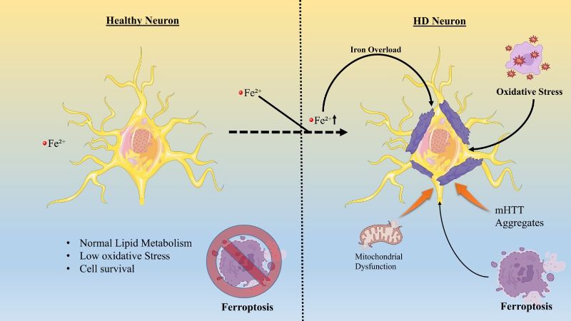
Figure 2: Schematic overview of Ferroptosis in Huntington’s disease- associated Neuronal degeneration.
The Nrf2 signalling pathway is essential for controlling lipid balance, management of iron and cellular defences against oxidative stress under redox circumstances. Because of this, it plays a crucial role in regulating both the neurodegenerative processes associated with Huntington’s disease and ferroptosis, a type of iron-dependent cell death (Song & Long, 2020). Keap1, which serves as an adapter for the Cul3-based E3 ubiquitin ligase, keeps Nrf2 anchored in the cytoplasm of the cell during normal, non-stressed conditions. Nrf2 is continuously ubiquitinated by Keap1, which causes the proteasome to break it down. This keeps Nrf2 levels low and prevents the over-activation of antioxidant genes.
When cells are exposed to oxidative damage, reactive electrophiles or iron-fueled lipid breakdown-hallmarks that initiate ferroptosis, certain cysteine positions on Keap 1 (such as Cys 151, Cys 273 and Cys 288). Because of these changes, keap1’s hold on Nrf2 is broken, allowing new Nrf2 to accumulate in the cytoplasm without being marked for destruction. After stabilization, Nrf2 translocate to the nucleus, where it receives additional support from keap1-independent pathways that are fuelled by kinases such as PKC, MAPKs and PI3K/Akt. Even when oxidative strain persists, defects in this kinase network frequently reduce Nrf2’s responses in Huntington’s disease (Kobayashi et al; 2004).
After forming heterodimers in the nucleus with tiny Maf proteins, Nrf2 binds to antioxidant response elements (AREs) in the promoters of several protective genes. The targets which include enzymes for producing and reusing glutathione(GCLC, GCLM, GSS) eliminating redox threats while replenishing NADPH ( NQO1, HO-1 TXNRD1) and most importantly-GPX4-the primary player that neutralizes dangerous lipid peroxides, directly protect against ferroptosis and promote neuron health (Gelerstein-Claro et al; 2025). In order to reduce the free iron pool that fuels Fenton reactions and lipid degradation, Nrf2 simultaneously regulates iron-related genes such as ferritin heavy/light chains and ferroportin. Nrf2 becomes the body’s primary defence against ferroptosis as a result of this coordinated gene activation (Anandhan et al; 2023).
Growing research on Huntington’s disease shows that at-risk brain regions like the striatum have impaired Nrf2 activation in the face of ongoing oxidative stress and iron excess. Transcriptional regulation, mitochondrial function and signalling cascades essential for Nrf2 induction and its migration to the nucleus are all hampered by aberrant huntingtin. In HD models and impacted tissues, thus reduces the ARE-regulated synthesis of glutathione, iron-binding proteins and antioxidants. The deficiency causes glutathione depletion, reduced GPX4, elevated iron levels and excessive lipid peroxidation all of which are key characteristics of ferroptosis (Tucci et al; 2022).
The Nrf2-2-keap1-ARE system’s disruption in Huntington’s disease creates an environment that is favourable for ferroptosis-mediated neuron loss, increasing the striatum’s vulnerability and promoting the continuous loss of brain cells. A significant overlap between ferroptosis mechanism and HD progression is formed by disrupted Nrf2 function, which connects oxidative damage, iron mishandling and lipid breakdown. Therefore, medication or gene-based technique to restore Nrf2 function stands out as promising strategies to prevent ferroptosis-related neuronal damage in HD (Xiang et al; 2024).
Heme oxygenase- 1 (HO-1); a Nrf2- ARE signaling pathway downstream target, is an inducible stress- responsive enzyme involved in redox homeostasis, ferroptosis, and the pathogenesis of HD. HO-1 degrades pro- oxidant heme into Fe2+, carbon monoxide, and biliverdin, which is quickly transformed into bilirubin, a powerful endogenous antioxidant. HO-1 decreases oxidative load, inhibits lipid peroxidation, and has anti- inflammatory and anti- apoptotic actions, particularly under situations of acute or moderate cellular stress, by removing heme and producing cytoprotective metabolites. Under physiological conditions, coordinated overexpression of HO-1 and iron handling proteins including ferritin and ferroportin promotes safe iron sequestration and export, preserving redox balance and neuronal integrity (Zou et al; 2025). However, dysregulated or persistent overexpression of HO-1 can upset this equilibrium by increasing the intracellular labile iron pool. Excess Fe2+ stimulates fenton chemistry, resulting in lipid peroxidation and ferroptotic cell death in the absence of appropriate iron buffering or export. This dual role of HO-1 is especially relevant in HD, where striatal neurons exhibit iron buildup, mitochondrial dysfunction, impaired glutathione homeostasis, decreased GPX4 activity, and increased sensitivity to lipid peroxidation (Liu et al; 2023). These neurons are particularly sensitive to iron- induced oxidative damage due to their high metabolic requirement, polyunsaturated lipid- rich membranes, and relatively low antioxidant capacity. In this scenario, brief HO-1 activation may provide neuroprotection by reducing oxidative stress and mutant huntingtin toxicity, but prolonged or excessive HO-1 activity may worsen iron overload and make neurons more susceptible to ferroptosis (Song & Long, 2020).
Overall, HO-1 is a key regulator of heme metabolism, iron homeostasis, and lipid redox biology. Its overall effect in HD is governed by the illness stage, the effectiveness of cellular iron management systems, and the capability of complementing antioxidant defenses.
Be carefully, controlling HO-1 in conjunction with the GPX4- glutathione (GSH) system, Nrf2 function as the primary regulator of antioxidant responses, iron management and ferroptosis resistance. Nrf2 separates from Keap1 in response to oxidative or chemical stress travels to the nucleus and activates ARE- linked genes such as HMOX1 (HO-1), SLC7A11, GCLC/GCLM, ferritin component and several redox protectors. In addition to releasing ferrous iron that can increase free iron reserves, HO-1 breaks down hazardous heme into protective biliverdin/bilirubin and carbon monoxide for cell defence. In order to maintain GPX4, which is essential for removing lipid hydroperoxides from membranes and halting ferroptosis, Nrf2 simultaneously increases cysteine import and GSH synthesis (Yan et al; 2023). In steady states, this configuration establishes a shielding redox circuit where Nrf2-boosted GSH/GPX4 and iron storage counterbalance HO-1’s iron output, preventing lipid damage. However, if GSH falls, GPX4 weakens or iron buffering fails, HO-1’s iron surge initiates fenton reaction, overloads GPX4 and causes ferroptotic cell death. Iron accumulation, mitochondrial malfunctions and defective GSH pathways in striatal neurons, reverses the effects of Nrf2-HO-1 from brain protection to ferroptosis susceptibility – and highlights the need for treatment that support GPX4/GSH while stabilize iron in this delicate interplay (Song & Long, 2020).
Mutant huntingtin (mHTT), the central driver and hallmark of HD, undergoes N-terminal truncation through proteolytic cleavage, producing monomeric or small oligomeric fragments that adopt aberrant conformations like β-sheet structures. Cytoplasmic accumulation of these toxic mHTT fragments disrupts neuronal proteostasis machinery, impairing proteasome degradation, chaperone-mediated folding, and autophagic clearance pathways. Toxic mHTT fragments that translocate to neuronal nuclei disrupt transcription of antioxidant genes (Ferrari et al; 2019). Toxic mHTT fragments, whether localized in the cytoplasm or nuclei, trigger mitochondrial dysfunction characterised by ATP depletion and excessive ROS production. Oxidative stress serves as the initiating factor in HD pathogenesis, despite the condition’s complex pathology (Chen et al; 2007). Transgenic HD mouse models and patients lack evidence of nuclear condensation, cytoplasmic vesicles, apoptotic bodies, or DNA fragmentation suggesting a non-apoptotic cell death mechanism (Crotti et al; 2014). High levels of lipid peroxidation characterize HD, particularly evident in the R6/2 mouse model where it co-localizes with mHTT inclusions bodies in striatal neurons. Elevated lipid peroxidation appears in cortical organotypic brain slices from the N90Q73 HD mouse model, alongside detection in CSF of HD patients. Ferrostatin-1 (Fer-1) inhibition of lipid peroxidation markedly ameliorates neuropathology in the R6/2 HD mouse model (Skouta et al; 2014). HD patient’s exhibit significantly reduced GSH levels alongside diminished GPX4 protein activity compared to healthy controls. Kumar et al. demonstrated that 3-nitropropionic acid (3-NP)-induced HD mouse models exhibit GSH and glutathione-S-transferase depletion across striatum, cortex, and hippocampus, effects reversible by cysteine and cysteine and cysteamine supplementation (Kumar et al; 2010). Excessive iron accumulation drives neuronal hypoxic stress and serves as a key trigger in HD pathogenesis. MRI reveals excessive iron deposition in the occipital cortex, globus pallidus, and putamen of HD patients. Quantitative susceptibility mapping (QSM) demonstrates elevated iron content in the putamen, caudate nucleus, and globus pallidus of HD patients. Striatal ferritin-bound iron content and iron-related proteins significantly increase in HD (Van Bergen et al; 2016). In the R6/2 HD mouse model, ferritin levels rise in striatum and cortex, while IRP and TfR decline and ferroportin (Fpn) increases; Fe-S enzyme expression also alters in HD striatum. Iron supplementation exacerbates striatal atrophy and accelerates neurodegeneration in HD mouse models. Intraventricular deferoxamine (DFOA) injection improves striatal pathology and motor function in R6/2 HD mice. Ferroptosis inhibitor Fer-1 and iron chelator significantly reduce neuronal death in HD rat brain slices (Berggren et al; 2015).
The Nrf2/HO-1/GPX4 pathway is an important defence against ferroptosis and oxidative damage, but it fails in Huntington’s disease (HD). During stress, Nrf2, a redox shift sensor, escapes its suppressor KEAP1, enters the nucleus and activates antioxidant response elements (AREs) to boost defensive genes such as heme oxygenase-1 (HO-1) and those supporting glutathione pathways that power GPX4. To preserve equilibrium, HO-1 breaks down heme into biliverdin, carbon monoxide and liberated iron, whereas GPX4 directly addresses lipid hydroperoxides to stop the chain reaction that cause ferroptosis (EI-Gazar et al; 2024). Increased oxidative pressure surpasses Nrf2 activation in HD animal models and human samples, resulting in reduced glutathione production, poor clearance of lipid peroxides, weak expression of targets like SLC7A11 and GPX4 and neurons primed for ferroptotic breakdown and degradation. Broader investigation of HD oxidative pathway emphasize the advantages of Nrf2 signaling and pin defective responses, such as reduced HO-1 and GPX4 levels, as causes of neuronal fragility, even though HD-specific causal linkages are still being investigated. This system’s breakdown causes excessive ROS accumulation, widespread lipid damage, mitochondrial problems and increased ferroptotic death, linking poor redox regulation to the loss of neurons in HD.
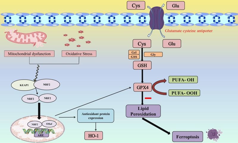
Figure 3: The Nrf2/HO-1/GPX4 axis as a regulator of Ferroptosis in Huntington’s disease.
Deferoxamine (DFOA), also called desferrioxamine, is a drug that chelates iron and aluminium. Deferoxamine treats acute iron poisoning, particularly in young children. This drug is commonly used to manage hemochromatosis, a condition involving iron buildup that may be hereditary or acquired. In addition to iron overload, Deferoxamine treats aluminium toxicity (excess aluminium accumulation) in certain patients (Kontoghiorghe & Kontoghiorghes, 2016).
Deferoxamine arises from stripping the Fe3+ from ferrioxamine B, a siderophore made by Streptomyces pilosus actinomycetes. Deferoxamine binds free iron in plasma and promotes its excretion via urine. By chelating excess iron in hemochromatosis patients, Deferoxamine limits organ and tissue damage, particularly to the liver. It also accelerates nerve injury recovery and limits recent nerve trauma severity. Deferoxamine can alter inflammatory mediator expression and release from certain cell types (Entezai et al; 2022).
Deferoxamine shows poor oral absorption from the GI tract, requiring IM, SC, or IV administration. Vitamin C enhances deferoxamine’s efficacy by releasing stored iron, raising levels available for chelation. Long- term Deferoxamine treatment may cause sensorineural hearing loss and retinopathy. Acute side effects may involve GI upset, anaphylaxis, skin discoloration, injection site irritation, and anaphylaxis. Deferoxamine has limited blood-brain barrier penetration due to its large molecular size and hydrophilic properties. Consequently, only minimal amounts reach the CNS, limiting its therapeutic efficacy against brain iron overload and reducing its promise as a treatment strategy for neurodegenerative disorders. Studies indicate that BBB disruption during neuroinflammation, trauma, or neurodegeneration enhances Deferoxamine penetration, enabling greater brain tissue diffusion (Cassinerio et al; 2014).
Deferiprone, an oral iron chelator, removes excess iron in thalassemia or sickle cell patients on frequent transfusions, preventing iron overload organ damage. Deferiprone, an oral bidentate iron chelator, binds iron at a 3:1 ratio to form a stable ferric complex primarily excreted in urine. It selectively chelates excess Fe3+, blocks iron-driven free radical production, and reduces oxidative damage linked to neurodegeneration. As a small, lipophilic molecule with strong membrane permeability, it outperforms deferoxamine in accessing intracellular iron pools. A major advantage is its ability to reach mitochondrial iron, where overload fuels ROS production and cell damage (Hider & Hoffbrand, 2018). Its low molecular weight and partial lipophilicity enable deferiprone to cross the blood-brain barrier more effectively than most chelators. It penetrates the brain primarily through passive diffusion and partly via transporter-mediated routes, reaching neuronal tissues with pathological iron buildup. Its ability to cross the blood-brain barrier, along with being taken by mouth, makes it a top treatment choice for brain diseases. It lowers harmful iron levels throughout the body and in the brain, helping balance metals and reduce brain cell death from oxidative stress (Cohen et al; 2003).
Deferasirox, an oral chelator, primarily treats chronic iron overload in patients on long-term transfusions for beta-thalassemia and other anemias. Deferasirox has a 8-16 hour half-life, enabling once-daily dosing. Two Deferasirox molecules bind one iron atom, which is then eliminated mainly via feces. Its low molecular weight and high lipophilicity enable oral administration, unlike deferoxamine which requires IV delivery (Bedford et al; 2013). Normally, little Deferasirox crosses the BBB, but in neurodegenerative conditions with inflammation, oxidative stress, or vascular issues increasing barrier permeability, neural tissue access improves. Evidence suggests plasma protein binding dynamics aid passive diffusion by slowly releasing deferasirox near endothelial barriers, allowing small amounts to cross. Even low brain can help when iron builds up slowly and clears steadily with long-term use (Kamalinia et al; 2013).
In HD, mutant huntingtin leads to disruption of iron regulatory proteins, which boosts Fe2+ levels in the striatum. This drives the Fenton reaction, generating hydroxyl radicals. These radicals target PUFA-phospholipids within neuronal membranes. Polyunsaturated fatty acid phospholipids (PUFA-PL) convert to phospholipid hydroperoxides (PL-OO-) upon peroxidation. There will be initiation of lipid peroxidation cascades by these reactive species, culminating in extensive membrane damage. Ultimately, this process leads to execution of ferroptosis. However, Ferrostatin-1 (Fer-1) acts a lipid radical scavenger. It embeds in lipid bilayers in neural models of HD, gives hydrogen atom to peroxyl radicals (LOO-) and produces stable Fer-1 radicals and non-propagating lipid hydroperoxides (LOOH). This leads to interruption of lipid peroxidation chains. Fer-1 stops lipid oxidation chains from rupturing neuronal membranes. In striatal neurons with HD, it reduces membrane stiffness and prevents the development of pores, maintains ATP production and the integrity of the mitochondrial cristae and prevents ferroptotic necrosis (Miotto et al; 2020).
Liproxstatin-1 (Lip-1), a synthetic spiroquinoxalinamine, functions as an antioxidant with structural divergence from Fer-1 but same radical-scavenging action. Liproxstatin-1 has more metabolic stability as well as in vivo potency relative to Fer-1, enhancing its efficacy in ferroptosis models. In HD, expression and activity of GPX4 decline, hindering the breakdown of phospholipid hydroperoxides (PL-OOH). In HD – affected neurons, Lip-1 maintains ability of GPX4’s to detoxify phospholipid hydroperoxides by strengthening its structural integrity under oxidative stress. Lip-1 also helps GPX4 in converting lipid hydroperoxides (LOOH) to harmless lipid alcohols, terminating peroxidation. Similar to Fer-1, Lip-1 traps lipid peroxyl radicals (LOO-), intercepting them directly in PUFA-rich membranes. It stops formation of harmful oxidized phosphatidylethanolamine (PE) species, like 15-HpETE-PE, the critical ferroptosis trigger (Fan et al; 2021).
Vitamin E (α-tocopherol and analogs), a fat-soluble chain-breaking antioxidant, directly quenches the final lipid peroxidation phase, aligning it squarely with ferroptosis blockers. Vitamin E embeds in phospholipid bilayers, the site of ferroptosis execution. Its chromanol ring donates hydrogen to lipid peroxyl radicals (LOO-), transforms them into stable lipid hydroperoxides (LOOH) and thus halts the lipid peroxidation chain reaction. The primary enzyme inhibitor of ferroptosis, GPX4, is less active in HD animals due to shortage of GSH and oxidative overload (Zhang et al; 2022). Vitamin E operates downstream of GPX4, functions without needing GSH and also safeguards membranes even when GPX4 is weakened or inactive. Mitochondrial dysfunction stands as a hallmark of HD. However, vitamin E blocks oxidation of lipids in mitochondrial membranes, maintains fluidity of membrane and structural wholeness and decreases the amplification of secondary mitochondrial ROS. It has been demonstrated that vitamin E inhibits the build-up of harmful lipid aldehydes (4-HNE, MDA), minimize oxidative damage that is dependent on iron and reduce the amount of neuroinflammatory signals that increases lipid oxidation (Kagan et al; 2024). Table 2 summarizes the preclinical experimental studies on therapeutic modulation of ferroptosis.
Table 2: Experimental investigations of Therapeutic modulators of Ferroptosis in Preclinical models.
|
S.No. |
Therapeutic candidate |
Experimental Model |
Dose |
Duration |
Outcomes |
References |
|
1. |
Deferoxamine |
APP/PS1 double transgenic mice watered with high-dose iron |
200 mg/kg (intranasal) |
90 days |
Reduced levels of Aβ and APP; improved long-term memory storage. |
(Guo et al; 2013) |
|
2. |
Deferoxamine |
Rotenone induced PD Wistar rats |
60 mg/kg (i.p.) |
8 weeks |
Blocked iron buildup in the substantia nigra, striatum, globus pallidus, hippocampus, and cerebellum; shielded dopaminergic neurons. |
(Xiong et al; 2012) |
|
3. |
Deferasirox |
Tau transgenic mice |
1.6 mg |
2 months |
Decreased hypophosphorylated tau levels and no effect on memory retention and motor ability. |
(Kwan et al; 2022) |
|
4. |
Liproxstatin-1 |
LPS-induced cognitive impairment in mice |
10 mg/kg (i.p.) |
1 Week |
Liproxstatin-1 alleviates LPS-induced learning/memory deficits by blocking hippocampal ferroptosis via reduced oxidative stress, inflammation, iron overload, and mitochondrial dysfunction. |
(Li et al; 2022) |
|
5. |
Vitamin E |
PTZ-induced kindling Epilepsy |
200 mg/kg (i.p.) |
29 days |
Vitamin-E attenuates PTZ-induced seizures by blocking neuronal ferroptosis, probably by inhibiting 15-LOX activity, limiting lipid peroxidation and iron buildup, and boosting GPX4 and GSH levels. |
(Zhang et al; 2022) |
Nrf2 stands out as a key endogenous defensive mechanism against ferroptosis. It coordinates antioxidant defenses, clearance of lipid peroxides, and maintenance of iron balance. Growing research show that Nrf2 signaling fails to activate adequately in HD. This makes neurons- especially striatal medium spiny neurons- extremely susceptible to damage caused by ferroptosis.
Nrf2 activation triggers a coordinated transcriptional program that inhibits key steps in the ferroptotic pathway. It boosts GPX4- the only enzyme that reduces hydroperoxides in membrane phospholipids- while also upregulate the expression of glutathione synthesis enzymes (GCLC, GCLLM) and the cysteine importer SLC7A11. This replenishes cellular glutathione stores. At the same time, Nrf2 controls iron handling by elevating ferritin (FTH1) and ferroportin levels. This limits the free iron pool that drives lipid radical production. Taken together, this Nrf2 driven system reestablishes redox equilibrium, decreases lipid peroxidation, and strengthens neurons against ferroptosis- mechanisms severely impaired in HD (Han et al; 2024).
Sulforaphane, an isothiocyanates from natural sources, activates Nrf2 by covalently altering cysteine sites on keap1. This promotes the buildup of Nrf2 in the nucleus and turns on heme- oxygenase-1 (HO-1) expression, which helps shield cells from inflammation and prevents apoptosis and oxidative damage (Zhang et al; 2023). Sulforaphane could strengthen Nrf2 activity, replenishes glutathione, and sharply reduces lipid peroxidation-shown by lower malondialdehyde and 4-hydroxynonenal buildup in HD. In HD models triggered by toxins, sulforaphane could decreases striatal neuron loss, enhances motor function, and safeguards mitochondrial health.
Synthetic triterpenoids like bardoxolone methyl (CDDO-Me) rank as the strongest known drugs for activating Nrf2. Through alkylation of keap1 cysteines, they drive strong upregulation of Nrf2 targets that handle antioxidant protection, iron storage, and lipid peroxide breakdown (Schiavoni et al; 2025). Though testing in genetic HD models is still limited, the clear parallels between CDDO-driven cell protection and ferroptosis blockade highlight their promise as regulators of ferroptotic risk in HD.
Dimethyl fumarate (DMF), an FDA-approved drug that activates Nrf2 and crosses into brain effectively, emerges as a top candidate for HD therapy. DMF triggers Nrf2 by electrophilically altering keap1, yielding long-lasting activation of genes for antioxidant defense and cell protection (Gao et al; 2024). DMF could raises GPX4 levels, rebuilds glutathione reserves, and strongly reduces buildup of lipid ROS. In HD models using toxins, DMF could enhance motor performance, limits striatal oxidative harm, and maintains neuronal structure. Crucially, these protective effects take place without caspase activation, which is consistent with preventing ferroptosis over apoptosis.
Iron chelation is a plausible way to prevent ferroptosis in HD, but long-term treatment poses serious safety risks because of systemic iron depletion, off-target toxicity, and interference with physiological functions that depend on iron. Sustained chelation can provoke anemia, weakened immunity, mitochondrial defects, and targeted organ damage- especially without underlying iron overload. These risks emphasize the necessity of careful dosage, close observation, and the creation of combination treatments that lessen the need for drastic iron removal. Combining iron chelation with Nrf2 activation or GPX4 enhancement could amplify neuroprotection, safeguard systemic iron balance, and boost the clinical viability of ferroptosis-focused treatments for HD.
Even though there is strong preclinical evidence linking ferroptosis to HD, a number of translational obstacles need to be removed before these approaches may be used in clinical settings. Major hurdles comprise efficient blood-brain barrier (BBB) penetration, long-term dosing refinement, and figuring out the best treatment window.
The BBB poses a primary barrier for ferroptosis inhibitors and iron chelators aimed at striatal neuron protection in HD. Numerous substances exhibiting strong in vitro ferroptosis inhibition show: Low lipophilicity, elevated molecular weight and efflux by transporters. For eg: Although having potent iron-chelating qualities, deferoxamine has restricted BBB penetration, limiting its central efficacy (Zhao et al; 2023).
Translational considerations
HD follows a chronic, gradual progression, requiring long-term treatment. However, prolonged medication exposure may be necessary for the ongoing suppression of ferroptosis. Extended dosing increases systemic toxicity risks, especially for iron chelators.
Translational considerations
Ferroptosis mechanisms could initiate early during HD onset, well before evident neuron death occurs. Majority of preclinical treatments begin at symptomatic stages, once significant striatal injury has already taken place. This raises the possibility that established neurodegeneration cannot be reversed by late intervention.
Translational considerations
Overcoming these translational obstacles proves vital for effectively advancing ferroptosis-targeted, disease-altering treatments in HD.
Future investigations should examine the effects of drug-based or gene-based stimulation of Nrf2 on boosting HO-1 and GPX4 levels to combat iron-induced lipid peroxidation in HD experimental systems. Research that contrasts Nrf2-activating drugs, inhibitors of KEAP1, and plant-derived agents could identify optimal agents for modulating defenses against ferroptosis.
Although HD involves disrupted iron buildup, the links between Nrf2 pathways and iron controllers such as transferrin receptor, ferritin, and ferroportin are not well defined. Upcoming studies need to unpack these connections and test if normalizing iron balance alone lessens sensitivity to ferroptosis.
Compounds that inhibit ferroptosis, such as liproxstatin-1, ferrostatin-1, and deferiprone, protect neurons in early-stage disease models, yet their sustained benefits and safety profiles in ongoing brain degeneration need thorough testing. Combining ferroptosis blockers with Nrf2 stimulators may enhance defense by addressing both acute lipid ROS and upstream antioxidant deficiencies. Head-to-head trials in HD animals should measure combined effects on GPX4 stability, neuronal survival, and behavioral outcomes.
Given that GPX4 is a fundamental anti-ferroptotic enzyme, advanced treatment approaches like as CRISPR-based upregulation or stem-cell transplantation modified to overexpress GPX4 could be investigated. Future research could determine whether GPX4 restoration improves behavioral outcomes and stops striatal neuronal loss.
Markers for lipid damage, iron dysregulation, or Nrf2 pathway engagement in clinics could sharpen early diagnosis and track treatment responses. Assays for blood malondialdehyde, 4-HNE, ferroptosis-linked miRNAs, or HO-1 might guide patient selection for antioxidants.
Studies should prioritize BBB-crossing versions of Nrf2/GPX4-boosting agents, nanoparticle carriers, and long-acting release methods to achieve sufficient CNS exposure. Launching phase 1 trials would clarify their potential for human application.
Huntington’s disease represents a multifaceted neurodegenerative condition where oxidative stress, iron buildup, and lipid peroxidation collectively drive ongoing neuronal degeneration. Growing data highlight ferroptosis as a central driver of neuronal fragility in HD, marked by iron overload, glutathione shortage, and defective clearance of lipid peroxides. The Nrf2/HO-1/GPX4 pathway functions as a core antioxidant shield, protecting neurons by preserving redox equilibrium, managing iron levels, and inhibiting lipid peroxidation. Disruption of this pathway undermines cellular defenses versus ferroptotic stress, promoting toxicity from mutant huntingtin and advancing disease course. Understanding the initiation and persistence of ferroptosis in HD, along with ways to modulate Nrf2, HO-1, and GPX4 against it, offers key perspectives on novel treatment options. Strengthening this pathway via drug activators, genetic modulation, or iron chelators shows potential to curb neurodegeneration and enhance patient outcomes, spotlighting ferroptosis blockade as a promising avenue for HD research ahead.
Acknowledgement
I want to extend my sincere thanks to the ISF College of Pharmacy for their invaluable assistance. I am also grateful to Professor Dr. YK Gupta, president of AIIMS Bhopal and AIIMS Jammu, for his insightful feedback and excellent recommendations.
Statements & Declarations
Funding
The authors declare that no funds, grants, or other support were received during the preparation of this manuscript.
Competing Interests
The authors have no relevant financial or non-financial interests to disclose.
Author’s contribution
Rohan Bansal: Paper writing, editing and data collection; Sanchi Jain, Mohin Modak: Paper editing and preparing figures and tables; Shamsher Singh: Conceptualization, Final review, Editing and Supervision.
Ethics approval
Not applicable
Abbreviations
HD: Huntington’s disease
BBB: Blood brain barrier
DMT1: Divalent metal transporter-1
GPX4: Glutathione peroxidase 4
GSH: Glutathione
PUFAs: Polyunsaturated fatty acid
ROS: Reactive oxygen species
MDA: Malondialdehyde
4-HNE: 4- hydroxynonenal
mHTT: Mutant huntingtin
MSNs: Medium spiny neurons
FPN1: Ferroportin
HO-1: Heme oxygenase-1
AREs: Antioxidant response elements
3-NP: 3-Nitropropionic acid
QSM: Quantitative susceptibility mapping
DFOA: Deferoxamine
Fer-1: Ferrostatin-1
Lip-1: Liproxstatin-1
LOOH: lipid hydroperoxides
PL-OOH: Phospholipid hydroperoxides
CDDO-Me: Bardoxolone methyl
Nrf2: Nuclear factor erythroid 2-related factor 2
RSL3: RAS-selective lethal 3
DMF: Dimethyl fumarate
KEAP1: Kelch-like ECH-associated protein 1.

Open Access By Aditum Open Access Journals id licensed under Creative Commons Attribution 4.0 International License. Based On a Work at aditum.org


