Pediatrics and Child Health Issues
OPEN ACCESS | Volume 4 - Issue 1 - 2025
ISSN No: 2836-2802 | Journal DOI: 10.61148/2836-2802/JPCHI


Sylvia Luen Yee Siu1*, Raymond Suen Bun Cheng2
1Postal address: Department of Paediatrics & Adolescent Medicine, Tuen Mun Hospital, Tsing Chung Koon Road, Tuen Mun, New Territories. Hong Kong Special Administrative Region, China.
2Institution: Department of Paediatrics & Adolescent Medicine, Tuen Mun Hospital, Tsing Chung Koon Road, Tuen Mun, New Territories. Hong Kong Special Administrative Region, China.
*Corresponding author: Sylvia Luen Yee Siu, Postal address: Department of Paediatrics & Adolescent Medicine, Tuen Mun Hospital, Tsing Chung Koon Road, Tuen Mun, New Territories. Hong Kong Special Administrative Region, China.
Received: May 04, 2025
Accepted: May 15, 2025
Published: May 26, 2025
Citation: Yee Siu SL, Raymond Suen B Cheng. (2025) “Neonatal Tension Pneumothorax.”, Pediatrics and Child Health Issues, 5(1); DOI: 10.61148/2836-2802/JPCHI /070.
Copyright: © 2025. Sylvia Luen Yee Siu. This is an open access article distributed under the Creative Commons Attribution License, which permits unrestricted use, distribution, and reproduction in any medium, provided the original work is properly cited.
A 690 grams male infant delivered at 24+2 weeks had pulmonary interstitial emphysema developed early on. His CXR showed right tension pneumothorax and pneumoperitoneum on day 6. Bowel perforation was once suspected because of the pneumoperitoneum but the baby improved dramatically after chest drain insertion with all leaks resolved. Milk feeding was successfully established subsequently.
Conclusion: This case highlights the urgent need to reposition ETT close to carina among neonates to avoid tension pneumothorax development from accidental one lung intubation.
newborn; tension pneumothorax; pneumoperitoneum; intubation case report
Neonatal Tension Pneumothorax
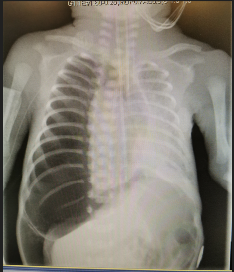
A 690g male infant delivered at 24+2 weeks had respiratory distress syndrome treated with Curosurf. Chest X-ray showed pulmonary interstitial emphysema (PIE) changes from day 5 onwards. On day 6 he suddenly developed desaturation with bradycardia, not responding to bagging. Auscultation revealed reduced air entry over right lung; endotracheal tube was changed for suspected blockage. Transient improvement noted. Then, the baby deteriorated again. Repeated auscultation showed markedly reduced air entry over right lung. Transillumination was positive. X-ray confirmed right tension pneumothorax but pneumoperitoneum was unexpected (figure 1). The baby’s condition did not improve upon repeated chest tapping and required chest compression and Adrenaline. A chest drain was inserted and the baby then improved. Bowel perforation was once suspected but the infant improved so dramatically after chest drainage and milk feeding was subsequently established, ruling out bowel perforation.
The commonest cause of pneumoperitoneum is perforated gut. Reviewing our case, the tension pneumothorax most likely arose from endotracheal tube (ETT) displaced into right main bronchus causing one lung ventilation in the emphysematous lung as latest X-ray before pneumothorax showed ETT just above carina. In PIE, elastance and compliance of diseased lungs are compromised; the resultant “double” ventilator pressure might then affect the vulnerable lung tissue and lead to tension pneumothorax. Under tension, air may track through the apertures where the aorta, esophagus, inferior vena cava, splanchnic nerves, roots of the azygous veins and the sympathetic chain traverse the diaphragm leading to pneumoperitoneum. This case highlights the urgent need to reposition ETT close to carina among neonates to avoid tension pneumothorax development from accidental one lung intubation.


Open Access By Aditum Open Access Journals id licensed under Creative Commons Attribution 4.0 International License. Based On a Work at aditum.org