
Neurosurgery and Neurology Research
OPEN ACCESS | Volume 7 - Issue 1 - 2025
ISSN No: 2836-2829 | Journal DOI: 10.61148/2836-2829/NNR


José Clayton Balo
Department of Neurosurgery, Main Military Hospital Institute Superior, Luanda - Angola.
*Corresponding author: José Clayton Balo, Department of Neurosurgery, Main Military Hospital Institute Superior, Luanda - Angola.
Received: September 10, 2026 | Accepted: September 26, 2026 | Published: October 14, 2026
Citation: José C Balo, (2026) “Acute Cervical Spinal Epidural Hematoma Following Cervical Massage: A Case Report” Neurosurgery and Neurology Research, 8(1); DOI: 10.61148/2836-2829/NNR/059.
Copyright: ©2026. José Clayton Balo. This is an open access article distributed under the Creative Commons Attribution License, which permits unrestricted use, distribution, and reproduction in any medium, provided the original work is properly cited.
Background: Cervical spinal epidural hematoma (CSEH) is a rare but potentially devastating cause of acute spinal cord compression. It may present with sudden severe cervical pain followed by rapidly progressive motor, sensory, or autonomic dysfunction. Although uncommon, cervical manipulation or massage has been reported in temporal association with epidural hematoma.
Case Description: A 75-year-old man with recurrent cervical pain developed acute, progressively worsening cervical pain radiating to the left arm after cervical massage at home. Over the following course, he developed severe weakness in all four extremities followed by urinary and fecal sphincter dysfunction. On admission, cervical pain was 8/10. Motor strength was 1/5 in the right upper limb, 2/5 in the left upper limb, and 0/5 in both lower limbs. Sensation was preserved and generalized osteotendinous hyperreflexia was present. Cervical computed tomography demonstrated an epidural hematoma extending from C3 to C6 with left-sided spinal cord compression. Magnetic resonance imaging confirmed the lesion. The patient underwent C3–C6 laminoplasty, evacuation of epidural clots, and fixation with microplates and microscrews.
Conclusion: CSEH should be considered in patients with acute severe cervical pain and rapidly progressive neurological deficits. The temporal association with cervical massage is clinically noteworthy, although causality cannot be established from a single case. Prompt imaging and urgent decompression are essential when significant neurological deterioration and spinal cord compression are present.
cervical spinal epidural hematoma; spinal cord compression; cervical spine; cervical massage; tetraparesis; spinal decompression
Spontaneous spinal epidural hematoma (SSEH) is an uncommon neurological emergency caused by bleeding into the spinal epidural space, producing compression of the spinal cord and/or nerve roots. Clinical presentation typically includes acute spinal pain, sometimes with radicular radiation, followed by progressive motor, sensory, or sphincter dysfunction.¹⁻⁶
Cervical and cervicothoracic hematomas are particularly important because a relatively short longitudinal lesion may produce profound neurological impairment. Contemporary series and systematic reviews indicate that preoperative neurological status and the timing of decompression are important determinants of outcome, although the evidence is derived mainly from retrospective cohorts and case reports.²⁻⁵
Magnetic resonance imaging is the preferred diagnostic modality because it delineates the location and longitudinal extent of the hematoma and its relationship with the spinal cord. Computed tomography may provide an important initial diagnostic clue in emergency settings or where MRI access is limited.⁷⁻⁹
Spinal epidural hematoma has multiple possible etiologies, including anticoagulation, antiplatelet therapy, vascular malformations, trauma, spinal procedures, coagulation abnormalities, and idiopathic bleeding. Cervical manipulation and massage have rarely been reported in temporal association with cervical epidural hematoma.⁹⁻¹⁴
We report a 75-year-old man with severe progressive tetraparesis and sphincter dysfunction associated with a C3–C6 cervical epidural hematoma occurring shortly after cervical massage. The patient underwent surgical decompression, hematoma evacuation, and cervical fixation.
Case Description
Clinical Presentation
A 75-year-old man with a history of recurrent cervical pain was apparently in good general health until 5 days before hospital admission, when he developed acute cervical pain with progressive worsening and radiation to the left upper limb. The acute episode was temporally associated with cervical massage performed at home.
His neurological condition subsequently deteriorated, with reduction in muscle strength involving all four extremities, followed by urinary and fecal sphincter dysfunction. His family brought him to the emergency department because of progressive neurological deterioration.
On assessment, the patient reported cervical pain rated 8/10 on the visual analogue scale. Neurological examination demonstrated severe motor impairment. Upper-limb strength was 1/5 on the right and 2/5 on the left, while both lower limbs had 0/5 strength. Sensory function was preserved, and generalized osteotendinous hyperreflexia was present.
The clinical findings were compatible with acute cervical spinal cord compression with severe motor and autonomic dysfunction. The available documentation did not provide a complete standardized International Standards for Neurological Classification of Spinal Cord Injury (ISNCSCI/AIS) examination; therefore, an AIS grade was not assigned retrospectively.
Imaging
Preoperative cervical CT demonstrated an epidural collection compatible with a hematoma extending from C3 to C6, producing left-sided spinal cord compression (Fig. 1). Cervical MRI subsequently confirmed the epidural hematoma and its compressive effect on the spinal cord (Fig. 2).
The combination of rapidly progressive neurological deterioration and imaging evidence of substantial spinal cord compression prompted urgent surgical management.
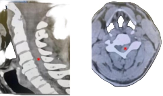
Fig. 1 Preoperative cervical spine computed tomography. (A) Sagittal image demonstrating an epidural hematoma extending from C3 to C6. (B) Axial image demonstrating the epidural collection with left-sided spinal cord compression (red asterisk).
Fig. 2 Preoperative cervical spine magnetic resonance imaging. Sagittal T1-weighted (A) and axial T2-weighted (B) images demonstrating the cervical epidural hematoma extending from C3 to C6 with left-sided spinal cord compression (red asterisk). The original MRI image should be inserted before submission.
Surgical Management
Because of the severe and progressive neurological deficit and radiologically confirmed spinal cord compression, the patient underwent surgical decompression. A C3–C6 laminoplasty was performed, followed by evacuation of the epidural blood clots. Cervical fixation was subsequently performed using microplates and microscrews.
The intraoperative material documented the evacuated epidural clots, the posterior cervical operative field with fixation, and intraoperative radiographic confirmation of the instrumentation (Fig. 3).
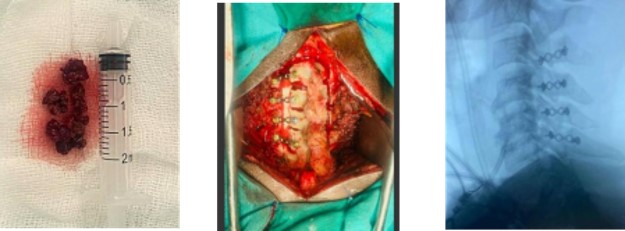
Fig. 3 Intraoperative findings and surgical stabilization. (A) Evacuated epidural blood clots. (B) Posterior cervical operative field with microplate fixation. (C) Intraoperative cervical radiograph demonstrating the fixation construct.
Postoperative Course
Postoperative cervical CT demonstrated postoperative changes following decompression and fixation, with sagittal, axial, and three-dimensional reconstructions documenting the surgical construct (Fig. 4).
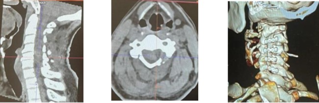
Fig. 4 Postoperative cervical spine computed tomography. (A) Sagittal reconstruction. (B) Axial image. (C) Three-dimensional reconstruction demonstrating the postoperative cervical fixation construct.
The patient is currently followed in the outpatient clinic and is undergoing physiotherapy. Detailed serial postoperative neurological examinations, standardized AIS grades, and quantitative functional outcome measures were not available in the clinical information supplied for this report.
Discussion
Cervical spinal epidural hematoma is rare but can cause rapidly progressive spinal cord dysfunction. The classic clinical sequence consists of sudden severe spinal pain followed by neurological deterioration, although presentations may vary.¹⁻⁶ In a 2024 surgical series of 26 patients with spontaneous spinal epidural hematoma, cervical and cervicothoracic lesions accounted for a substantial proportion of cases, and most surgically treated patients improved neurologically.²
A systematic review and meta-analysis by Vastani et al. analyzed 181 studies involving 295 surgically treated adult patients and identified preoperative neurological status and operative interval among factors associated with outcome.³ A multicenter retrospective study similarly evaluated prognostic factors and treatment efficacy and emphasized the importance of individualized management according to neurological status and hematoma characteristics.⁴
Nakamura et al. specifically investigated ultraearly evacuation within 12 hours and reported an association with better functional outcomes in symptomatic spontaneous spinal epidural hematoma.⁵ These findings are consistent with the principle that severe or progressive neurological deficits in the setting of significant spinal cord compression warrant rapid diagnosis and treatment.
The present patient had profound neurological impairment, including marked weakness in all four extremities and urinary and fecal sphincter dysfunction. The hematoma extended from C3 to C6 and compressed the spinal cord predominantly on the left. This combination of clinical and radiological findings supported urgent surgical decompression.
MRI is generally regarded as the most informative diagnostic examination for spinal epidural hematoma. Kato and Terae emphasized the diagnostic role of MRI in cervical epidural hematoma, while more recent reports demonstrate that CT can occasionally identify the lesion before MRI, particularly in emergency settings.⁷⁸
Etiological assessment is also important. A 2024 case series of 20 spontaneous spinal hematomas found frequent associations with anticoagulant therapy and vascular abnormalities, illustrating the importance of reviewing medications, coagulation status, and, where appropriate, investigating vascular causes.¹⁴ The available information in the present case does not document anticoagulant or antiplatelet exposure, coagulation studies, or vascular imaging; these data should be retrieved if available.
The temporal association between cervical massage and symptom onset is a notable feature. Ryu et al. reported a cervical epidural hematoma associated with cervical spine massage.⁹ Other published reports have described epidural hematoma following spinal manipulation or physiotherapy.¹⁰⁻¹³ A 2026 report described spontaneous cervicothoracic epidural hematoma following physiotherapy in an elderly patient receiving apixaban.¹⁰
These reports support awareness of the possibility of spinal epidural bleeding after cervical manipulation, but they do not establish a direct causal mechanism. In the present case, massage preceded the acute pain and neurological deterioration, but the absence of complete etiological investigation prevents attribution of causality. The appropriate description is therefore a temporal association rather than a proven causal relationship.
The surgical treatment in this case consisted of C3–C6 laminoplasty, evacuation of the epidural clots, and fixation with microplates and microscrews. There is no universally established operative technique for all spinal epidural hematomas; surgical exposure should be individualized according to hematoma location, longitudinal extension, spinal stability, and the need for adequate decompression.²⁻⁴
Conservative management has also been reported in selected patients with mild deficits or early neurological improvement. Recent case reports describe complete recovery without surgery in carefully selected patients, but these circumstances differ from the present case, in which neurological deficits were severe and progressive and sphincter dysfunction was present.¹⁵⁻¹⁷
Neurological outcome after spinal epidural hematoma depends on multiple factors, including initial neurological status, hematoma extent, and time to decompression.⁵˒¹⁸ The present patient's continued outpatient rehabilitation is therefore clinically important, although the lack of standardized postoperative examinations limits quantitative assessment of recovery.
This case highlights an important diagnostic principle: acute severe neck pain followed by rapidly progressive weakness and sphincter dysfunction should prompt immediate consideration of cervical spinal cord compression, including epidural hematoma. Early imaging and appropriate decompression may be decisive in preventing further neurological injury.
Conclusion
Cervical spinal epidural hematoma is an uncommon but potentially devastating cause of acute spinal cord compression. In a patient with acute severe cervical pain followed by rapidly progressive tetraparesis and sphincter dysfunction, urgent spinal imaging is essential.
In this case, CT and MRI demonstrated a C3–C6 epidural hematoma with left-sided spinal cord compression. The patient underwent C3–C6 laminoplasty, evacuation of the epidural clots, and cervical fixation. He remains under outpatient follow-up and physiotherapy.
The temporal relationship between cervical massage and symptom onset is clinically noteworthy, but causality cannot be established from this case alone. The report emphasizes early recognition, rapid imaging, and timely decompressive management in patients with severe neurological deterioration.
Statements and Additional Information
Conflict of Interest
The authors declare that they have no conflict of interest.
Funding
No external funding was received for this work.
Informed Consent
Written informed consent for publication of the case and anonymized clinical and radiological images should be obtained from the patient or his legally authorized representative before submission.
Ethical Approval
The authors should confirm whether institutional ethics committee approval or a formal waiver was required for this single case report according to local institutional policy.
Data Availability
The clinical information relevant to this case is contained within the manuscript. No directly identifying patient information should be submitted.
Author Contributions
José Clayton Balo: conceptualization, clinical case description, investigation, literature review, writing—original draft, writing—review and editing. Additional authors and their contributions should be added before submission if applicable.

Open Access By Aditum Open Access Journals id licensed under Creative Commons Attribution 4.0 International License. Based On a Work at aditum.org


