
Neurosurgery and Neurology Research
OPEN ACCESS | Volume 7 - Issue 1 - 2025
ISSN No: 2836-2829 | Journal DOI: 10.61148/2836-2829/NNR


José Clayton Balo1*, Ladislau Nele2
1Neurosurgeon and Master of Main Military Hospital / Superior Institute, Director of Neurosurgical Department.
2Neurosurgeon at Josina Machel Hospital.
*Corresponding author: José Clayton Balo, Neurosurgeon and Master of Main Military Hospital / Superior Institute, Director of Neurosurgical Department.
Received: September 10, 2026 | Accepted: September 30, 2026 | Published: October 14, 2026
Citation: José C Balo, Nele L, (2026) “Cerebellar Pilocytic Astrocytoma Presenting With Obstructive Hydrocephalus in an Adolescent: A Case Report” Neurosurgery and Neurology Research, 8(1); DOI: 10.61148/2836-2829/NNR/058.
Copyright: ©2026. José Clayton Balo. This is an open access article distributed under the Creative Commons Attribution License, which permits unrestricted use, distribution, and reproduction in any medium, provided the original work is properly cited.
Background
Pilocytic astrocytoma is a circumscribed central nervous system glioma classified as CNS WHO grade 1 and is one of the most common pediatric brain tumors. Cerebellar pilocytic astrocytomas may produce symptoms of increased intracranial pressure and cerebellar dysfunction, particularly when compression of the fourth ventricle results in obstructive hydrocephalus.
Case Presentation
A 15-year-old right-handed male patient presented with holocranial headache, projectile vomiting, gait disturbance, and progressive cerebellar dysfunction. Neurological examination revealed drowsiness, mixed appendicular and truncal ataxia, dysdiadochokinesia, nystagmus, and intention tremor. Non-contrast computed tomography demonstrated a 3 × 3 cm cystic and nodular lesion in the right cerebellar hemisphere, producing mass effect on the fourth ventricle, dilatation of the ventricular system, and periventricular edema. The Evans index was 0.40, and the third ventricle measured 1.6 cm. Emergency ventriculoperitoneal shunt placement was initially performed to treat the obstructive hydrocephalus. Subsequently, the patient underwent bilateral posterior fossa craniectomy, evacuation of the cystic component, and complete removal of the mural tumor nodule. Histopathological examination demonstrated pilocytic astrocytoma, CNS WHO grade 1.
Outcome
The postoperative course was favorable, and the patient was discharged 15 days after surgery. Follow-up cranial CT performed one year after surgery showed no evidence of tumor recurrence. At the most recent clinical follow-up, the patient was attending school normally and participating in regular academic activities without functional limitations or significant neurological disability.
Conclusion
Cerebellar pilocytic astrocytoma may present with significant obstructive hydrocephalus despite its generally indolent biological behavior. Prompt management of hydrocephalus followed by maximal safe tumor resection can result in favorable neurological and functional outcomes. Long-term clinical and radiological surveillance remains essential.
Cerebellar pilocytic astrocytoma; Pediatric low-grade glioma; Hydrocephalus; Posterior fossa tumor; Gross-total resection; Cerebellar tumor
1. Introduction
Pilocytic astrocytoma (PA) is a circumscribed glioma classified as CNS WHO grade 1 and is one of the most common primary brain tumors in children. Although generally associated with a favorable prognosis, its clinical presentation varies according to anatomical location and tumor size. Pediatric low-grade gliomas are increasingly regarded as chronic diseases in which long-term functional and neurocognitive outcomes are important in addition to tumor control. [1,2,9,10]
The cerebellum is a common site of pediatric pilocytic astrocytoma. Tumors arising in the posterior fossa may produce neurological symptoms through direct cerebellar involvement or obstruction of cerebrospinal fluid circulation. Consequently, patients may present with headache, vomiting, gait disturbance, ataxia, and signs of raised intracranial pressure. Hydrocephalus is particularly relevant because compression of the fourth ventricle can compromise cerebrospinal fluid circulation.
Recent pediatric series have demonstrated that hydrocephalus is frequent in posterior fossa pilocytic astrocytoma. In a cohort of 57 children with posterior fossa PA, 47.3% had hydrocephalus at diagnosis and 33.3% ultimately required ventriculoperitoneal shunting. [5] A separate cerebellar PA cohort also demonstrated a high prevalence of hydrocephalus at presentation. [4]
Surgery remains the principal treatment for a surgically accessible cerebellar PA. Current evidence supports maximal safe resection when it can be achieved without unacceptable neurological morbidity. Recent data also suggest that stable residual disease can often be monitored rather than subjected automatically to repeated surgery. [2,3,4]
We report the case of an adolescent with a cystic and nodular cerebellar pilocytic astrocytoma presenting with symptomatic obstructive hydrocephalus, successfully managed with staged cerebrospinal fluid diversion followed by complete removal of the mural tumor nodule.
2. Case Presentation
2.1 Clinical Presentation
A 15-year-old right-handed male patient presented with holocranial headache associated with projectile vomiting and gait disturbance.
On neurological examination, the patient was drowsy and demonstrated mixed cerebellar ataxia involving both appendicular and truncal coordination. Dysdiadochokinesia, nystagmus, and intention tremor were also observed.
The clinical findings were suggestive of significant posterior fossa dysfunction associated with increased intracranial pressure.
2.2 Initial Computed Tomography
A non-contrast computed tomography (CT) scan of the head demonstrated a hypodense cystic and nodular expansive lesion involving the right cerebellar hemisphere, measuring approximately 3 × 3 cm in its longitudinal and anteroposterior dimensions.
The lesion produced a significant mass effect on the fourth ventricle, resulting in dilatation of the ventricular system and periventricular edema. The Evans index was 0.40, and the third ventricle measured 1.6 cm in diameter (Figure 1). These findings were consistent with obstructive hydrocephalus secondary to compression of the fourth ventricle by the posterior fossa lesion.
2.3 Initial Management
Given the presence of symptomatic obstructive hydrocephalus and altered level of consciousness, emergency cerebrospinal fluid diversion was performed. A ventriculoperitoneal shunt was placed as the first stage of treatment.
2.4 Magnetic Resonance Imaging After Ventriculoperitoneal Shunting
Non-contrast magnetic resonance imaging (MRI) of the brain performed after ventriculoperitoneal shunt placement demonstrated a reduction in ventricular size, with the catheter positioned within the right lateral ventricle.
A septated cystic lesion was identified in the right cerebellar hemisphere, associated with a mural nodule measuring approximately 3 × 3 cm. The lesion exerted mass effect on the fourth ventricle and adjacent brainstem (Figure 2).
2.5 Definitive Surgical Treatment
The patient subsequently underwent bilateral posterior fossa craniectomy. The cystic component was evacuated, followed by complete removal of the mural tumor nodule. The surgical objective was gross-total removal of the solid tumor component while relieving compression of the fourth ventricle and surrounding posterior fossa structures.
2.6 Histopathological Examination
Histopathological examination demonstrated pilocytic astrocytoma, CNS WHO grade 1. The submitted photomicrograph (Figure 4) shows an H&E-stained glial neoplastic proliferation. Molecular characterization was not available in the clinical information provided for this report.
2.7 Postoperative Course and Follow-up
The postoperative course was favorable. The patient was discharged from the hospital 15 days after surgery. A pre-discharge cranial CT scan was obtained and demonstrated postoperative changes in the posterior fossa (Figure 3).
The patient was subsequently followed in the outpatient clinic. At one-year follow-up, surveillance cranial CT demonstrated no evidence of tumor recurrence.
At the most recent clinical follow-up, the patient was attending school normally and participating in regular academic activities without functional limitations or significant neurological disability.
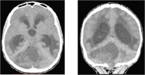
Figure 1. Preoperative non-contrast computed tomography (CT) of the brain. Axial and coronal images demonstrate marked ventricular dilatation consistent with obstructive hydrocephalus and a cystic and nodular expansive lesion involving the right cerebellar hemisphere, with mass effect on the fourth ventricle and associated periventricular edema.
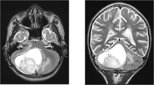
Figure 2. Brain magnetic resonance imaging (MRI) after ventriculoperitoneal shunt placement. Axial and coronal T2-weighted images demonstrate a cystic and nodular lesion involving the right cerebellar hemisphere, measuring approximately 3 × 3 cm, with mass effect on the fourth ventricle and adjacent brainstem. The ventricular system shows reduced dilatation after cerebrospinal fluid diversion.
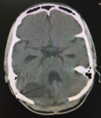
Figure 3. Postoperative non-contrast computed tomography (CT) of the brain. Axial image demonstrates postoperative changes in the posterior fossa following resection of the right cerebellar tumor, with improvement of the mass effect on the fourth ventricle.
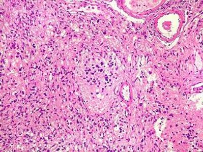
Figure 4. Histopathological examination of the cerebellar tumor. Hematoxylin and eosin (H&E)-stained photomicrograph demonstrating a glial neoplastic proliferation composed of relatively uniform cells with elongated nuclei and a fibrillary background, with associated vascular structures. The histopathological findings, in conjunction with the original pathology report, were consistent with pilocytic astrocytoma, CNS WHO grade 1.
3. Discussion
3.1 Clinical presentation and hydrocephalus
The patient presented with headache, projectile vomiting, drowsiness, gait disturbance, and multiple cerebellar signs, including truncal and appendicular ataxia, dysdiadochokinesia, nystagmus, and intention tremor. This presentation is consistent with the anatomical location of the lesion and its effect on the fourth ventricle.
Hydrocephalus is a well-recognized complication of pediatric cerebellar PA. Santos et al. reported hydrocephalus in 47.3% of children with posterior fossa PA, with 33.3% ultimately requiring ventriculoperitoneal shunting. [5] A recent cerebellar PA series similarly emphasizes hydrocephalus as a frequent presenting feature. [3] The marked ventricular enlargement in our patient, together with drowsiness and fourth-ventricle compression, supported urgent CSF diversion.
3.2 Staged management of hydrocephalus
In this patient, ventriculoperitoneal shunting was performed before definitive tumor surgery. The subsequent reduction in ventricular size demonstrates the effectiveness of CSF diversion. Management of hydrocephalus associated with posterior fossa tumors remains individualized: some patients improve after tumor removal, whereas others require permanent CSF diversion. The decision depends on neurological status, ventricular enlargement, fourth-ventricle obstruction, and the timing and feasibility of definitive tumor surgery. [3,5]
3.3 Imaging characteristics
The lesion demonstrated a cystic and nodular configuration with a mural nodule, a recognized imaging pattern of pilocytic astrocytoma. A 2024 single-center study of 42 children with cerebellar PA found that 66.6% had cystic tumors, 73.8% achieved gross-total resection, and progression occurred in 11 cases. The authors concluded that surgery should aim for gross-total resection of the solid-enhancing tumor component. [3]
MRI is important for characterization and postoperative surveillance. Contemporary imaging work has also explored whether contrast-free MRI can be used for long-term surveillance in selected pediatric PA patients, which may be relevant in resource-limited settings. In a 2025 study, non-contrast MRI retained high specificity for detecting growth, although sensitivity was lower than perfect. [6]
3.4 Extent of resection and recurrence
The principal therapeutic objective in a surgically accessible PA is maximal safe resection. The present patient underwent complete removal of the mural tumor nodule and had no radiological evidence of recurrence at one-year follow-up.
Recent evidence supports an association between gross-total resection and reduced progression or recurrence. Grigorian et al. reported a hazard ratio of 0.27 for recurrence/progression after GTR in a pediatric PA cohort, with GTR remaining associated with longer progression-free intervals in patients with KIAA1549-BRAF fusion. [4] A separate cohort of 315 pediatric PA patients found progression/recurrence in 8.6% after GTR compared with 41.4% after non-GTR. [7]
However, current evidence also cautions against aggressive reoperation for every residual lesion. Kameda-Smith et al. analyzed 200 pediatric cerebellar PAs and concluded that stable residual disease can often be followed with clinicoradiological surveillance rather than automatically undergoing a second resection. Multiple operations were associated with greater treatment-related morbidity in their cohort. [2] Santos et al. similarly reported that repeated surgery for residual disease increased the risk of postoperative cranial nerve deficits. [5]
Thus, the contemporary principle is not radical resection at any cost, but maximal safe resection with preservation of neurological function.
3.5 Functional outcome
The favorable functional outcome in this case is particularly relevant. The patient initially presented with substantial cerebellar dysfunction, yet currently attends school normally and performs his academic activities without functional limitations. This clinical outcome complements the radiological finding of no recurrence at one year.
Posterior fossa surgery can be associated with neurological complications, including cranial nerve dysfunction, motor deficits, and cerebellar mutism syndrome. The long-term posterior fossa series by Santos et al. reported favorable functional outcomes in 91.2% of patients but also identified repeated surgery as a risk factor for postoperative cranial nerve deficits. [5] The present case had no reported significant persistent postoperative neurological disability.
3.6 Long-term surveillance
Although there was no evidence of recurrence at one year, longer follow-up is necessary. Pediatric PA can recur after prolonged intervals, particularly when residual tumor remains. A long-term pediatric cerebellar series with follow-up extending to 20 years documented generally favorable outcomes but also reported recurrence in a subset of patients. [8]
Postoperative imaging surveillance is therefore important. Earlier surveillance literature supports early postoperative MRI and subsequent interval imaging, while newer data suggest that selected non-contrast MRI protocols may reduce cost and burden while retaining useful diagnostic accuracy. [6,16] In the present setting, however, access to serial imaging was limited by resource constraints, representing an important limitation of this report.
3.7 Molecular classification and targeted therapy
Modern classification and management of pediatric low-grade gliomas increasingly incorporate molecular alterations in the RAS/MAPK pathway. BRAF fusions, BRAF V600E mutations, and other kinase alterations have important diagnostic and therapeutic implications. [1,9,10,11]
The 2025 study by Grigorian et al. found KIAA1549-BRAF fusion in 58 of 112 pediatric PA patients and reported that GTR was associated with lower recurrence/progression rates. [4] Molecular testing was not available in the present case. If archived tissue is accessible, molecular characterization could strengthen the final report and potentially provide clinically relevant information should progression occur.
For recurrent or progressive molecularly selected pLGG, targeted treatments have expanded considerably. Reviews published in 2024-2026 describe increasing roles for MEK, BRAF, and RAF inhibitors. [9,10,11,12,13,14] Tovorafenib, a type II RAF inhibitor, has become an important option for selected relapsed or refractory BRAF-altered pLGG, while selumetinib and other MAPK-directed agents have demonstrated activity in selected patients. [12,13]
These therapies are not relevant to the current patient because he underwent successful surgical treatment without documented progression or recurrence, but they are important considerations in the contemporary management framework.
3.8 Role of radiotherapy
Radiotherapy was not administered in this patient and should not be described as part of his treatment. Contemporary pediatric low-grade glioma management has moved toward more selective use of radiotherapy because of concerns about late neurocognitive, endocrine, vascular, and secondary malignancy effects. Recent consensus guidelines emphasize individualized indications, timing, and age-related considerations. [14,15]
For a completely resected, CNS WHO grade 1 pilocytic astrocytoma without evidence of residual or progressive disease, observation after surgery is consistent with contemporary management principles. Radiotherapy remains an option in selected unresectable or progressive/recurrent cases, particularly when other treatments are unsuitable. [15,17]
3.9 Relevance in resource-limited settings
This case also highlights the practical challenge of managing pediatric brain tumors where access to serial MRI, molecular testing, and advanced neuro-oncology services may be limited. The favorable outcome in this patient was achieved with prompt CSF diversion, definitive surgery, histopathological diagnosis, and clinical follow-up.
Nevertheless, resource limitations should not be interpreted as evidence that prolonged imaging surveillance is unnecessary. Rather, they represent a limitation that must be transparently reported and should encourage pragmatic follow-up strategies when resources permit.
4. Limitations
The main limitation of this report is the limited availability of resources for serial neuroimaging during follow-up. Consequently, long-term radiological surveillance was less extensive than would ideally be recommended for a pediatric pilocytic astrocytoma. The documented one-year CT showed no evidence of recurrence, and the patient remained clinically well with normal school attendance and no functional limitations, but longer MRI-based follow-up would provide stronger evidence of durable tumor control.
A second limitation is the absence of molecular characterization of the tumor. BRAF and other MAPK-pathway alterations are now recognized as clinically relevant in pediatric low-grade glioma. If tumor tissue remains available, molecular testing could strengthen the diagnostic and prognostic characterization of the case.
5. Conclusion
Cerebellar pilocytic astrocytoma should be considered in the differential diagnosis of cystic and nodular posterior fossa lesions in children and adolescents. Although generally characterized by favorable biological behavior, cerebellar PA can produce severe neurological symptoms through fourth-ventricle compression and obstructive hydrocephalus.
In this case, urgent management of hydrocephalus followed by complete removal of the mural tumor nodule resulted in a favorable clinical and radiological outcome. At one-year follow-up there was no evidence of tumor recurrence. At the latest clinical assessment, the patient was attending school normally and participating in academic activities without functional limitations.
The case supports maximal safe resection, individualized management of hydrocephalus, and prolonged clinical and radiological surveillance. Molecular characterization and targeted therapies are increasingly relevant for patients with residual, recurrent, or progressive disease.
6. Declarations
Conflict of Interest: The authors declare no conflicts of interest.
Funding: No external funding statement was provided; this should be completed by the authors before submission if applicable.
Patient Consent: Because the patient was a minor at the time of treatment, written informed consent for publication of the case and accompanying anonymized images should be obtained from the parent or legal guardian before submission.
Data Availability: The clinical information presented in this report is contained within the manuscript. Additional patient-identifying information should not be submitted.
Ethics: The authors should confirm whether institutional ethics committee approval or a formal exemption/waiver was applicable to this single case report according to local policy.
7. Figure Legends
Figure 1. Preoperative non-contrast computed tomography (CT) of the brain. Axial and coronal images demonstrate marked ventricular dilatation consistent with obstructive hydrocephalus and a cystic and nodular expansive lesion involving the right cerebellar hemisphere, with mass effect on the fourth ventricle and associated periventricular edema.
Figure 2. Brain magnetic resonance imaging (MRI) after ventriculoperitoneal shunt placement. Axial and coronal T2-weighted images demonstrate a cystic and nodular lesion involving the right cerebellar hemisphere, measuring approximately 3 × 3 cm, with mass effect on the fourth ventricle and adjacent brainstem. The ventricular system shows reduced dilatation after cerebrospinal fluid diversion.
Figure 3. Postoperative non-contrast computed tomography (CT) of the brain. Axial image demonstrates postoperative changes in the posterior fossa following resection of the right cerebellar tumor, with improvement of the mass effect on the fourth ventricle.

Open Access By Aditum Open Access Journals id licensed under Creative Commons Attribution 4.0 International License. Based On a Work at aditum.org


