
Neurosurgery and Neurology Research
OPEN ACCESS | Volume 7 - Issue 1 - 2025
ISSN No: 2836-2829 | Journal DOI: 10.61148/2836-2829/NNR

José Clayton Balo1*, Danilson Kidy1, Mateus Tomás1, Edna Amaral2
1Department of Neurosurgery, Main Military Hospital Superior Institute, Luanda – Angola.
2Department of Pathology, Main Military Hospital Superior Institute, Luanda - Angola1.
*Corresponding author: José Clayton Balo, Department of Neurosurgery, Main Military Hospital Superior Institute, Luanda – Angola.
Received: September 10, 2026 | Accepted: September 26, 2026 | Published: October 13, 2026
Citation: José C Balo, Kidy D, Tomás M, Amaral E, (2026) “Giant Supratentorial Cystic Astroblastoma In A 59-Year-Old Man: Complete Neurological Recovery After Gross Total Macroscopic Resection Followed By Fatal Postoperative Pulmonary Thromboembolism” Neurosurgery and Neurology Research, 8(1); DOI: 10.61148/2836-2829/NNR/057.
Copyright: ©2026. José Clayton Balo. This is an open access article distributed under the Creative Commons Attribution License, which permits unrestricted use, distribution, and reproduction in any medium, provided the original work is properly cited.
Astroblastoma is a rare primary central nervous system neoplasm characterized by astroblastic perivascular pseudorosettes. The 2021 WHO classification introduced the molecular entity astroblastoma, MN1-altered; contemporary studies demonstrate substantial molecular heterogeneity. We report a 59-year-old man with hypertension who presented with three months of progressive headache, dysarthria and right-sided weakness, followed by impaired consciousness. CT demonstrated a large left frontoparietal cystic lesion. MRI showed a 7.5 × 5.0 × 5.4 cm cystic-nodular lesion, approximately 10 mm of midline shift and compression of the lateral ventricles. A left frontoparietal osteoplastic craniotomy was performed, with aspiration of brown cystic fluid and gross total macroscopic resection of the mural solid component. Histopathology demonstrated rounded/oval tumor cells with perivascular pseudorosette-like arrangements and very low mitotic activity, interpreted as astroblastoma with low proliferative activity. The immediate postoperative course was excellent, with complete neurological recovery and Karnofsky Performance Status of 100. On postoperative day 3, the patient developed acute respiratory failure and cardiac arrest. Autopsy demonstrated pulmonary thromboembolism, establishing the cause of death. This case is unusual because of the patient's advanced age, male sex, large cystic supratentorial tumor and fatal early systemic postoperative complication. Molecular confirmation was not available; therefore, the contemporary diagnosis astroblastoma, MN1-altered cannot be asserted. The case emphasizes integrated radiological, histopathological and molecular diagnosis and the importance of postoperative vigilance for venous thromboembolism.
Astroblastoma; MN1; BEND2; cystic brain tumor; supratentorial tumor; perivascular pseudorosettes; gross total resection; pulmonary thromboembolism; case report
Astroblastoma is an uncommon primary CNS tumor, traditionally recognized by a circumscribed growth pattern, astroblastic perivascular pseudorosettes and variable vascular hyalinization. The 2021 WHO CNS classification incorporated astroblastoma, MN1-altered as a molecularly defined entity within circumscribed astrocytic gliomas. Histology remains important, but recent literature demonstrates that morphology alone may overlap with ependymoma and other neuroepithelial tumors. Molecular profiling has consequently become central to contemporary classification.
Recent molecular work has shown that the group previously classified as astroblastoma, MN1-altered is heterogeneous. A 2026 cohort of 200 tumors identified three fusion-defined groups, including MN1::BEND2, EWSR1::BEND2 and MN1::CXXC5, with clinically different outcomes. This makes precise molecular characterization particularly important when interpreting older histopathological diagnoses.
The present case is clinically unusual because it occurred in a 59-year-old man with a giant left frontoparietal cystic lesion. The patient recovered completely after gross total macroscopic resection but died suddenly on postoperative day 3 from autopsy-confirmed pulmonary thromboembolism.
2. Case Presentation
A 59-year-old man with known arterial hypertension presented with a three-month history of progressive headache, dysarthria and progressive reduction in strength of the right hemibody. He subsequently developed depressed consciousness. On admission, the Glasgow Coma Scale was 14/15, with right hemiplegia. Karnofsky Performance Status was greater than 70. There was no documented previous intracranial neoplasm.
2.1. Neuroimaging
Non-contrast CT demonstrated a large left frontoparietal hypodense lesion with a central cystic component and discrete enhancement of the solid component after contrast administration. MRI demonstrated a predominantly cystic and nodular left frontoparietal lesion, hypointense on T1 and hyperintense on T2, measuring approximately 7.5 × 5.0 × 5.4 cm, with approximately 10 mm of midline shift and compression of the lateral ventricles.
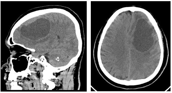
Figure 1. Preoperative CT (1 and 2) and MRI (3 and 4) demonstrating the large left frontoparietal cystic-nodular lesion with mass effect and midline shift.
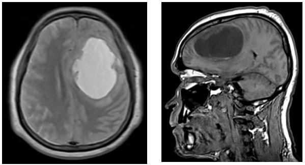
Figure 1. Preoperative CT (1 and 2) and MRI (3 and 4) demonstrating the large left frontoparietal cystic-nodular lesion with mass effect and midline shift.
2.2. Surgical Procedure
A left frontoparietal osteoplastic craniotomy was performed using five burr holes, followed by an inferiorly based curvilinear durotomy. The cystic component was punctured and aspirated, yielding brown-colored fluid. Corticotomy was performed at the parietal level while avoiding the primary motor cortex. The solid mural component was dissected and resected circumferentially in all directions, aiming for gross total macroscopic removal. After confirmation of no visible residual tumor, the operative cavity showed apparently healthy surrounding brain. Hemostasis was achieved with bipolar coagulation and Spongostan. The cavity was irrigated with 0.9% saline, the dura was closed hermetically, the bone flap was replaced and an epidural drain was positioned. No intraoperative complication was reported.
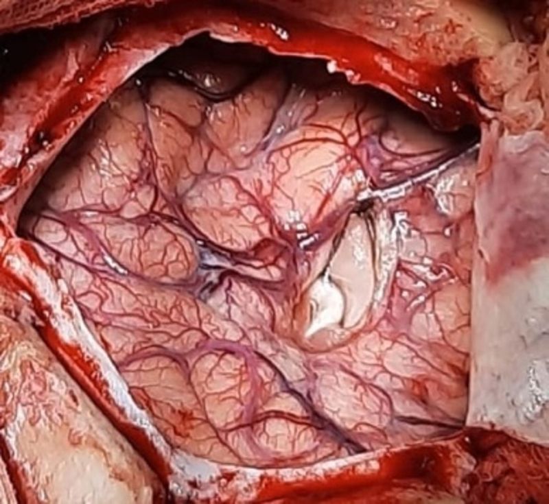
Figure 2. Intraoperative photograph showing the left frontoparietal exposure during tumor surgery.
2.3. Histopathology
Histological examination demonstrated blood elements and a neoplastic proliferation composed of rounded and oval cells. Areas of small cells arranged radially around blood vessels formed perivascular pseudorosette-like structures. Mitotic figures were very uncommon. The morphology was interpreted as compatible with astroblastoma with low proliferative activity. No molecular testing for MN1/BEND2/CXXC5 or DNA methylation profiling was available.
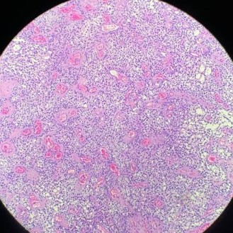
Figure 3. Histopathological examination (hematoxylin and eosin) showing a cellular neoplastic proliferation with numerous vascular structures and perivascular architectural patterns compatible with astroblastic pseudorosettes.
2.4. Postoperative Course and Autopsy
The immediate postoperative course was excellent, with complete improvement of the preoperative neurological deficits. The patient's Karnofsky Performance Status reached 100. As thromboprophylaxis, enoxaparin (0.3 mL subcutaneously once daily) was administered. However, on postoperative day 3, the patient developed acute respiratory failure followed by cardiac arrest. Cardiopulmonary resuscitation, including the administration of vasoactive drugs, was unsuccessful. Autopsy demonstrated findings consistent with pulmonary thromboembolism, which was considered the cause of death despite the administration of pharmacological thromboprophylaxis.
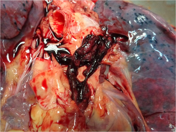
Figure 4. Autopsy findings demonstrating pulmonary thromboembolism.
3. Discussion
The present case expands the clinical spectrum of astroblastoma because of the patient's age and sex. Contemporary literature indicates that astroblastoma predominantly affects children, adolescents and young adults, with a female predominance. A 2024 series of eight molecularly tested cases had a median age of 35.1 years and a female-to-male ratio of 1.6:1, while a systematic review of adults with MN1-altered CNS tumors also emphasized marked demographic and clinical heterogeneity. The present 59-year-old male is therefore unusual, although elderly molecularly confirmed cases have been reported.
The imaging phenotype is nevertheless compatible with the recognized spectrum. Astroblastomas are often circumscribed supratentorial tumors with solid, cystic or mixed morphology. Radiology-pathology correlation studies describe well-defined cystic-solid masses, sometimes with a bubbly or mural-nodular appearance. Our lesion was very large, measured 7.5 cm in its greatest dimension and produced 10 mm of midline shift and ventricular compression, explaining the progressive motor deficit and impaired consciousness.
Histologically, the presence of perivascular pseudorosette-like architecture and low mitotic activity supported the original diagnosis. However, contemporary studies show that the diagnosis cannot always be reliably established by morphology alone. MN1::BEND2-positive tumors may lack classic astroblastoma morphology, while EWSR1::BEND2, YAP1::BEND2, MAMLD1::BEND2 and TCF3::BEND2 rearrangements have been reported in astroblastoma-like CNS tumors. Therefore, the most defensible wording for this case is 'astroblastoma based on characteristic histopathological findings, with low proliferative activity; molecular characterization was not available.'
The 2026 study of 200 tumors is particularly relevant. DNA methylation and sequencing identified three groups: MN1::BEND2-enriched supratentorial Group A, EWSR1::BEND2-enriched Group B, and MN1::CXXC5 Group C. Five-year progression-free survival was 47%, 14% and 75%, respectively, while 10-year overall survival was 89%, 54% and 89%. These data show that astroblastoma is not biologically uniform and that molecular subgrouping may become central to risk-adapted treatment.
Surgery remains the principal treatment for accessible symptomatic tumors. Individual-patient meta-analysis and recent series support gross total resection as the main modality associated with disease control, although the evidence is observational. In our patient, complete macroscopic resection was followed by complete neurological recovery and KPS 100, consistent with the favorable immediate neurological outcomes reported after extensive resection. Recent pediatric cases with MN1::BEND2 fusion have likewise shown stability after gross total resection.
The fatal event in our patient was not evidence of tumor progression. The patient developed acute respiratory failure on postoperative day 3, and autopsy established pulmonary thromboembolism. A 2024 meta-analysis of patients undergoing craniotomy for brain tumors reported a clinically relevant prevalence of postoperative venous thromboembolism. Current perioperative neurosurgical guidance emphasizes individualized prophylaxis because clinicians must balance thrombotic risk against intracranial bleeding risk. The available case data do not document the exact prophylaxis strategy; therefore, no conclusion should be drawn about adequacy or causality.
The case also demonstrates the value of autopsy in sudden postoperative deterioration. In a neurosurgical patient, acute respiratory collapse may have several possible causes, and autopsy can establish the diagnosis when clinical circumstances are uncertain. Here, it separated the systemic postoperative complication from the neurological and oncological outcome.
A further implication is the importance of molecular testing. If archival paraffin-embedded material remains available, MN1/BEND2/CXXC5 fusion testing, RNA sequencing or DNA methylation profiling could be considered. Such testing could potentially refine the diagnosis and permit comparison with current molecular subgroups. The lack of molecular testing is the main limitation of this report.
Overall, the case demonstrates a striking divergence between immediate neurosurgical outcome and short-term systemic outcome: complete neurological recovery followed by fatal pulmonary thromboembolism. This combination, together with the patient's advanced age and giant cystic frontoparietal lesion, constitutes the principal contribution of the report.
|
Domain |
Published evidence |
Present case |
|
Age/sex |
Mostly children/young adults; female predominance |
59-year-old male |
|
Location |
Supratentorial hemispheric lesions common |
Left frontoparietal |
|
Imaging |
Circumscribed solid/cystic or mixed lesions |
7.5 cm cystic-nodular lesion; 10 mm shift |
|
Histology |
Perivascular pseudorosettes; variable hyalinization |
Perivascular pseudorosette-like pattern; very low mitotic activity |
|
Molecular diagnosis |
Increasingly essential for current classification |
Not available |
|
Surgery |
Maximal safe/GTR commonly used |
Gross total macroscopic resection |
|
Immediate outcome |
Often favorable after resection |
Complete neurological recovery; KPS 100 |
|
Long-term outcome |
Recurrence can occur, including late recurrence |
Not assessable because of early death |
|
Fatal event |
Not characteristic of tumor biology |
Autopsy-confirmed pulmonary thromboembolism, POD 3 |
5. Study Limitations
The present case report has several limitations. Postoperative CT and MRI were not available because of equipment downtime at our institution during the postoperative period. Therefore, radiological confirmation of the extent of tumor resection could not be obtained, and the assessment of gross total resection was based on intraoperative findings. In addition, molecular characterization, including MN1/BEND2/CXXC5 fusion testing and DNA methylation profiling, was not available. Consequently, the diagnosis was established on the basis of the characteristic histopathological findings and low proliferative activity. Finally, long-term oncological follow-up was not possible because of the patient's death on postoperative day 3 from autopsy-confirmed pulmonary thromboembolism.
6. Conclusion
This case describes an unusual giant supratentorial cystic astroblastoma in a 59-year-old man. Gross total macroscopic resection resulted in complete neurological recovery and KPS 100. Nevertheless, fatal pulmonary thromboembolism occurred on postoperative day 3 and was confirmed at autopsy. The case reinforces that astroblastoma should remain in the differential diagnosis of a large circumscribed supratentorial cystic-nodular lesion even in older adults. This case also illustrates a diagnostic challenge faced in resource-limited settings. Although contemporary classification increasingly relies on molecular characterization, such testing was not locally available in Angola at the time of diagnosis. This limitation highlights the importance of strengthening access to molecular neuropathology and developing referral pathways for molecular testing of rare central nervous system tumors.
The early fatal thromboembolic complication also highlights the need for individualized postoperative VTE surveillance and prophylaxis in patients undergoing intracranial tumor surgery.
7. Declarations
Ethics approval: Institutional approval or waiver should be obtained according to local regulations.
Consent for publication: Written informed consent for publication of clinical information and anonymized imaging/pathological/autopsy material should be obtained from the legally authorized representative or next of kin, according to institutional requirements.
Conflict of interest: The authors declare no conflict of interest related to this manuscript.
Funding: No specific funding was received for this work.
Author contributions: [Insert final author contributions before submission].
Data availability: Additional anonymized information may be made available by the corresponding author subject to ethical and privacy restrictions.

Open Access By Aditum Open Access Journals id licensed under Creative Commons Attribution 4.0 International License. Based On a Work at aditum.org


