
Journal of Urology and Nephrology Research
OPEN ACCESS | Volume 3 - Issue 1 - 2026
ISSN No: 3065-6699 | Journal DOI: 10.61148/3065-6699/JUNR


Marwan Abdulaziz Ibrahim
Department of Urology, Teaching Al-Diwaniyah Hospital, Al- Diwaniyah, Iraq.
*Corresponding author: Marwan Abdulaziz Ibrahim, Department of Urology, Teaching Al-Diwaniyah Hospital, Al- Diwaniyah, Iraq.
Received: June 02, 2026 | Accepted: June 08, 2026 | Published: June 15, 2026
Citation: Marwan A Ibrahim. (2026) “Conservative Management of Life-Threatening Delayed Hemorrhage After Percutaneous Nephrolithotomy Despite Negative Angiography: A Case Report”. Journal of Urology and Nephrology Research, 3(1); DOI: 10.61148/ 3065-6699/JUNR/055
Copyright: © 2026 Marwan Abdulaziz Ibrahim. This is an open access article distributed under the Creative Commons Attribution License, which permits unrestricted use, distribution, and reproduction in any medium, provided the original work is properly cited.
Background:
Percutaneous nephrolithotomy (PCNL) is the standard treatment for large renal calculi. Although generally safe, hemorrhage remains one of its most serious complications. In severe delayed cases, vascular injuries such as arteriovenous fistula, pseudoaneurysm, or arterial laceration are usually identified on angiography and managed with selective embolization. Management becomes more challenging when angiography is negative despite significant bleeding.
Case Presentation: A 29-year-old man with a single 22-mm right renal pelvic stone underwent uneventful prone PCNL under ultrasound guidance through a lower posterior calyceal puncture. On postoperative day 1, following mobilization, he developed intermittent severe bleeding from the nephrostomy site associated with a hemoglobin drop from 13.5 g/dL to 8.0 g/dL, without hematuria. The patient was admitted to the intensive care unit and received aggressive resuscitation with intravenous fluids, packed red blood cells, and fresh frozen plasma. After initial stabilization, abdominal computed tomography revealed a large right retroperitoneal hematoma. Urgent renal angiography subsequently demonstrated no pseudoaneurysm, arteriovenous fistula, or active arterial contrast extravasation. Conservative management was continued in the intensive care unit under close observation.
Outcome: The patient showed gradual clinical and hematological improvement. Daily serial hemoglobin measurements demonstrated no further significant decline, and repeated bedside ultrasonography showed a stable retroperitoneal hematoma without expansion. On postoperative day 6, he developed reactive pleural effusion, likely secondary to fluid resuscitation, which was managed conservatively after internal medicine consultation. Hemoglobin increased to 11.1 g/dL before discharge, and he was discharged in good condition on postoperative day 12. At one-month follow-up, he developed a mild upper urinary tract infection that responded to antibiotic therapy, and the double-J stent was removed uneventfully.
Conclusion: In carefully selected hemodynamically stable patients with severe delayed post-PCNL hemorrhage and negative angiographic findings, conservative management may achieve favorable outcomes under close monitoring.
percutaneous nephrolithotomy; delayed hemorrhage; negative angiography; retroperitoneal hematoma; conservative management
Percutaneous nephrolithotomy (PCNL) remains the standard treatment for large and complex renal calculi because of its high stone-free rates and lower morbidity compared with open surgery [1]. Despite its safety and effectiveness, serious complications may occur, including sepsis, hemorrhage, and visceral injury [2–4].
Because PCNL requires tract creation through the renal parenchyma, bleeding remains a recognized and clinically important complication requiring prompt assessment and management [12,13]. Delayed bleeding typically occurs within the first three weeks after surgery [5,6]. Overall postoperative bleeding manifestations, including transient hematuria and minor bleeding, have been reported in up to 25% of patients, whereas severe life-threatening hemorrhage requiring urgent intervention is uncommon, occurring in approximately 0.8% of cases [7–11].
Most severe delayed bleeding events are related to vascular lesions such as pseudoaneurysm, arteriovenous fistula, or segmental arterial injury [14,15]. In these situations, renal angiography is considered the reference diagnostic modality, identifying the bleeding source in more than 90% of cases while simultaneously allowing selective angioembolization [16].
However, a small subset of patients may develop severe delayed hemorrhage despite negative renal angiography, creating a challenging management dilemma regarding repeat intervention versus conservative treatment[17]. Evidence guiding management in this scenario remains scarce.
We report a rare case of life-threatening delayed post-PCNL hemorrhage with negative angiography that was successfully managed conservatively, highlighting that carefully selected hemodynamically stable patients may avoid unnecessary invasive intervention.
Case Report
A 29-year-old man presented with a 22-mm right renal pelvic stone. Full preoperative laboratory investigations, including coagulation profile, were within normal limits.
A right ureteric stent was inserted cystoscopically in the lithotomy position, and the bladder was filled with normal saline to induce artificial hydronephrosis. PCNL was then performed in the prone position through a posterior lower calyceal puncture under ultrasound guidance using a 3.5-MHz Philips probe. Initial access was obtained with a 12-Fr nephrostomy set through the papillary zone while avoiding vascular injury with Doppler assistance. Tract dilatation was subsequently performed using metallic Alken dilators over a guidewire, and a 30-Fr working sheath was advanced into the collecting system. The stone was visualized using a 26-Fr rigid nephroscope (Storz) and fragmented with pneumatic lithotripsy. At the end of the procedure, clear urine was observed through the working sheath, and a 20-Fr nephrostomy tube was placed for tract tamponade.
On postoperative day 1, following early mobilization, bleeding was noted on the dressing around the nephrostomy site. After changing the dressing and opening the nephrostomy tube for routine drainage, severe bleeding was observed and was exacerbated by changes in patient position. The patient developed mild to moderate abdominal distension, pallor, and tachycardia. Hemoglobin decreased from 13.5 g/dL to 8.1 g/dL. Four units of packed red blood cells were transfused after admission to the intensive care unit.
After hemodynamic stabilization, computed tomography of the abdomen and pelvis demonstrated a large right retroperitoneal hematoma with no intraperitoneal bleeding or visceral injury (Figures 1-4).
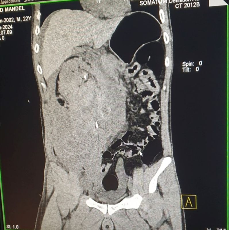
Figure (1) illustrates a retroperitoneal hematoma.
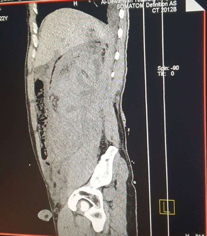
Figure (2) depicts the progression of the hematoma.
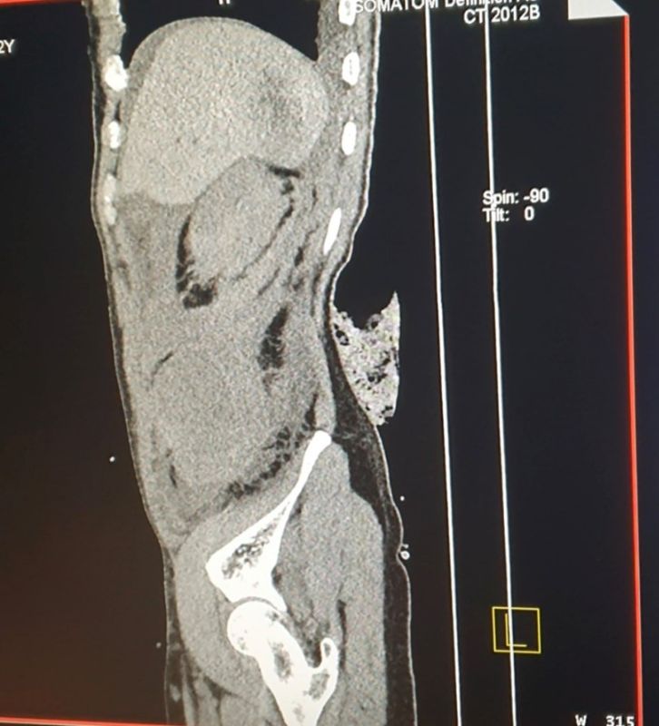
Figure (3) presents the inferior extension of the hematoma
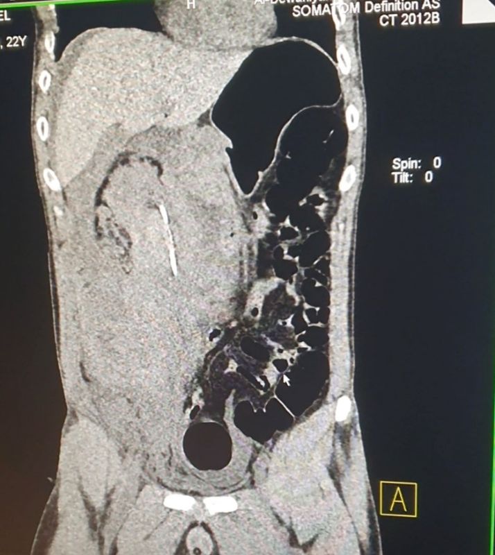
Figure (4) shows the right kidney, which is free of stones, along with a visible ureteric stent.
Selective right renal angiography via right femoral access demonstrated normal renal vascularity with no evidence of pseudoaneurysm, arteriovenous fistula, or active contrast extravasation; therefore, no embolization was performed.
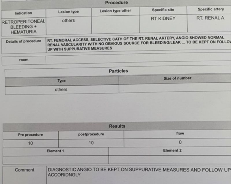
Figure (5) illustrates the selective angiography of the right renal artery.
The patient was admitted to the intensive care unit for close observation, serial monitoring of vital signs, and repeated hemoglobin assessment every 6 hours while conservative management was continued. On postoperative day 4, ecchymosis developed over the right flank and lower subcostal region, with old dark oozing blood noted on the nephrostomy dressing (Figure 6)
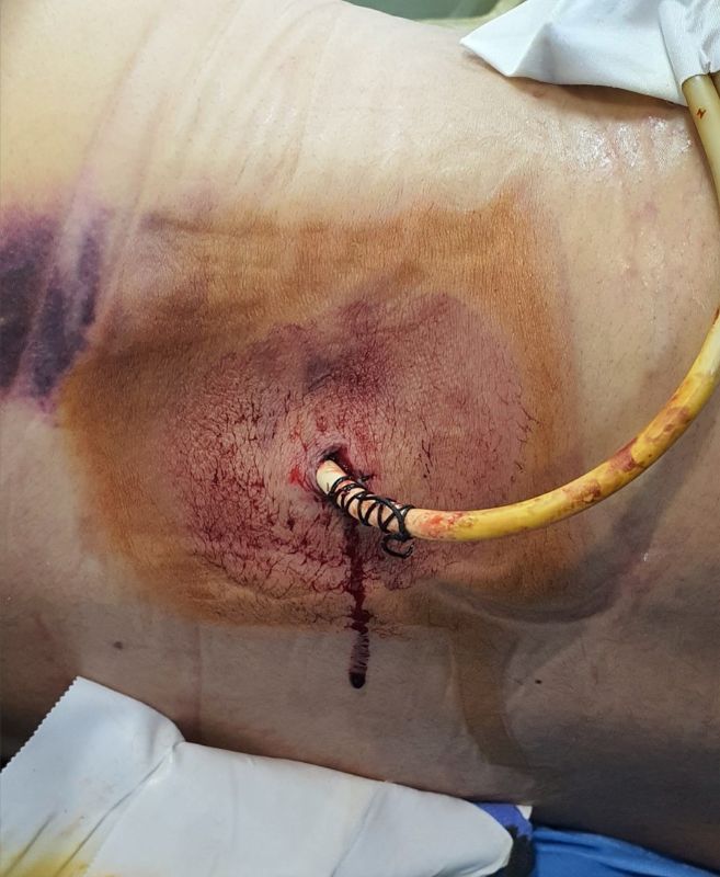
Figure (6) displays a right nephrostomy tube accompanied by bruising in the costal and lower costal regions.
Serial bedside ultrasonography was used to assess and monitor the retroperitoneal hematoma, in conjunction with repeated CT scans, which demonstrated a stable, non-expanding hematoma. Repeat angiography was not performed, as the patient remained hemodynamically stable and no radiological findings indicated active bleeding.
On postoperative day 7, the nephrostomy tube was removed. Serial hemoglobin levels on postoperative days 3, 4, 5, 6, 7, 8, 9, and 12 were 8.0, 7.5, 7.8, 8.0, 8.5, 9.2, 10.1, and 11.1 g/dL, respectively, demonstrating progressive recovery without further major bleeding.
On postoperative day 9, the patient developed reactive pleural effusion, likely secondary to fluid resuscitation and transfusion-related volume overload, and was managed conservatively after internal medicine consultation. By postoperative day 12, the patient was clinically stable and discharged in good condition.
Several days after discharge, the patient re-presented with mild fever (38.5°C) and mild right loin pain. He was treated for presumed acute pyelitis with antibiotics and improved clinically.
One month later, the ureteric stent was removed by flexible cystoscopy under local anesthesia. The patient recovered uneventfully and resumed normal daily activities.
Discussion
Delayed hemorrhage after percutaneous nephrolithotomy (PCNL) is an uncommon but potentially life-threatening complication that requires prompt recognition and appropriate management. While minor postoperative bleeding is relatively frequent and usually self-limited, severe hemorrhage requiring transfusion or intervention remains rare, with reported rates ranging from 0.3% to 1% [19–21]. Most cases of significant delayed bleeding are attributed to vascular injuries such as pseudoaneurysm, arteriovenous fistula, or segmental arterial laceration, which are typically identifiable on renal angiography and amenable to selective angioembolization [23,24].
Renal angiography is widely regarded as the gold standard diagnostic and therapeutic modality in this setting, with reported detection rates exceeding 90% [16]. However, a subset of patients presents with clinically significant hemorrhage despite negative angiographic findings, posing a diagnostic and therapeutic challenge. Previous studies have reported that approximately 6.1% to 22.2% of patients evaluated for delayed post-PCNL bleeding demonstrate negative angiography [25–28]. Notably, these patients are generally described as having less severe hemorrhage, reflected by relatively higher nadir hemoglobin levels compared with those with confirmed vascular lesions [26].
In contrast, the present case represents an uncommon clinical scenario in which severe delayed hemorrhage occurred despite negative angiographic findings. The patient experienced a substantial hemoglobin decline requiring multiple blood transfusions, highlighting that negative angiography does not necessarily exclude clinically significant or even life-threatening bleeding. This discrepancy underscores the limitations of angiography and challenges the assumption that negative findings are synonymous with low-risk bleeding.
Several mechanisms may explain the occurrence of significant hemorrhage in the absence of angiographic evidence. First, bleeding may originate from extra-renal or atypical vascular sources not routinely visualized during selective renal angiography, such as lumbar, subcostal, or capsular arteries. Previous reports by Srivastava et al. [29], Choi et al. [30], and Ding et al. [31] have documented non-renal vascular injuries as potential sources of post-PCNL hemorrhage. In the present case, the distribution of flank ecchymosis extending toward the subcostal region may support the possibility of an extra-renal bleeding source, although this could not be definitively confirmed.
Second, angiography has inherent sensitivitylimitations and typically requires a minimum bleeding rate of approximately 0.5 mL/min to detect active contrast extravasation [32]. Intermittent bleeding, temporary clot formation, or vasospasm may further reduce the likelihood of detecting active hemorrhage at the time of imaging, resulting in false-negative findings despite clinically significant bleeding [17]. In the present case, the requirement for multiple blood transfusions likely reflected cumulative blood loss rather than continuous bleeding at a rate detectable during angiography.
Third, venous bleeding or hemorrhage from small accessory vessels may not be readily visualized on arterial angiographic studies [26]. Although venous bleeding is often self-limited and may respond to tamponade, the severity of bleeding observed in this case suggests a more complex or multifactorial mechanism.
Beyond these pathophysiological considerations, several risk factors for delayed post-PCNL bleeding have been described in the literature, including prior ipsilateral renal surgery, increased stone complexity (such as staghorn calculi), multiple access tracts, prolonged operative time, and comorbid conditions such as diabetes and hypertension [36]. Studies by Said et al. [33], Yesil et al. [34], Turna et al. [35], and Valecha et al. [38] have highlighted the contribution of these factors to increased bleeding risk. However, the present case did not exhibit most of these established risk factors, as the patient had a solitary pelvic stone and underwent an otherwise uncomplicated procedure. This observation reinforces that severe delayed hemorrhage may occur even in the absence of classical risk factors, highlighting both the unpredictable nature of this complication and the limited predictive value of these variables on an individual patient basis[36,37]
Although selective angioembolization remains the standard of care for severe delayed hemorrhage, it is not without risks, including non-target embolization, renal function impairment, and procedure-related complications, as reported by Nouralizadeh et al. [39]. Additionally, embolization may fail in cases of unidentified or anatomically complex bleeding sources, potentially necessitating repeated interventions, as described by Ali et al. [40]. Furthermore, Xiao et al. reported that patients requiring embolization had longer hospital stays and higher treatment costs, with some requiring repeat procedures, thereby imposing additional clinical and economic burdens [26].These limitations further support the role of conservative management in carefully selected patients
Conservative management has been suggested in selected cases of delayed post-PCNL bleeding, particularly in patients who are hemodynamically stable, exhibit self-limited or minor hemorrhage, have negative or inconclusive imaging findings, present contraindications to angiography (such as contrast hypersensitivity or renal insufficiency), or in whom superselective angioembolization is not feasible due to inability to localize the bleeding source[17,26].
In the present case, conservative management was justified for several reasons. The patient remained hemodynamically stable following initial resuscitation, serial hemoglobin measurements demonstrated gradual recovery without further significant decline, and imaging confirmed a stable, non-expanding retroperitoneal hematoma. These findings supported continued non-invasive management.
The favorable outcome observed in this case highlights the potential role of conservative management even in cases of severe hemorrhage with negative angiographic findings. This approach may help avoid unnecessary invasive procedures and their associated risks while preserving renal function.
Importantly, this case challenges the commonly held notion that negative angiography is typically associated with mild bleeding. Instead, it demonstrates that severe hemorrhage can occur in the absence of detectable vascular lesions, thereby underscoring the need for individualized, clinically guided management strategies rather than reliance solely on imaging findings.
Conclusion
Severe delayed hemorrhage following percutaneous nephrolithotomy may occur despite negative angiographic findings, representing a challenging clinical scenario. This case highlights that, in carefully selected hemodynamically stable patients, conservative management with close clinical and radiological monitoring may be considered an alternative to immediate angioembolization. Such an approach may help avoid unnecessary invasive procedures, reduce associated risks, and preserve renal function.
These findings emphasize the importance of individualized, clinically guided decision-making rather than reliance solely on angiographic results, particularly in cases where imaging fails to identify a clear bleeding source.
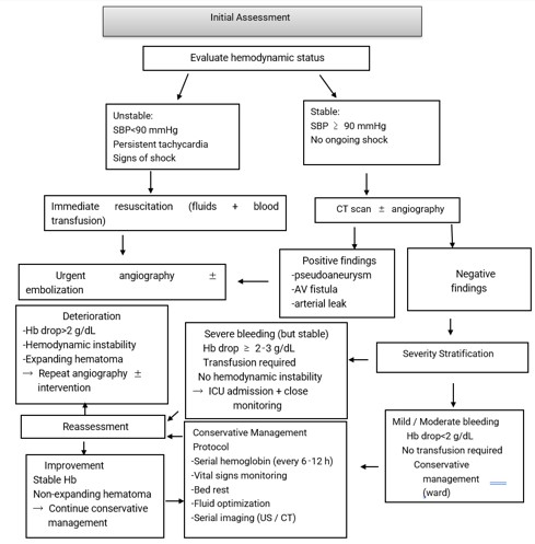
Figure (7) Proposed decision-making algorithm for management of delayed post-PCNL hemorrhage.
Ethical Approval and Patient Consent
This case report was conducted in accordance with institutional ethical standards. Formal ethical approval was waived according to local regulations.
Written informed consent was obtained from the patient for publication of this case report and any accompanying images. A copy of the written consent is available for review by the Editor upon request.
This study was conducted in accordance with the Declaration of Helsinki.











Open Access By Aditum Open Access Journals id licensed under Creative Commons Attribution 4.0 International License. Based On a Work at aditum.org


