
International Journal of Telehealth and Digital Medicine
OPEN ACCESS | Volume 1 - Issue 1 - 2026
ISSN No: - | Journal DOI: 10.61148/IJTDM


Tasawar Iqbal
Institute of Physiology and Pharmacology, University of Agriculture, Faisalabad.
Corresponding author: Tasawar Iqbal, Institute of Physiology and Pharmacology, University of Agriculture, Faisalabad.
Received: September 20, 2026 | Accepted: October 05, 2026 | Published: October 08, 2026
Citation: Iqbal T. (2026) “Overview of Snakebite Antivenom: Production, Mechanisms, Clinical Applications, and Future Perspectives” International Journal of Telehealth and Digital Medicine, 1(1); DOI: 10.61148/2766-8614/JCCRCS/004.
Copyright: © 2026 Tasawar Iqbal. This is an open access article distributed under the Creative Commons Attribution License, which permits unrestricted use, distribution, and reproduction in any medium, provided the original work is properly cited.
Snakebite envenomation is a neglected tropical disease that poses a significant public health challenge worldwide, particularly in tropical and subtropical regions. Each year, millions of people suffer snakebites, resulting in substantial mortality, permanent disability, and socioeconomic burden. Snake venoms are complex mixtures of proteins, peptides, enzymes, and toxins that produce neurotoxic, hemotoxic, cytotoxic, myotoxic, and cardiotoxic effects, leading to severe local and systemic complications. Antivenom remains the only specific and effective treatment capable of neutralizing circulating venom toxins and preventing life-threatening outcomes. This review provides a comprehensive overview of snakebite Antivenom, covering venom composition and mechanisms of toxicity, venom collection and processing, horse immunization, antibody production, purification methods, quality control, storage, and clinical applications. The review also discusses the different types of Antivenoms, including monovalent and polyvalent formulations, their mechanisms of action, indications for clinical use, dosing strategies, and management of adverse reactions. Furthermore, recent advances in Antivenom development, including recombinant antibodies, monoclonal antibodies, synthetic antibody fragments, and next-generation immunotherapies, are highlighted as promising approaches to improve treatment efficacy and safety. Despite considerable progress in manufacturing technologies, challenges such as venom variability, limited accessibility, high production costs, cold-chain requirements, and the risk of hypersensitivity reactions continue to limit the global effectiveness of Antivenoms. Strengthening research, improving manufacturing capacity, enhancing quality standards, and expanding equitable access to effective Antivenoms are essential to reduce the global burden of snakebite envenomation. Continued innovation in biotechnology and immunotherapy is expected to transform future Antivenom development and improve clinical outcomes for snakebite victims worldwide.
1. Introduction
Snakebite envenomation is a major yet often neglected public health problem, particularly in tropical and subtropical regions where human populations frequently come into contact with venomous snakes. It causes substantial morbidity and mortality, especially among agricultural workers, rural communities, and children. According to the World Health Organization, an estimated 5.4 million snakebites occur annually, resulting in approximately 1.8–2.7 million cases of envenomation and 81,000–138,000 deaths worldwide, with many survivors suffering permanent physical disabilities. The burden is greatest in South Asia, Southeast Asia, sub-Saharan Africa, and parts of Latin America, where limited access to timely medical care and effective antivenom contributes to poor clinical outcomes [1].
Snake venoms are complex mixtures of biologically active proteins, peptides, enzymes, and toxins that disrupt multiple physiological systems. Depending on the snake species, venom may exert neurotoxic, hemotoxic, cytotoxic, myotoxic, or cardiotoxic effects, leading to paralysis, coagulopathy, tissue necrosis, organ failure, and death if left untreated. The severity of envenomation depends on several factors, including the snake species, venom composition, amount of venom injected, bite location, and the victim's age and health status [2].
Antivenom remains the only specific and effective treatment for systemic snakebite envenomation. Conventional antivenoms are produced by immunizing large mammals, primarily horses, with gradually increasing doses of snake venom to stimulate the production of neutralizing antibodies. These antibodies are subsequently purified and processed into whole immunoglobulin G (IgG) or antibody fragments such as Fab or F(ab′)₂ to improve safety and therapeutic efficacy. Timely administration of appropriate antivenom significantly reduces mortality, prevents irreversible tissue damage, and improves patient outcomes [3].
Despite its life-saving role, antivenom therapy faces several challenges, including limited availability, high production costs, species-specific efficacy, cold-chain storage requirements, and the risk of hypersensitivity reactions. Recent advances in biotechnology, recombinant antibodies, monoclonal antibody therapies, and synthetic antibody fragments are paving the way for safer, more effective, and broadly neutralizing next-generation antivenoms[4]. This review provides a comprehensive overview of snakebite envenomation, the mechanisms of snake venom toxicity, conventional antivenom production, mechanisms of action, clinical applications, current challenges, and future perspectives in antivenom development.
2. Types of Snakebite
2.1 Venomous Snakes
Venomous snakes possess specialized venom glands and hollow or grooved fangs that inject venom into their prey or a victim during a bite. Snake venom is a complex mixture of enzymes, proteins, peptides, and other bioactive molecules that immobilize prey and facilitate digestion. In humans, these toxins can cause local tissue destruction, neurotoxicity, coagulopathy, hemorrhage, myotoxicity, and systemic organ dysfunction. The severity of envenomation depends on factors such as snake species, venom composition, the amount of venom injected, bite location, and the victim's age and health status. Prompt administration of the appropriate antivenom is the cornerstone of treatment for venomous snakebites [5].
The medically important venomous snake families include Elapidae, Viperidae, Atractaspididae, and selected members of Colubridae. Elapid snakes, such as cobras, kraits, mambas, coral snakes, and taipans, primarily produce neurotoxic venom that affects the nervous system and may lead to respiratory paralysis. Vipers and pit vipers possess predominantly hemotoxic and cytotoxic venoms that cause coagulopathy, bleeding, edema, tissue necrosis, and kidney injury. Some rear-fanged colubrids can also cause clinically significant envenomation, although severe cases are relatively uncommon [6].
2.2 Non-Venomous Snakes
Non-venomous snakes lack specialized venom delivery systems capable of causing systemic envenomation in humans. They capture prey primarily through constriction or by swallowing prey alive rather than injecting venom. Although their bites are generally not life-threatening, they may produce pain, superficial lacerations, bleeding, and secondary bacterial infections if not properly managed. Wound cleaning, tetanus prophylaxis when indicated, and infection prevention are usually sufficient, and antivenom is not required for bites from non-venomous snakes [7].
2.3 Classification of Medically Important Snake Species
Medically important snakes are those capable of causing significant illness or death in humans. The World Health Organization classifies these species according to their medical importance and geographic distribution. The principal families include:
Family Elapidae: Cobras (Naja spp.), kraits (Bungarus spp.), mambas (Dendroaspis spp.), coral snakes (Micrurus spp.), and taipans (Oxyuranus spp.), which predominantly produce neurotoxic envenomation.
Family Viperidae: True vipers (Vipera spp.) and pit vipers (Bothrops, Crotalus, Trimeresurus, and related genera), whose venoms commonly produce hemotoxic, cytotoxic, and myotoxic effects.
Family Atractaspididae: Burrowing asps (Atractaspis spp.), which possess highly specialized fangs and venom capable of producing severe local injury and systemic toxicity.
Family Colubridae (selected species): Certain rear-fanged snakes, including Dispholidus typus (boomslang) and Thelotornis spp. (twig snakes), can cause severe coagulopathy and life-threatening hemorrhage [8].
In South Asia, including Pakistan and India, the "Big Four" snakes Indian cobra (Naja naja), common krait (Bungarus caeruleus), Russell's viper (Daboia russelii), and saw-scaled viper (Echis carinatus) are responsible for the majority of clinically significant snakebite cases.
3. Snakebite Envenomation
3.1 Epidemiology and Global Burden
Snakebite envenomation is recognized by the World Health Organization as a Category A Neglected Tropical Disease (NTD) because it disproportionately affects impoverished rural populations with limited access to healthcare. Globally, approximately 5.4 million snakebites occur each year, resulting in 1.8–2.7 million cases of envenomation and an estimated 81,000–138,000 deaths annually. In addition, nearly 400,000 survivors experience permanent disabilities, including amputations, chronic ulceration, contractures, renal impairment, and psychological trauma [1]. The highest incidence of snakebite occurs in South Asia, Southeast Asia, sub-Saharan Africa, Latin America, and Oceania, where agriculture is the primary occupation and encounters between humans and snakes are common. Delayed access to healthcare, inadequate availability of effective antivenom, poor transportation infrastructure, and reliance on traditional healers contribute substantially to increased mortality and morbidity. In Pakistan, medically important snake species are widely distributed, and snakebite remains a significant public health concern, particularly in rural farming communities [9].
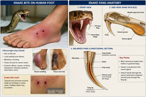
Figure 1. Anatomy of Snake Fangs and Venom Delivery Following Snakebite
3.2 Clinical Manifestations of Snakebite
Clinical manifestations of snakebite depend on the snake species, venom composition, amount of venom injected, and patient characteristics. Local manifestations commonly include pain, swelling, edema, erythema, blister formation, ecchymosis, tissue necrosis, and regional lymphadenopathy. Systemic manifestations may involve neurotoxicity (ptosis, diplopia, dysphagia, muscle weakness, and respiratory paralysis), hemotoxicity (coagulopathy, spontaneous bleeding, thrombocytopenia, and disseminated intravascular coagulation), myotoxicity (muscle pain and rhabdomyolysis), nephrotoxicity resulting in acute kidney injury, cardiovascular complications such as hypotension and arrhythmias, and, in severe cases, anaphylaxis and multiorgan failure. Prompt clinical assessment, laboratory investigations, supportive care, and early administration of appropriate antivenom are critical for improving patient outcomes and reducing mortality [10].
4. Mechanism of Action of Snake Venom
Snake venom is a highly complex biological secretion composed of proteins, peptides, enzymes, and non-protein toxins that act synergistically to immobilize prey and facilitate digestion. Major venom components include phospholipase A₂ (PLA₂), snake venom metalloproteinases (SVMPs), serine proteases (SVSPs), three-finger toxins (3FTxs), L-amino acid oxidases (LAAOs), hyaluronidases, and disintegrins. These toxins interfere with the nervous, hematological, muscular, cardiovascular, and immune systems, resulting in a wide range of local and systemic manifestations. The composition and toxicity of venom vary considerably among snake species, geographical regions, and even individual snakes [11].
4.1 Neurotoxic Effects
Neurotoxic venoms are predominantly produced by elapid snakes, including cobras (Naja spp.), kraits (Bungarus spp.), mambas (Dendroaspis spp.), and coral snakes (Micrurus spp.). Neurotoxins interfere with normal neuromuscular transmission by acting at either the presynaptic or postsynaptic level. Presynaptic neurotoxins, primarily phospholipase A₂ enzymes, damage motor nerve terminals and inhibit the release of acetylcholine, whereas postsynaptic α-neurotoxins bind competitively to nicotinic acetylcholine receptors at the neuromuscular junction, preventing receptor activation. Consequently, skeletal muscle contraction is impaired, leading to progressive paralysis. Clinically, neurotoxic envenomation presents with ptosis, blurred vision, diplopia, dysarthria, dysphagia, generalized muscle weakness, and respiratory muscle paralysis. Without timely respiratory support and antivenom administration, respiratory failure may become fatal [12].
4.2 Hemotoxic Effects
Hemotoxic venoms are characteristic of vipers and pit vipers. These venoms contain metalloproteinases, serine proteases, phospholipases, and anticoagulant proteins that disrupt normal hemostasis and vascular integrity. Snake venom metalloproteinases damage endothelial cells and basement membranes, causing capillary leakage and hemorrhage. Venom serine proteases activate or consume clotting factors, resulting in venom-induced consumption coagulopathy (VICC). Platelet dysfunction, fibrinogen depletion, and activation of fibrinolysis further contribute to spontaneous bleeding. Patients commonly develop local bleeding, gingival bleeding, hematuria, gastrointestinal hemorrhage, intracranial bleeding, hypotension, disseminated intravascular coagulation (DIC), and acute kidney injury due to hypoperfusion, hemolysis, or pigment nephropathy [13].
4.3 Cytotoxic Effects
Cytotoxic venoms cause direct destruction of cells and surrounding tissues. Cytotoxicity is mediated primarily by phospholipase A₂ enzymes, cytotoxins, and metalloproteinases, which disrupt cell membranes, degrade extracellular matrix proteins, and induce inflammatory responses.
Local tissue injury develops rapidly after envenomation and is characterized by severe pain, swelling, edema, erythema, blister formation, hemorrhage, and progressive necrosis. Extensive tissue destruction may result in compartment syndrome, chronic ulceration, secondary bacterial infection, permanent disability, or limb amputation if treatment is delayed [14].
4.4 Myotoxic and Cardiotoxic Effects
Certain snake venoms possess potent myotoxins that directly damage skeletal muscle fibers by disrupting cell membrane integrity and increasing intracellular calcium concentrations. This process leads to muscle fiber necrosis, rhabdomyolysis, and release of intracellular proteins such as creatine kinase and myoglobin into the circulation. Myoglobin released during rhabdomyolysis can obstruct renal tubules and contribute to acute kidney injury. Cardiotoxic venom components may directly impair cardiac muscle function, alter ion channel activity, and induce oxidative stress. Clinical manifestations include hypotension, arrhythmias, reduced myocardial contractility, myocarditis, and circulatory shock. Severe cardiotoxicity may lead to cardiovascular collapse and death if untreated [15].
4.5 Pathophysiological Changes Following Envenomation
Following injection into the victim, venom rapidly diffuses through lymphatic and vascular systems into local tissues and systemic circulation. Hyaluronidase, commonly referred to as the "spreading factor," facilitates venom dissemination by degrading connective tissue components.
Venom toxins initiate multiple pathological processes simultaneously, including endothelial injury, inflammation, coagulation abnormalities, complement activation, oxidative stress, cellular apoptosis, and direct organ toxicity. Local inflammation is characterized by infiltration of neutrophils and macrophages, release of pro-inflammatory cytokines such as tumor necrosis factor-α (TNF-α), interleukin-1β (IL-1β), and interleukin-6 (IL-6), together with increased production of reactive oxygen species. Systemically, these events may result in coagulopathy, neurotoxicity, rhabdomyolysis, acute kidney injury, hepatic dysfunction, respiratory failure, cardiovascular instability, and multiorgan failure. Early administration of appropriate antivenom effectively neutralizes circulating venom toxins before irreversible tissue injury occurs [16].
5. Snake Venom Collection
5.1 Venom Extraction (Milking) Techniques
Snake venom used for research and antivenom production is obtained from healthy adult venomous snakes maintained in licensed serpentariums or specialized venom production facilities. Venom extraction, commonly known as milking, is performed only by trained personnel using strict biosafety procedures to minimize the risk of injury. During the procedure, the snake is gently restrained while allowing its fangs to penetrate a sterile collection vessel covered with a parafilm or latex membrane. Gentle pressure is applied to the venom glands located behind the eyes, causing venom to flow through the fangs into the collection container. The collected venom is immediately inspected to remove debris and contaminants before further processing. The quantity and composition of venom vary depending on species, age, sex, nutritional status, geographic origin, season, and interval between milkings. To maintain snake welfare and ensure venom quality, extraction is performed at appropriate intervals, typically every 2–6 weeks depending on the species [17].
5.2 Venom Handling and Biosafety
Fresh venom is an extremely hazardous biological material and must be handled under stringent biosafety conditions. Personnel should wear appropriate personal protective equipment (PPE), including gloves, protective eyewear, laboratory coats, and face shields when necessary. Following collection, venom is transferred into sterile tubes, centrifuged to remove cellular debris, and stored at low temperatures until further processing. Each sample is carefully labeled with species identification, collection date, geographic origin, and batch number to ensure traceability. Standard operating procedures, emergency protocols, availability of species-specific antivenom, and proper training are essential for all personnel involved in venom collection and handling. Compliance with institutional animal welfare regulations and biosafety guidelines is mandatory throughout the process [18].
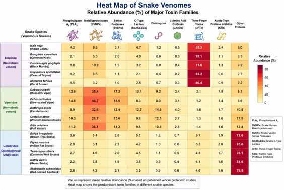
Figure 2. Comparative Heat Map of Major Snake Venom Toxin Families Across Different Snake Species
6. Venom Processing and Potency Assessment
6.1 Determination of Venom Potency
Following collection, venom undergoes physicochemical and biological characterization to ensure consistency and suitability for antivenom production. Venom potency is commonly assessed by determining its median lethal dose (LD₅₀) using standardized laboratory animal models. Additional biological assays evaluate hemorrhagic, neurotoxic, myotoxic, phospholipase, proteolytic, and coagulant activities. Modern analytical techniques such as high-performance liquid chromatography (HPLC), liquid chromatography mass spectrometry (LC–MS/MS), enzyme-linked immunosorbent assay (ELISA), sodium dodecyl sulfate polyacrylamide gel electrophoresis (SDS–PAGE), and proteomic analyses are widely employed to characterize venom composition and batch consistency [19].
6.2 Freeze-Drying (Lyophilization) of Venom
To preserve biological activity and improve long-term stability, freshly collected venom is rapidly frozen and subsequently subjected to lyophilization (freeze-drying). During this process, frozen water is removed by sublimation under vacuum, producing a dry, stable powder while minimizing protein degradation. Lyophilized venom retains its toxic and immunogenic properties for extended periods and is easier to transport, store, and standardize than liquid venom. This processing step is essential for producing high-quality venom suitable for research, diagnostic applications, and antivenom manufacturing [20].
6.3 Venom Storage and Quality Control
Lyophilized venom should be stored in airtight, moisture-resistant containers at −20°C or lower and protected from heat, humidity, and direct light to maintain stability. Under appropriate storage conditions, venom may remain biologically active for many years. Quality control procedures include sterility testing, moisture determination, protein concentration measurement, biochemical profiling, toxin composition analysis, microbiological examination, and biological activity assays. Batch-to-batch consistency is critical because variations in venom composition directly influence the efficacy and specificity of the resulting antivenoms [21].
7. Horse Immunization for Antivenom Production
Horse immunization is the cornerstone of conventional antivenom production. Horses are the preferred host animals because of their large body size, ability to produce high concentrations of antibodies, ease of handling, long productive lifespan, and well-established safety record in antivenom manufacturing. During immunization, horses are repeatedly exposed to small, gradually increasing doses of snake venom, stimulating the immune system to produce high-affinity neutralizing antibodies against venom toxins. The resulting hyperimmune plasma serves as the primary source of immunoglobulins used for antivenom production [22].
7.1 Selection of Healthy Horses
Healthy adult horses aged between 3 and 10 years are generally selected for immunization. Before inclusion in the production program, each horse undergoes comprehensive veterinary examination to ensure good health and freedom from infectious diseases. Hematological and biochemical analyses are performed to evaluate liver and kidney function, nutritional status, and overall physiological condition. Selected horses should possess good temperament, adequate body weight, and no previous history of chronic illness. Throughout the immunization program, animals are maintained under appropriate housing conditions with balanced nutrition, continuous veterinary supervision, and compliance with institutional animal welfare regulations [23].
7.2 Gradual Immunization Using Increasing Doses of Venom
The immunization protocol begins with administration of a very small quantity of venom, usually mixed with an immunological adjuvant such as Freund's Complete Adjuvant (FCA) for the primary immunization. Subsequent booster injections are administered with Freund's Incomplete Adjuvant (FIA) or other approved adjuvants to enhance antibody production while minimizing tissue irritation. Venom doses are gradually increased over several weeks or months according to the horse's immune response and clinical condition. Injections are typically administered by the subcutaneous or intramuscular route at multiple anatomical sites to reduce local tissue damage and improve antigen presentation. Repeated exposure stimulates B lymphocytes to proliferate and differentiate into plasma cells that produce high concentrations of venom-specific immunoglobulin G (IgG). Booster immunizations continue until antibody titers reach the desired level for plasma collection [24].
7.3 Monitoring Antibody Production
The immune response is monitored regularly by collecting small blood samples throughout the immunization schedule. Serum antibody titers are measured using immunological techniques such as enzyme-linked immunosorbent assay (ELISA), immunodiffusion assays, and venom-neutralization tests. In addition to antibody quantification, horses are routinely examined for body weight, appetite, body temperature, injection-site reactions, and general health. Hematological and biochemical parameters are periodically evaluated to ensure that repeated immunization does not adversely affect animal welfare. Once sufficient neutralizing antibody levels have been achieved, the horse is considered suitable for large-scale plasma collection [25].
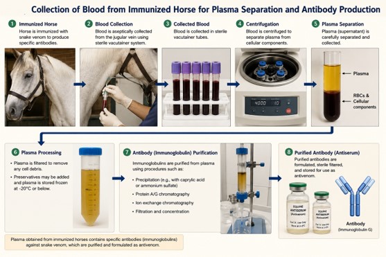
Figure 3. Production of Snake Antivenom: Blood Collection, Plasma Separation, and Immunoglobulin Purification
8. Antivenom Production Process
The production of antivenom involves a series of standardized procedures designed to obtain highly purified venom-specific antibodies while ensuring product safety, potency, and consistency. Modern manufacturing follows Good Manufacturing Practices (GMP) and strict quality control guidelines recommended by the World Health Organization [26].
8.1 Venom Injection Protocol in Horses
Following selection of healthy animals, horses are immunized with carefully measured doses of one or more snake venoms. The initial dose is intentionally low to avoid toxicity and is progressively increased through multiple booster injections administered over 3–6 months, depending on the desired antibody response. For monovalent antivenom, horses are immunized with venom from a single snake species. For polyvalent antivenom, mixtures of venoms from several medically important snake species are administered to induce broad-spectrum antibody production. Throughout immunization, horses are monitored closely for adverse reactions and overall health status [23].
8.2 Collection of Horse Blood
Once adequate antibody titers are confirmed, whole blood is collected under sterile conditions by trained veterinary personnel. Approximately 6–8 liters of blood may be obtained from a healthy adult horse during a single bleeding session without compromising animal welfare. Blood collection is performed at carefully controlled intervals, allowing sufficient recovery time between collections. Immediately after collection, anticoagulants such as sodium citrate may be added to prevent coagulation during processing [27].
8.3 Separation of Plasma from Whole Blood
Collected blood is processed promptly to separate plasma from cellular components. Separation is usually achieved by refrigerated centrifugation or plasmapheresis. Plasma contains the venom-specific immunoglobulins, whereas erythrocytes, leukocytes, and platelets are either discarded or returned to the horse during plasmapheresis procedures. Plasma is then stored at low temperatures until antibody purification begins [28].
8.4 Isolation of Immunoglobulins from Plasma
The hyperimmune plasma contains predominantly immunoglobulin G (IgG) antibodies capable of specifically binding and neutralizing venom toxins. These antibodies are isolated using established fractionation methods such as caprylic acid precipitation, ammonium sulfate precipitation, enzyme digestion, and chromatographic purification. Following purification, antibody preparations are further processed to produce either intact IgG molecules or enzymatically generated antibody fragments, depending on the intended antivenom formulation [29].
9. Antibody Structure and Fragmentation
Antibodies used in antivenom production are primarily IgG molecules, which possess high specificity for venom toxins. Understanding antibody structure is essential because selective removal of certain antibody regions improves the safety and tolerability of antivenom [24].
9.1 Structure of IgG Antibody
The immunoglobulin G (IgG) molecule is a Y-shaped glycoprotein composed of four polypeptide chains linked by disulfide bonds. It consists of two identical heavy chains (H1 and H2) and two identical light chains (L1 and L2). Each arm of the IgG molecule contains a variable region that specifically recognizes and binds snake venom antigens. The Fc (fragment crystallizable) region mediates interactions with immune cells and complement proteins, whereas the Fab (fragment antigen-binding) regions are responsible for specific antigen recognition and neutralization of venom toxins during antivenom therapy [30].
9.2 Production of Antibody Fragments (Fab and F(ab′) ₂)
To improve the safety and pharmacological properties of antivenoms, purified IgG molecules are enzymatically digested into smaller antibody fragments. Papain cleaves IgG above the hinge region, producing two Fab fragments and one Fc fragment, whereas pepsin cleaves below the hinge region to generate a single F(ab′)₂ fragment with two linked antigen-binding sites. Fab fragments penetrate tissues rapidly but have a shorter plasma half-life, while F(ab′)₂ fragments circulate longer and provide more sustained neutralization of circulating venom toxins [24].
9.3 Removal of the Fc Region to Reduce Adverse Immune Reactions
The Fc region is primarily responsible for activating complement proteins and interacting with Fc receptors on immune cells. Although these functions are beneficial during normal immune responses, they may contribute to adverse reactions following administration of equine-derived Antivenoms. Therefore, most modern Antivenoms remove the Fc portion through enzymatic digestion. The resulting Fab or F(ab′)₂ fragments exhibit reduced immunogenicity and are associated with lower incidences of serum sickness, complement activation, and immediate hypersensitivity reactions while maintaining effective venom-neutralizing capacity [31].
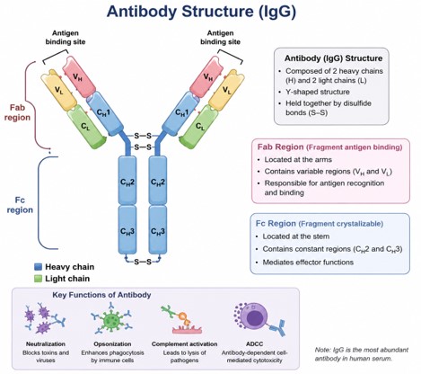
Figure 4. Structure of Immunoglobulin G (IgG) Antibody
10. Purification of Antivenom
Purification is one of the most critical stages of antivenom manufacturing because it removes unwanted plasma proteins, contaminants, and potentially immunogenic substances while preserving venom-neutralizing antibodies. High-purity antibody preparations improve product safety, efficacy, and stability [32].
10.1 Purification Techniques
Several complementary purification methods are employed during antivenom production.
Caprylic Acid Precipitation
Caprylic acid precipitation is among the most widely used purification methods because of its simplicity, efficiency, and cost-effectiveness. Caprylic acid selectively precipitates non-IgG plasma proteins while leaving immunoglobulins in solution. This technique produces highly purified antibody preparations with minimal protein aggregation and excellent recovery [33].
Ammonium Sulfate Precipitation
Ammonium sulfate precipitation separates proteins according to their solubility. Controlled addition of ammonium sulfate precipitates immunoglobulins while allowing unwanted proteins to remain in solution. Although effective, this method generally requires additional purification steps to remove residual salts and contaminants [32].
Chromatography (HPLC)
High-performance liquid chromatography (HPLC) and other chromatographic techniques, including ion-exchange, affinity, and size-exclusion chromatography, are employed to further improve antibody purity. These methods efficiently remove protein aggregates, enzyme residues, endotoxins, and degraded proteins while maintaining biological activity [34].
Ultrafiltration
Ultrafiltration utilizes semipermeable membranes to concentrate antibody preparations, exchange buffers, remove low-molecular-weight impurities, and adjust the final protein concentration. This process also improves product stability before sterile filtration and formulation [24].
10.2 Quality Assessment of Purified Antibody Fragments
Purified Fab, F(ab′)₂, or IgG preparations undergo rigorous quality control before formulation and packaging. Quality assessment includes evaluation of protein concentration, purity, molecular integrity, sterility, endotoxin content, residual enzyme activity, aggregate formation, physicochemical stability, and venom-neutralizing potency. Analytical techniques such as SDS-PAGE, HPLC, ELISA, Western blotting, mass spectrometry, and in vivo neutralization assays are commonly used to verify antibody identity, purity, and biological activity. Only batches meeting predefined specifications for safety, potency, sterility, and stability are approved for clinical use [35].
11. Types of Antivenom
Antivenoms are biological products containing venom-specific antibodies or antibody fragments that neutralize the toxic components of snake venom. They are classified according to the range of snake species against which they are effective. The two principal categories are monovalent and polyvalent antivenoms. The choice between these formulations depends on the identification of the offending snake, regional snake distribution, availability of diagnostic facilities, and local treatment guidelines [24].
11.1 Monovalent Antivenom
Monovalent antivenom is produced by immunizing horses or other host animals with the venom of a single snake species. Consequently, it contains antibodies that specifically neutralize toxins from that particular species or closely related species with similar venom composition. Because of its high specificity, monovalent antivenom generally requires lower therapeutic doses, minimizes unnecessary exposure to foreign proteins, and is associated with a lower risk of hypersensitivity reactions. It is most effective when the offending snake has been accurately identified either visually or through reliable diagnostic methods. However, its use is limited in regions where multiple medically important snake species coexist or where identification of the snake is uncertain.
Advantages: High species-specific efficacy, lower antivenom dose requirements, reduced exposure to heterologous proteins, and a lower risk of adverse immune reactions.
Limitations: Requires accurate snake species identification, is ineffective against unrelated snake venoms, and has limited usefulness in regions with diverse venomous snake populations [36].
11.2 Polyvalent Antivenom
Polyvalent antivenom is prepared by immunizing horses with a mixture of venoms from several medically important snake species. It contains antibodies capable of neutralizing toxins from multiple venomous snakes and is therefore the most commonly used formulation in many tropical and subtropical countries. Polyvalent antivenoms are particularly valuable in rural settings where snake identification is difficult or impossible. In South Asia, for example, polyvalent antivenoms are typically formulated to neutralize the venoms of the "Big Four" snakes: Indian cobra (Naja naja), common krait (Bungarus caeruleus), Russell's viper (Daboia russelii), and saw-scaled viper (Echis carinatus).
Advantages: Provides broad-spectrum venom neutralization, is effective when the offending snake species is unknown, and is suitable for regions with multiple medically important venomous snakes.
Limitations: Requires larger treatment doses, increases exposure to foreign immunoglobulins, carries a higher risk of allergic reactions and serum sickness, and involves greater manufacturing complexity and cost [37].
Table 1. Commonly Used Snake Antivenoms Around the World
|
S. No. |
Antivenom (Brand/Generic Name) |
Manufacturer/Country |
Target Snake Species |
Region of Use |
|
1 |
Inoserp® Pan-Africa |
Inosan Biopharma, Spain |
African vipers and elapids |
Sub-Saharan Africa |
|
2 |
Fav-Afrique®* |
Sanofi Pasteur (France)* |
Multiple African venomous snakes |
Africa |
|
3 |
EchiTAb-Plus-ICP® |
Instituto Clodomiro Picado, Costa Rica |
Echis spp., Bitis spp., Naja spp. |
West Africa |
|
4 |
SAIMR Polyvalent Antivenom |
South African Vaccine Producers, South Africa |
Cobras, mambas, puff adders, rinkhals |
Southern Africa |
|
5 |
Bharat Serums Polyvalent Antivenom |
Bharat Serums and Vaccines, India |
Indian cobra, common krait, Russell's viper, saw-scaled viper |
India |
|
6 |
VINS Polyvalent Snake Venom Antiserum |
VINS Bioproducts Ltd., India |
"Big Four" Indian snakes |
India, Nepal, Sri Lanka |
|
7 |
Premium Serums Polyvalent Antivenom |
Premium Serums & Vaccines Pvt. Ltd., India |
Indian medically important snakes |
India |
|
8 |
Snake Venom Antiserum I.P. |
Haffkine Bio-Pharmaceutical Corporation, India |
Indian cobra, krait, Russell's viper, saw-scaled viper |
India |
|
9 |
Thai Red Cross Polyvalent Antivenom |
Queen Saovabha Memorial Institute, Thailand |
Cobra, krait, pit vipers |
Thailand |
|
10 |
Thai Red Cross Monovalent Antivenoms |
Queen Saovabha Memorial Institute, Thailand |
Species-specific antivenoms |
Southeast Asia |
|
11 |
CSL Snake Antivenoms |
CSL Seqirus, Australia |
Brown snakes, tiger snakes, taipans, death adders |
Australia |
|
12 |
CroFab® |
BTG International (now SERB Pharmaceuticals), USA |
North American pit vipers |
United States |
|
13 |
Anavip® |
Laboratorios Silanes, Mexico |
Rattlesnakes and pit vipers |
USA and Mexico |
|
14 |
Antivipmyn® |
Laboratorios Silanes, Mexico |
Pit vipers and coral snakes |
Mexico and Latin America |
|
15 |
Coralmyn® |
Laboratorios Silanes, Mexico |
Coral snakes (Micrurus spp.) |
Mexico |
|
16 |
Bothrofav® |
Instituto Butantan, Brazil |
Bothrops species |
Brazil |
|
17 |
Crotalic Antivenom |
Instituto Butantan, Brazil |
Crotalus species |
Brazil |
|
18 |
Bothrops Antivenom |
Instituto Clodomiro Picado, Costa Rica |
Bothrops asper and related species |
Central America |
|
19 |
Polyvalent Snake Antivenom |
Instituto Nacional de Salud, Colombia |
Regional venomous snakes |
Colombia |
|
20 |
Polyvalent Snake Antivenom |
Instituto Nacional de Salud, Peru |
South American pit vipers and coral snakes |
Peru |
12. Quality Control of Antivenom
Quality control is a critical component of antivenom manufacturing to ensure that each batch is safe, effective, sterile, and consistent. International regulatory agencies and the World Health Organization (WHO) recommend rigorous quality assurance procedures throughout production, purification, formulation, packaging, and distribution [38].
12.1 Sterility Testing
Sterility testing confirms that the final antivenom preparation is free from bacterial, fungal, and other microbial contamination. Samples are cultured in appropriate microbiological media under standardized conditions according to international pharmacopeial guidelines. In addition to sterility testing, manufacturers routinely evaluate endotoxin levels using validated assays such as the Limulus Amebocyte Lysate (LAL) test to ensure that pyrogenic contaminants are absent. Only sterile, pyrogen-free products are approved for clinical use [39].
12.2 Safety and Potency Testing
Safety testing evaluates the absence of unacceptable toxicity and confirms that residual enzymes, preservatives, and impurities remain within acceptable limits. Physicochemical analyses assess protein concentration, purity, pH, osmolality, aggregate formation, and product stability. Potency testing determines the ability of antivenom to neutralize venom toxicity. Traditionally, potency has been assessed using standardized in vivo neutralization assays that determine the amount of antivenom required to neutralize a defined quantity of venom. Increasingly, validated in vitro methods such as enzyme-linked immunosorbent assays (ELISA), antivenomics, and functional toxin-neutralization assays are being incorporated to complement or reduce animal testing [40].
12.3 Packaging and Storage Conditions
Following quality approval, antivenom is aseptically filled into sterile glass vials or ampoules and sealed under Good Manufacturing Practice (GMP) conditions. Both liquid and lyophilized formulations are commercially available. Each product label includes information regarding snake species covered, protein content, batch number, manufacturing date, expiry date, storage requirements, dosage recommendations, and manufacturer details. Packaging materials should protect the product from moisture, excessive heat, and mechanical damage during transportation and storage [17].
13. Shelf Life and Stability of Antivenom
The stability of antivenom is influenced by formulation type, purification method, storage temperature, and packaging conditions. Appropriate storage is essential to preserve antibody activity and therapeutic efficacy throughout the product's shelf life [41].
13.1 Storage Recommendations
Lyophilized antivenoms should be stored in a cool, dry environment protected from direct sunlight and excessive humidity. Most manufacturers recommend storage at temperatures below 25°C, although refrigeration at 2–8°C is preferred where feasible. Liquid antivenoms generally require continuous refrigeration between 2°C and 8°C and should never be frozen, as freezing may denature antibodies and reduce therapeutic effectiveness. Reconstituted lyophilized antivenom should be used promptly and discarded if not administered within the manufacturer's recommended time [21].
13.2 Shelf Life
When manufactured and stored under recommended conditions, lyophilized antivenoms typically remain stable for approximately 3–5 years, while some products may retain potency for even longer following formal stability testing. Liquid antivenoms generally have a shorter shelf life because antibodies gradually lose biological activity during storage. Regular stability studies evaluate protein integrity, sterility, neutralizing potency, physicochemical characteristics, and moisture content to ensure that antivenom remains effective throughout its stated shelf life [42].
14. Mechanism of Action of Antivenom
Antivenom is the only specific treatment capable of neutralizing snake venom toxins. It acts by binding venom components with high specificity, thereby preventing their interaction with physiological targets and facilitating their elimination from the body. Early administration is essential because antivenom is most effective before irreversible tissue injury develops [43].
14.1 Neutralization of Circulating Venom Toxins
Following intravenous administration, venom-specific antibodies rapidly circulate throughout the bloodstream and bind free venom toxins. This interaction forms stable antigen antibody complexes that prevent toxins from interacting with critical physiological systems. Neutralization is most effective against toxins that remain in the circulation. Consequently, early administration significantly improves clinical outcomes and reduces mortality [11].
14.2 Prevention of Toxin Binding to Target Tissues
Venom toxins exert their pathological effects by binding to receptors, enzymes, cell membranes, coagulation factors, or extracellular matrix proteins. Antivenom antibodies competitively bind these toxins before they reach their target tissues, thereby preventing neurotoxicity, coagulopathy, myotoxicity, cytotoxicity, and other systemic complications. Although antivenom effectively neutralizes circulating toxins, it cannot reverse irreversible tissue damage that has already occurred. Therefore, prompt treatment remains the most important determinant of therapeutic success [44].
14.3 Formation and Clearance of Venom–Antibody Complexes
Once antibodies bind venom toxins, immune complexes are formed and subsequently removed from the circulation by the reticuloendothelial system, primarily in the liver and spleen. Phagocytic cells recognize and eliminate these complexes, thereby reducing circulating venom concentrations and limiting ongoing tissue injury. Supportive medical care, including respiratory support, fluid management, dialysis, blood product replacement, and wound management, may still be required because antivenom does not immediately reverse established organ damage [45].
15. Clinical Applications of Antivenom
Antivenom is indicated for patients with clinically significant envenomation and remains the standard treatment for systemic snakebite poisoning. Administration should be based on clinical manifestations rather than the bite alone [46].
15.1 Indications for Administration
Antivenom is indicated for patients with systemic or progressive local envenomation, including extensive swelling, neurotoxicity, coagulopathy, spontaneous bleeding, cardiovascular instability, acute kidney injury, severe myotoxicity or rhabdomyolysis, and rapidly progressive tissue necrosis. Patients without clinical evidence of envenomation generally do not require antivenom therapy [47].
15.2 Route and Dosage
Antivenom is administered intravenously, usually by slow infusion after appropriate dilution according to the manufacturer's recommendations. Intravenous administration ensures rapid systemic distribution and effective neutralization of circulating venom. The dose depends primarily on the quantity of venom injected rather than patient age or body weight. Additional doses may be administered if systemic manifestations persist or recur after the initial treatment [46].
15.3 Management of Adverse Reactions
Despite advances in purification, equine-derived antivenoms may cause early and delayed immune reactions. Early adverse effects include urticaria, pruritus, fever, bronchospasm, hypotension, and anaphylaxis, requiring prompt treatment with intramuscular adrenaline (epinephrine), antihistamines, corticosteroids when indicated, oxygen, and supportive care. Delayed reactions (serum sickness) usually occur 5–14 days after treatment, presenting with fever, rash, arthralgia, and lymphadenopathy, and are generally managed with oral antihistamines and corticosteroids [48].
Table 2. Comparison of Monovalent and Polyvalent Antivenoms Used in Snakebite Management
|
Feature |
Monovalent Antivenom |
Polyvalent Antivenom |
|
Definition |
Antivenom produced against the venom of a single snake species or closely related species. |
Antivenom produced against the venoms of multiple medically important snake species. |
|
Spectrum of Activity |
Effective against one specific snake species or closely related species. |
Effective against several venomous snake species. |
|
Specificity |
High species-specific neutralizing activity. |
Broad-spectrum neutralizing activity. |
|
Clinical Use |
Preferred when the offending snake has been accurately identified. |
Preferred when the snake species is unknown or difficult to identify. |
|
Dose Required |
Usually requires a lower dose due to high specificity. |
Often requires a higher dose because it targets multiple venoms. |
|
Risk of Adverse Reactions |
Lower risk due to reduced exposure to foreign proteins. |
Slightly higher risk because of greater protein content. |
|
Advantages |
High efficacy, lower dose requirement, reduced allergic reactions, and cost-effective when the snake species is known. |
Broad coverage, suitable for mixed snake populations, and useful in emergency settings without species identification. |
|
Limitations |
Ineffective against unrelated snake species and requires accurate snake identification. |
May have lower potency against individual venoms, higher production cost, and increased risk of hypersensitivity reactions. |
|
Examples |
Monovalent Cobra Antivenom, Monovalent Russell's Viper Antivenom, Monovalent Taipan Antivenom. |
Indian Polyvalent Antivenom (Big Four), SAIMR Polyvalent Antivenom, Inoserp® Pan-Africa, CroFab® (North American pit vipers). |
|
Common Regions of Use |
Australia, Thailand, and regions where snake species can be accurately identified. |
India, Pakistan, Africa, Latin America, and other regions with diverse venomous snake species. |
16. Challenges and Future Perspectives
Although conventional antivenoms have saved countless lives, their effectiveness remains limited by variable venom composition, adverse immune reactions, production costs, dependence on animal immunization, cold-chain requirements, and restricted availability in rural and resource-limited regions. Next-generation antivenom research is therefore focusing on safer, more specific, and broadly effective therapeutic approaches. Recombinant and humanized monoclonal antibodies can target clinically important venom toxins while reducing immunogenicity and improving manufacturing consistency. Engineered Fab fragments, nanobodies, single-domain antibodies, aptamers, peptide inhibitors, and synthetic binding proteins may further improve tissue penetration and toxin-specific neutralization. Combination therapies incorporating antibodies with small-molecule inhibitors against phospholipase A₂, metalloproteinases, and serine proteases could provide broader protection against complex venoms. Future development should also emphasize affordable production, regional manufacturing, heat-stable formulations, improved diagnostics, technology transfer, and stronger regulatory systems. These advances could increase accessibility, reduce treatment delays, and improve clinical outcomes for snakebite victims worldwide [49].
17. Conclusion
Snakebite envenomation remains a major global health challenge, particularly in low- and middle-income countries where access to timely diagnosis and effective treatment is limited. Antivenom remains the only specific therapy capable of neutralizing venom toxins and reducing morbidity and mortality. Advances in venom characterization, horse immunization, antibody purification, and quality control have significantly improved the safety and efficacy of modern Antivenoms. However, challenges such as venom variability, limited availability, high production costs, and adverse reactions continue to hinder optimal treatment. Future research on recombinant antibodies, monoclonal antibodies, synthetic antibody fragments, and next-generation therapeutics offers promising opportunities to develop safer, more effective, affordable, and broadly protective Antivenoms for improved global snakebite management.
Author Contributions: Tasawar Iqbal: Conceptualization, methodology, writing – original draft, and writing – review & editing.
Funding: This research received no external funding. The author conducted and completed the review independently without financial support from any organization or funding agency.
Institutional Review Board Statement: Not applicable.
Data Availability Statement: No original data were generated or analyzed in this review. All information is derived from previously published sources, which are properly cited within the manuscript.
Conflicts of Interest: The authors declare no conflicts of interest.
Generative AI statement: The manuscript was refined using Grammarly as an AI-based writing support tool to improve grammatical accuracy, clarity, readability, and overall linguistic quality.

Open Access By Aditum Open Access Journals id licensed under Creative Commons Attribution 4.0 International License. Based On a Work at aditum.org


