International Journal of Medical Case Reports and Medical Research
OPEN ACCESS | Volume 4 - Issue 1 - 2025
ISSN No: 2994-6905 | Journal DOI: 10.61148/2994-6905/IJMCRMR


Berhe Tesfai1,2*, Hailemichael Gebremariam3, Dawit Sereke1, Okbu Frezgi1,2
1Orotta National Referral Maternity Hospital, Ministry of Health, Asmara, Eritrea.
2Department of Obstetrics and Gynaecology, Orotta College of Medicine and Health Sciences, Asmara, Eritrea.
3Dekemhare Hospital, Zoba Debub, Ministry of Health, Dekemhare, Eritrea.
*Corresponding author: Berhe Tesfai, Orotta National Referral Maternity Hospital, Ministry of Health, Asmara, Eritrea.
Received: June 20, 2025
Accepted: June 28, 2025
Published: July 08, 2025
Citation: Tesfai B, Gebremariam H, Sereke D, Frezgi O., (2025) “Diagnosis Challenge of Massive Parasitic Leiomyoma, Case Report, 2025.” International Journal of Medical Case Reports and Medical Research, 4(1); DOI: 10.61148/2994-6905/IJMCRMR/169.
Copyright: © 2025 Berhe Tesfai. This is an open access article distributed under the Creative Commons Attribution License, which permits unrestricted use, distribution, and reproduction in any medium, provided the original work is properly cited.
Parasitic leiomyoma is an extremely rare variant of uterine leiomyoma occurring outside uterus. Due to its atypical clinical presentation and unusual location, these tumors pose a preoperative diagnostic challenge. A multiparous patient presented with abdominal pain and swelling of 6 months. Upon abdominal physical examination a hard, mobile, huge mass was identified. Transabdominal ultrasound and MRI revealed a massive adnexal mass. Intraoperatively, a hard mass attached to the mesentery was found without attachment to the ovaries, uterus and broad ligament. The mass was respected and histopathology revealed leiomyoma. Postoperative time was uneventful, and the patient discharged after 4 days. Parasitic leiomyoma has atypical clinical presentation, unusual location, and challenge clinicians to reach correct diagnosis.
parasitic leiomyoma, myoma, myomectomy
Parasitic or wandering leiomyoma are very rare form of uterine leiomyoma (1-9). Parasitic leiomyoma is a pedunculated sub-serosal fibroid that undergoes torsion and detaches from the uterus (9) and has no myometrial connections. There are several hypotheses about the origin of parasitic fibroids, including the iatrogenic seeding of fibroid pieces after morcellation in laparoscopic myomectomy, and pedunculated sub-serosal fibroid separating from its stalk and joining other abdominopelvic structures. (6) They are usually reported in women who underwent hysterectomy or myomectomy and present with abdominal pain and distention. (3)
Due to its rarity, atypical clinical presentation and unusual location, these tumors pose a diagnostic challenge (1, 5, 10) Patients usually present with atypical clinical presentation with a large, calcified mass (parasitic fibroid) (2) and acute abdomen (1). These fibroids are challenging to diagnose by imaging. (3, 6) A parasitic fibroid was mimicking an ovarian tumor on clinical examination, turned out to be sub-serous fibroid on ultrasonography and finally diagnosed as parasitic fibroid. (5)
Broad ligament myoma also presents unique diagnostic and management challenges due to their anatomical location, overall low incidence rate, radiological difficulty in differentiating with an ovarian tumor and potential for varied clinical manifestations. (11-13) The most common treatment of parasitic leiomyoma involves an abdominal or laparoscopic surgery in order to completely remove the mass is the best treatment of abdominal wall myoma in order to reduce the recurrence. (3, 14)
Case Report:
A 34-year-old para 4 mother presented with abdominal pain and swelling for 6 months. She had regular menses, and all her pregnancy and deliveries were uneventful. The patient had no alteration in bowel habit or history of gynecologic surgery. Upon abdominal physical examination, a huge 10x10cm mass, hard, mobile, mass with rough surface and irregular border was identified. Transabdominal ultrasound and MRI of the pelvis revealed a big mass on the left adnexa. With the diagnosis of pedunculated myoma, laparotomy was decided and intraoperatively, a huge, whitish mass attached to intestine and omentum was found floating. The mass had no attachment to the ovaries and uterus. (a, b, c) The mass was respected, and histopathology analysis revealed leiomyoma and postoperative time and follow up was uneventful.
a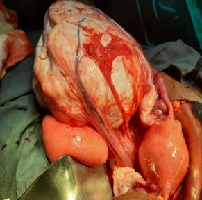 b
b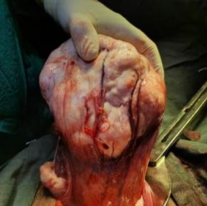 c
c 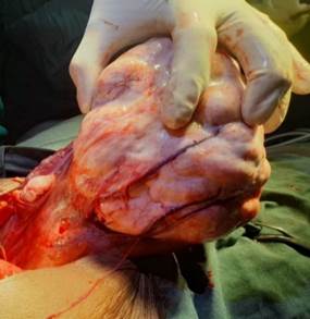
Figure 1: hard, huge mass attached to the mesentry with healthy uterus and ovaries
Discussion :
Parasitic leiomyoma is an extremely rare variant of uterine leiomyoma in our setting and occurs rarely in clinical practice. This was consistent with many literatures that parasitic leiomyoma is an extremely rare variant of uterine leiomyoma occurring outside uterus. (2-7) This patient presented with abdominal pain and swelling, which is nonspecific, and diagnosis was made intra-operatively. This was consistent with other studies that patient presented with vague symptoms, (7) acute abdomen (1), and they are challenging to diagnose by imaging. (5, 6) Primary parasitic fibroid was mimicking like an ovarian tumor on clinical examination (5) and can also be misdiagnosed as broad ligament myoma. (8, 12, 13) High index of suspicion and including as differential in abdominal-pelvic mass or on patients after myomectomy is recommended.
Parasitic leiomyoma is believed to be commonly occurred by torsion and detachment of pedunculated sub-serosal fibroid from the uterus or after laparoscopic resection of myoma and peritoneal seeding. This patient had no previous surgery for myoma and had no clinical signs of sub-serosal myoma. This was consistent with previous literatures that parasitic myomas typically occur after a pedunculated sub-serosal fibroid loses its uterine blood supply (6, 8) or after laparoscopic myomectomy. (8)
Imaging studies can give clues for the diagnosis of abdominopelvic mass, but it may not clearly diagnose parasitic myoma, consistent with other reports (3, 7, 9) The treatment option of parasitic myoma is complete resection of the mass by laparotomy or laparoscopy with complete abdominal washing after the procedure to decrease recurrence. As she had not completed her fertility, only mass resection was done and hysterectomy was deferred but the abdomen was washed, consistent to other report that thorough inspection and washing of the abdominopelvic cavity at the end of surgery is vital. (8)
Conclusion :
Parasitic leiomyoma is an extremely rare variant of uterine leiomyoma with atypical clinical presentation, unusual location, and challenge to clinicians to reach correct diagnosis preoperatively. Therefore, having clinical suspicion and including as differential in any abdomino-pelvic mass is of utmost importance in making diagnosis.
Declaration:
Acknowledgments: Authors acknowledges for the hospital staff for management of the case
Informed consent: The patient gave written informed consent for publication and use of the data for educational purposes.
Competing of interest: Authors have no competing of interest to disclose
Author Contributions: All authors have contributed to writing the case report and all reviewed and approved the final version of the case report.


Open Access By Aditum Open Access Journals id licensed under Creative Commons Attribution 4.0 International License. Based On a Work at aditum.org