
International Journal of Interventional Radiology and Imaging
OPEN ACCESS | Volume 4 - Issue 1 - 2026
ISSN No: 3065-6702 | Journal DOI: 10.61148/3065-6702/IJIRI


Sami Smerat1*, Ahmad Abu Arrah2
1School of Computed Tomography and MRI Sciences, Arab American University (AAUP), Ramallah, Palestine.
2Department of Medical Imaging, Arab American University (AAUP), Jenin, Palestine.
*Corresponding author: Sami Smerat, School of Computed Tomography and MRI Sciences, Arab American University (AAUP), Ramallah, Palestine.
Received: July 05, 2026 | Accepted: July 15, 2026 | Published: July 20, 2026
Citation: Smerat S, Ahmad A Arrah., (2026) “Acute encephalopathy presented with status epilepticus: Study Case.” International Journal of Interventional Radiology and Imaging, 4(1); DOI: 10.61148/3065-6702/IJIRI/050.
Copyright: © 2026 Sami Smerat. This is an open access article distributed under the Creative Commons Attribution License, which permits unrestricted use, distribution, and reproduction in any medium, provided the original work is properly cited.
Background: Acute necrotizing encephalopathy of childhood (ANEC) is an uncommon form of acute brain inflammation in children. It is distinguished by a fast and progressive deterioration in the patient's overall condition. Through our presentation of the case, our aim is to highlight the need of early identification and aggressive treatment of ANEC in order to achieve a better outcome. This report details the case of a 5-year-old female patient who first experienced flu-like symptoms, but subsequently developed a rapidly worsening brain disorder characterized by symmetrical damage to the thalamus and a lesion in the brainstem.
Case report: Researchers present a case of a 5-year-old child who was previously in good health. Following her bout with the illness, she experienced a swiftly advancing encephalopathy and status epilepticus. The first electroencephalogram (EEG) showed severe encephalopathy, while the magnetic resonance imaging (MRI) displayed pronounced symmetrical bilateral thalamic enhancement with brainstem lesions, consistent with the diagnosis of ANEC. Despite receiving rigorous ANEC therapy, the patient's prognosis was unfavorable.
Conclusion: ANEC is a rare and lethal disease within the field of pediatrics. Prompt initiation of assertive care and timely diagnosis are crucial to mitigate the elevated mortality risk associated with the illness. Moreover, it is crucial to acquire a prompt neuroradiological image, such as a brain MRI, in order to verify the diagnosis of this rare condition. This emphasizes the importance of the patient's detailed medical background, which indicates a swift and continuous deterioration in consciousness and the occurrence of status epilepticus following a viral infection. Furthermore, we want to enhance the awareness of this disorder among general practitioners and healthcare workers.
Acute necrotizing encephalopathy of childhood, Encephalitis, electroencephalogram, encephalopathy, Epilepsy, epilepticus, acute ischemic stroke patients, acute stroke treatment
ANEC, an uncommon kind of acute encephalopathy in children, is often caused by a viral infection. Patients afflicted with this disease typically have localized neurological symptoms, seizures, and changes in their state of consciousness [1]. Fulminant acute necrotizing encephalopathy is characterized by bilateral symmetrical involvement of the thalami, as shown in a magnetic resonance imaging (MRI) of the patient's brain. This finding is indicative of a poor prognosis [2], [3]. If there is no brainstem involvement, using large doses of steroids early in the treatment can lead to improved outcomes [4]. Early clinical diagnosis of ANEC may lead to a better prognosis, since significant involvement of the brainstem area indicates a severe and rapidly worsening course of the condition [5]. To enhance the prognosis, we are sharing this case to emphasize the significance of promptly diagnosing and treating this uncommon condition in young individuals. Infants and young children are impacted by ANEC, a condition that is predominantly prevalent in countries such as Taiwan and Japan [6]. According to a separate research, 13 patients underwent treatment and 28 cases were officially recorded, indicating a high prevalence of ANEC in India [7].
Case report
A previously asymptomatic five-year-old child presented to our emergency clinic with a three-day duration of progressively deteriorating level of awareness. The parents also reported that the kid regularly had episodes of generalized tonic-clonic convulsions, characterized by eye rolling, frothy mouth secretions, and lasting for a few minutes. The day prior to her clinical presentation, the family also noted that she experienced a mild increase in body temperature and a slight cough, like symptoms of influenza [8].
The patient's vital signs and temperature were within the normal range during the initial physical examination. She displayed a decreased state of consciousness and a Glasgow Coma Scale (GCS) score of 12 out of 15. The patient's deep tendon reflexes were rapid and without clonus, and she showed a positive bilateral Babinski sign. The distribution of her pupils was equal, they responded to light, and at times showed horizontal nystagmus. The results of the tone assessment were within the expected range. Due to the patient's encephalopathy, it was not possible to conduct a power and gait examination. The systemic assessment did not reveal any significant findings for the other results [9].
The patient's initial complete blood count revealed a white blood cell count of 3.30×110/L, a hemoglobin level of 11.9g/L, and a platelet count of 167×109/L. The liver function test showed elevated levels of aspartate aminotransferase (447 IU/L; normal range 0-34 IU/L), alanine aminotransferase (59 IU/L; normal range 0-34 IU/L), and Glutamyl transferase (430 IU/L). The sample exhibited several abnormalities: urea (6.25 mmol/L), potassium (4.07 mmol/L), chloride (100.2 mmol/L), abnormal carbon dioxide (14.3 mEq/L) (normal range: 23–29 mEq/L), creatinine (51.2 umol/L), calcium (2.35 mmol/L), magnesium (0.99 mmol/L), phosphate (1.33 mmol/L), and C-reactive protein (21.3 mg/L) (normal range: less than 10.0 mg/L).
A lumbar puncture was not performed for research on cerebrospinal fluid (CSF) due to the patient's fragile clinical condition. The first electroencephalogram (EEG) showed a pattern consistent with encephalopathy. However, a brain MRI performed immediately after revealed symmetrical bilateral areas of increased signal intensity in the thalami, with involvement of the brainstem (Figure 1) and (Figure 2).
The patient's initial prescriptions consisted of ceftriaxone, vancomycin, and acyclovir, which were administered for a duration of seven days in conjunction with antibiotics and antivirals. The patient's medical background and the findings from the brain MRI led to the diagnosis of acute necrotizing encephalopathy.
Within eight hours after her initial clinical presentation, she received intravenous pulse methylprednisolone at a dosage of 30 mg/kg/d for three consecutive days starting on the first day of her hospital admission. The patient's clinical state deteriorated further, displaying increased encephalopathy and unresponsiveness. According to the information provided, the patient was given a single dose of intravenous immunoglobulin, with a dosage of 2 grams per kilogram of body weight, on the second day of her stay [10]. Upon her first admission, she was prescribed oseltamivir for a duration of five days, since the respiratory gene Xpert test confirmed her H1N1 illness [11].
Regrettably, the patient's clinical condition worsened fast, as indicated by her GCS score of 8 out of 15 and poor respiratory effort. Consequently, she needed mechanical ventilation and intubation. Her neurological state underwent additional examination, revealing the presence of fixed and dilated pimples. An immediate brain computed tomography scan Figure 3.A showed the presence of a tonsillar herniation and severe cerebral edema [12].
Upon the patient's failure to show improvement by the fifth day of admission, she underwent five separate sessions of plasma exchange, in addition to receiving a 5% albumin solution, on her seventh day of hospitalization. On the tenth day, a follow-up brain MRI showed a significant lesion in the brainstem, accompanied by hemorrhaging and a cystic change Figure 3.B.
Considering the patient's current clinical condition and the worsening brain MRI, there is evidence of brainstem investigation confirmed the absence of any brainstem reactions. An EEG with isoelectric findings was confirmed by the data shown in Figure 3.C.
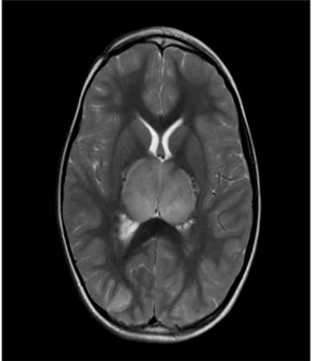
Figure 1. Brain MRI axial T2 sequence shows extensive symmetrical bilateral thalamic swelling.
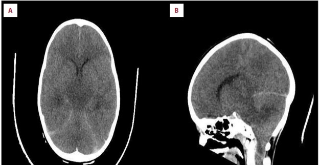
Figure 2. Brain CT scan soft tissue window showing significant edema with tonsillar herniation: (A): Axial view (B): Sagittal view.
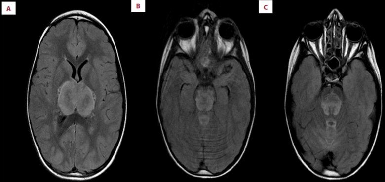
Figure 3. Brain MRI FLAIR Sequence showing widespread brainstem lesion (midbrain) with microhemorrhage with necrosis.
Discussion
The disease referred to as ANEC is lethal. Early diagnosis and rapid treatment can lead to a more favorable outcome for patients. ANEC is a rare clinical condition that affects the brain in children. Without early identification and diagnosis, patients may have a fast development to a severe and acute form of encephalopathy. The primary virus associated with ANEC is human herpes virus-6. ANEC is commonly preceded by a viral infection. However, it is possible that several viruses, such as the herpes simplex virus, coxsackievirus, and influenza A virus, may have a role in causing this sickness. Typical clinical manifestations of ANEC include focused neurological symptoms, status epilepticus, and a rapid fall in consciousness [13].
The patient exhibited a sudden and severe decrease in her state of consciousness, accompanied by a prolonged seizure activity. This was followed by an infection in the upper respiratory tract and a positive confirmation of influenza A virus. Based on research conducted in 2015, doctors have established specific diagnostic criteria for acute necrotizing encephalopathy (ANE) that they may depend on when treating patients who fulfill these criteria. For instance, a viral disease is commonly an early indication of Acute Necrotizing Encephalopathy (ANE). Subsequently, there are neurological symptoms like seizures and a gradual decline in consciousness. CSF shows elevated protein levels without an increase in white blood cells. There is also an increase in aminotransferase levels without high ammonia levels. Radiological imaging reveals multiple, symmetrical brain lesions that affect specific areas such as the thalami, cerebral periventricular white matter, upper brainstem tegmentum, putamen, and cerebellar medulla, while sparing other regions of the brain [14].
It is important to exclude any diseases that might result in a similar look. The differential diagnosis can be classified into two categories: clinical and radiological. Heat stroke, hemolytic uremic syndrome, toxic shock syndrome, Reye syndrome, encephalopathy syndrome, and hemorrhagic shock are examples of clinical disorders that belong to this category. Similarly, Wernicke encephalopathy, Leigh encephalopathy, glutaric acidemia, and severe disseminated encephalomyelitis are instances of radiological (pathological) variations. Additional criteria for acute necrotizing encephalopathy type 1 (ANE1) encompass the following: The MRI scans reveal alterations in the insular cortices, medial temporal lobes, amygdala, external capsule, mammillary, hippocampi, and the spinal cord. It is important to note that these changes are often observed in individuals with a positive family history or neurological symptoms after an acute bout of infection [15]
According to research, Asian children, particularly those in Taiwan and Japan, were the most impacted by this sickness. The afflicted patients were between the ages of five months and eleven years, with the highest number of cases occurring between six and eighteen months of age [16].
A 6-year-old child in a state of well-being suffered neurological symptoms subsequent to an upper viral respiratory tract infection. The neurological imaging showed bilateral thalami lesions in the first instance of ANEC at the Bahrain Defense Force Hospital, without any involvement of the brainstem. Thankfully, she made a complete recovery following the administration of intravenous pulse steroid treatment [14]. However, the patient's brainstem was impacted. Patients with ANEC often exhibit MRI findings of bilateral symmetrical thalamic lesions, characterized by many areas of necrosis and edema, including the basal ganglia and cerebral deep white matter regions. In cases of fulminant progressive ANEC, the brainstem may be affected by these lesions [17]. The patient's brain MRI revealed typical features of ANEC, including symmetrical bilateral thalamic lesions and severe lesions in the brainstem showing hemorrhagic necrosis and microhemorrhage.
For patients diagnosed with acute necrotizing encephalopathy of childhood (ANEC), administering intravenous pulse methylprednisolone at a dosage of 30 mg/kg/day within the initial 24 hours of symptom onset may enhance the lack of brainstem lesions [5]. However, as stated by Gon Lee et al., individuals with ANE who exhibit extensive brainstem involvement are at a higher risk of developing a rapidly worsening form of the disease, with a mortality rate of 40% [18]. The patient's rapid and severe sickness, characterized by the lack of brainstem reflexes, was attributed to the hallmark findings of ANE on her brain MRI. These results revealed extensive damage to the brainstem, including regions of necrosis and hemorrhages. ANE1, a hereditary form of acute necrotizing encephalopathy, can be caused by a missense mutation [19].
Recurrent acute necrotizing encephalitis in juvenile children is the result of a genetic mutation in the RAN binding protein 2 (RANBP2) gene [20]. It is highly recommended to test for RANBP2 missense mutation in individuals who show typical clinical and neurological features of ANE, as stated by Levine et al [21]. Researchers conducted a whole-exome sequencing test on our patient to examine the presence of RANBP2 missense mutations, however the findings yielded no positive results. The MRI showed that both sides of the thalamus in the brainstem were affected in a symmetrical manner, which is consistent with the findings reported by Huang et al [22]. Furthermore, they verified that the distinguishing characteristic of ANEC is the presence of brain lesions that exhibit bilateral symmetry, are multifocal, and impact the brainstem [23].
Conclusion
This case study highlights the critical importance of prompt neuroradiological imaging for pediatric patients with unexpected neurological symptoms and signs. Since ANEC is an uncommon ailment, brain imaging and the patient's history of a recent viral illness are important factors in the diagnosing process. It is advised to start therapy as soon as possible for better results and to prevent fatality.

Open Access By Aditum Open Access Journals id licensed under Creative Commons Attribution 4.0 International License. Based On a Work at aditum.org


