International Journal of Interventional Radiology and Imaging
OPEN ACCESS | Volume 4 - Issue 1 - 2026
ISSN No: 3065-6702 | Journal DOI: 10.61148/3065-6702/IJIRI

Sami Smerat1*, Ahmad Abu Arrah2
1School of Computed Tomography and MRI Sciences, Arab American University (AAUP), Ramallah, Palestine.
2Department of Medical Imaging, Arab American University (AAUP), Jenin, Palestine.
*Corresponding author: Sami Smerat, School of Computed Tomography and MRI Sciences, Arab American University (AAUP), Ramallah, Palestine.
Received: January 05, 2026 | Accepted: January 15, 2026 | Published: January 25, 2026
Citation: Smerat S, Ahmad A Arrah., (2026) “Advances in CT Imaging: Present Innovations and Future Outlook.” International Journal of Interventional Radiology and Imaging, 4(1); DOI: 10.61148/3065-6702/IJIRI/049.
Copyright: © 2026 Sami Smerat. This is an open access article distributed under the Creative Commons Attribution License, which permits unrestricted use, distribution, and reproduction in any medium, provided the original work is properly cited.
Since its debut into clinical use, computed tomography (CT) has undergone significant technological advancements. Significant clinical applications and a significant impact on patient care have resulted from the engineering advancements. This essay examines the trends in computer technology development.
Since its inception, tomography has used these patterns to shed light on potential future developments. Significantly more advancements in dose, speed, and spatial resolution are anticipated. in the upcoming ten years, efficiency is anticipated
Computed Tomography; image quality; technology trends
Using x-rays to assess an object's projection from every angle, computed tomography is a diagnostic imaging technique that reconstructs the linear attenuation coefficient across the object(Smerat et al., 2025). The end product is a three-dimensional representation of the anatomy, even though the images are often obtained as a collection of parallel axial slices(Smerat et al., 2025). The advancement of multi-detector row CT and helical scanning over the past 20 years is a significant breakthrough(Smerat et al.; Sraheen et al., 2025). The speed at which the three-dimensional volume may be scanned has significantly increased as a result of these developments, and routine spatial resolution in the slice direction has significantly improved(Smerat et al.).
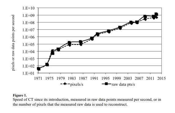
Together with other developments since the creation of CT, as seen in Figure 1, this led to a remarkable improvement in CT imaging speed since its launch in the early 1970s(Hjouj et al., 2021). Over this time, imaging speed has grown by more than seven orders of magnitude and is essentially exponential(Mansour et al., 2022). The technology has grown much more reliable thanks to this speed increase as well as advancements in low-contrast detectability and image quality(Dwaik & Smerat, 2023). As a result, CT has become widely used in medical care worldwide. Primary care doctors considered it to be among the most significant medical technological advancements (Dwaik & Smerat, 2023).
Clinical use of the technology has expanded as a result of improvements in image quality and speed as well as the technique's robustness and usefulness,more recently, the goal of lowering radiation exposure has surfaced as another technological motivator(Hoeijmakers et al., 2025). As a consequence of the higher usage, even though the radiation dose for each scan has decreased in recent years, the radiation dose associated with CT has increased(Sraheen et al., 2025). For instance, out of the overall amount of ionizing radiation exposure to the U.S(Sartoretti et al., 2022).
CT imaging was accountable for around 50% of the artificial radiation exposure, 24% of the ionizing radiation exposure to the population of the United States(Hjouj et al., 2021; Sartoretti et al., 2022). For any single patient, the advantages from CT significantly surpass the risk, yet the worry regarding the population dose has led to dose decrease to emerge as a key technology catalyst(Sraheen et al., 2025). Advancements in CT technology have been made in the upcoming decade is likely to be influenced by the same factors that have influenced the advancements in CT since its beginning: image clarity, speed, enhancements and innovations usage, and reduction in dosage(Smerat et al.).
The remainder of this document will outline the possible enhancements in CT resulting from advancements. in the imaging system itself, especially the enhancements that can be anticipated in imaging velocity, spatial clarity, and dose effectiveness(Lell & Kachelrieß, 2020). Enhancements that have emerged because the advancement of sophisticated image reconstruction algorithms is being addressed. Fresh Reconstruction techniques have, consequently, presented a new challenge that must be tackled, the demand for image quality evaluation tools for non-linear imaging systems(Rajendran et al., 2022). Possible novel applications are assessed, accompanied by a short conversation regarding whether the system architectures of Various vendors will come together or separate and offer a diverse array of imaging platforms(Sekhar et al., 2025).
Temporal accuracy and imaging velocity
Figure 1 illustrates that the imaging speed of CT has risen by 9 orders of magnitude since then. in 40 years, this significant growth has been achieved through two methods. One exists enhancement of scan duration, meaning shortening the time required to gather the data for any individual piece. The second involves boosting the count of slices assessed simultaneously. despite the application of multi-detector row technology.
Figure 2 illustrates the minimum scanning duration (the key factor influencing this for traditional systems with the rotation speed of the CT Gantry) following the introduction of CT. During the initial ten years of CT's availability, there was a swift decline in its accessibility, and typically a significantly slower decrease since approximately 1980. Certain exceptions exist that are marked in figure 2. One type is the electron-beam CT (EBCT) system, which featured a distinctive design. specifically to achieve an extremely brief scan duration, with cardiac imaging as the focus applications (Kulkarni et al., 2021).
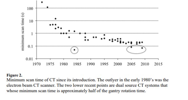
Favored systems utilizing mechanical rotation, there are two recent aspects that are, approximately a twice as fast as their peers. These are the dual-source scanners that will be examined in more detail below(Schmidt & Flohr, 2020). It is reasonable to inquire how much quicker CT systems might become in the upcoming years. if ever? Dual-source technology, illustrated in Figure 2, can attain a significant discrete decrease in imaging duration by utilizing two x-ray sources and two detectors mounted on the gantry functioning concurrently. This minimizes the least rotation required to achieve the necessary projections that range from approximately 180° to half that amount, consequently decreasing the minimum scan duration for a specified gantry rotation speed, by a multiple of two(Schmidt & Flohr, 2020).
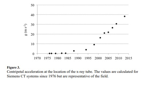
elements that are attached to the spinning frame, particularly on the x-ray tube. Illustration 3 illustrates the pattern of g-forces on the CT x-ray tube. There have been notable rises since around the mid-1990s. It is a remarkable feat of engineering to have parts on the gantry endure g-forces that approach forty times the Earth's gravitational pull(Schmidt & Flohr, 2020). It is also remarkable that the trend has not indicated any flattening, strongly implying that additional Further rises are probable(Lell & Kachelrieß, 2020). Because the centripetal force grows with the square of the rotational speed, additional rises in the tolerance to g forces will lead to a diminishing impact on the rotation time. However, decreases from the present minimum rotation duration of 0.25 s to 0.2 s or fewer results can be anticipated in the next ten years(Lell & Kachelrieß, 2020; Rajendran et al., 2022).
Higher temporal resolution in cardiac imaging aims to enhance clarity. Multi-cycle acquisitions improve resolution but have long exam times and high radiation doses(Smerat et al., 2025). Recent developments in reconstruction algorithms allow for motion estimation and correction, reducing blurring and artifacts, benefiting cardiac imaging significantly(Lossau et al., 2019).
In summary, the minimum rotation time of CT systems in this section on temporal resolution In the upcoming ten years, it could decline from the present levels of approximately 0.250 s to between 0.15 and 0.2 s(Lossau et al., 2019). This would result in minimal imaging durations of approximately 0.040 s in dual source systems and approximately 0.075 seconds in traditional single-source systems. Additional enhancements in efficiency Temporal resolution in cardiac imaging will be attainable through reconstruction techniques. algorithms that adjust for remaining movement(Lossau et al., 2019).
Detector technology
The spatial resolution of CT scanners is significantly influenced by the detector aperture, with substantial improvements observed over time, particularly in slice thickness(Sraheen et al., 2025). However, advances in in-plane detector resolution have plateaued since the mid-1980s due to the reliance on scintillator photo diode detectors(Sraheen et al., 2025). These detectors have approximately 90% geometric efficiency in one dimension but have limitations in improving resolution due to increased reflector area, which hinders dose efficiency. Enhancements in detector aperture could lead to improved limiting spatial resolution and detective quantum efficiency (DQE), especially at mid to high frequencies(McCollough et al., 2025; Sraheen et al., 2025). Noise, particularly electronic noise, also poses challenges, disproportionately affecting low signal levels, necessitating increased radiation doses or enhanced detector technology(McCollough et al., 2025; Rehani et al., 2025). Proposals for direct conversion photon counting detectors promise solutions, achieving better spatial resolution and avoiding geometric inefficiencies. These detectors eliminate electronic noise by counting individual photons and enabling spectral imaging without altering protocols(Ren et al., 2025). Nonetheless, technical challenges, such as handling high x-ray flux, remain, delaying their integration into mainstream CT systems, with an initial focus likely on specialized rather than general-purpose applications(Ren et al., 2025).
Volumetric coverage
CT systems, introduced in the late 1970s, initially faced limitations in volumetric coverage due to single-slice imaging and slow rates(Wei et al., 2024). However, the advent of helical scanning and multi-detector row technology revolutionized this, enabling faster, thin-slice volumetric imaging(Wei et al., 2024). Despite advantages like rapid whole-organ imaging, these advanced systems face drawbacks such as cone-beam artifacts, higher x-ray scatter, and cost, limiting their general-purpose use(Yoo et al., 2024).
Radiation dose, advanced reconstruction methods, and special purpose systems
Recent advancements in CT technology have significantly reduced radiation doses to patients through optimized imaging protocols, efficient x-ray beam collimation, and advanced reconstruction algorithms. Initial helical scanners faced issues like "helical over-dosing," where radiation exposure in extreme anatomical ranges was wasted(Smerat et al., 2025). New collimators prevent this by avoiding unnecessary radiation(Larson, 2024). Controlling x-ray flux enhances the precision of reconstructed images, with techniques like mA modulation and "bowtie filters" leading to improved dose efficiency(Arfat et al., 2024; Larson, 2024). Moreover, new architectures like Inverse Geometry CT and dynamic pre-patient attenuators offer personalized illumination patterns, potentially doubling efficiency(Sabiniewicz-Ziajka & Szarmach, 2024). Statistical and model-based iterative reconstruction methods have emerged, significantly enhancing image quality, especially under low-dose conditions(Szarmach et al., 2025). These non-linear reconstructions challenge traditional image quality metrics, requiring new evaluation standards. Furthermore, the market is shifting towards specialized CT systems, such as those for breast and orthopedic imaging, indicating a trend towards tailored applications.
Conclusion
Computed tomography (CT) has advanced significantly since the 1970s, improving image quality and becoming widespread in clinical medicine. Future developments include enhanced temporal resolution and iterative reconstruction methods, alongside finer x-ray detectors for better spatial resolution and lower radiation doses. Diverse CT systems will emerge, optimized for various clinical needs, leading to new applications in diagnosis.

Open Access By Aditum Open Access Journals id licensed under Creative Commons Attribution 4.0 International License. Based On a Work at aditum.org