International Journal of Hematology Research and Reports
OPEN ACCESS | Volume 2 - Issue 1 - 2025
ISSN No: - | Journal DOI: 10.61148/IJHRR

Ramjeela S1*, Dr Sukesh2, Dr Swathi D3
1Research Scholar, Srinivas University, Mangalore, Karnataka, India.
2Professor, Department of Pathology, Srinivas Institute of Medical Sciences and Research Centre, Mangalore, Karnataka, India.
3Assistant Professor, Department of Forensic Sciences, Institute of Allied Health Sciences, Srinivas University, Mangalore, Karnataka, India.
*Corresponding Author: Ramjeela S, Research Scholar, Srinivas University, Mangalore, Karnataka, India.
Received Date: June 24, 2024
Accepted Date: July 01, 2024
Published Date: July 26, 2024
Citation: Ramjeela S, Dr Sukesh, Dr Swathi D. (2024) “Comparison Of Platelet Rich Fibrin Over Platelet Rich Plasma: A Current Review.”, International Journal of Hematology Research and Reports, 1(1); DOI: 10.61148/ICRCT/001.
Copyright: © 2024. Ramjeela S. This is an open access article distributed under the Creative Commons Attribution License, which permits unrestricted use, distribution, and reproduction in any medium, provided the original work is properly cited.
Clinical applications have been made of platelet rich fibrin (prf), a platelet concentrate that can promote tissue regeneration. It's interesting to note that despite numerous techniques being put forth, not much information has been found about the final cell counts after centrifugation. There are several centrifuges that are commercially available with their corresponding procedures using a new technique for cell quantification. Platelet concentrates, such as platelet-rich fibrin (prf) and platelet-rich Plasma (prp), have been used extensively as a scaffold to promote tissue regeneration in numerous medical specialties. Prp was initially presented as a specialized restorative technique meant to concentrate entire blood platelets. The aim of this study was to compare the effectiveness of prp and prf.
platelet concentrates; platelet rich plasma; platelet rich fibrin; secretory proteins; leucocyte-poor platelet rich plasma; advanced rich plasma plus
1. Introduction
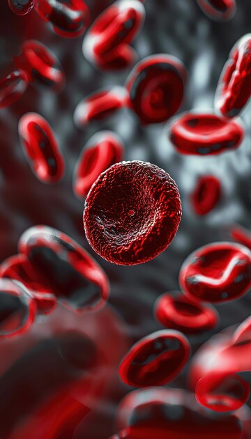
Platelets are fragments of megakaryocytes' cytoplasm released by Bone marrow. More than 30 bioactive proteins are present in them, many of which are essential for tissue repair or hemostasis. All wound healing processes are initiated by seven basic protein growth factors that are actively released by platelets [1]. In regenerative medicine, various platelet concentrates have been employed as surgical adjuvants to promote tissue regeneration and wound healing. It's even unclear exactly how a wound heals, while it's commonly known that platelets are involved in inflammation, angiogenesis, homeostasis, and tissue regeneration. Platelets have bioactive factors (growth factors, immune system messengers, and enzymes) are extracted from peripheral blood [2]. Additionally, the platelets' growth factors promote the need for and development of mesenchymal stem cells as well as other target cells [3].
In PRP growth factors are released by the activated platelets and are essential for the quick healing of wounds [4]. PRP functions as both a hemostatic and an adhesive graft substance [5]. The platelet concentration in PRP varies between 500,000 and 1 million µL.PRP also contains vitronectin, fibronectin, and fibrin, three blood proteins that are known to function as cell adhesion agents. After activation, the platelet granules fuse to the cell membrane (a process known as degranulation), where histones and carbohydrate side chains are added to the secretory proteins (such as TGF-β, PDGF, and so forth) to make them bioactive [6]. Following their secretion, the active proteins attach to target cells' transmembrane receptors, including mesenchymal stem cells, osteoblasts, fibroblasts, endothelial cells, and epidermal cells. After these agonists bind to trans membrane receptors, they activate an intracellular signal protein, which triggers the expression of a gene sequence that controls the growth of cells, the development of matrices, the synthesis of collagen, the production of osteoid tissue, and other processes leading to tissue regeneration and repair[7].
When comparing PRF to PRP, the main benefits of PRF are its ease of use, affordability, lack of biochemical processing of patient blood samples, immune system support, and, because of its lower degree of polymerization, somewhat better potential for wound healing [8]. Furthermore, because PRF is more effective at promoting cell migration and proliferation and lacks bovine thrombin and anticoagulants, it carries fewer dangers than PRP [9].
2. Platelet Concentrates
Platelet concentrates were only utilized to stop bleeding when the patient had significant thrombocytopenia. Because of the enormous numbers of growth factors that are stored inside platelets and the adhesive qualities of fibrin matrix, which is the final product of coagulation cascade, there has been an increase in the application of platelet concentrates for the regeneration of hard and soft tissues. Fibrin glue or sealant was first used in the production of platelet concentrates to promote tissue repair [10].
According to the study conducted by Bielecki t et al classification of platelet concentrate based on two essential criteria: the presence of cell content (mostly leukocytes) and the fibrin architecture. This division made it possible to classify the products into four major families [11]. Leukocyte-Poor Platelet-Rich Plasma (P-PRP) products are preparations that have a low-density fibrin network following activation but do not contain leukocytes [12]. All of the items in this family, by definition, can be utilized as liquid solutions or as activated gel. As a result, it can be injected (for instance, in sports medicine) or applied (much like fibrin glues) to a skin incision or suture while it gels [13].
2.1. Platelet-Rich Plasma
Platelet-rich plasma (PRP) has been extensively employed as a first generation platelet concentrate Leukocyte-Poor Platelet-Rich Plasma (P-PRP) has a low-density fibrin network following activation but do not contain leukocytes[14]. Platelet-rich plasma (PRP) has been extensively employed as a first generation platelet concentrate. All of the items in this family, by definition, can be utilized as liquid solutions or as activated gel. As a result, it can be sutured or injected into a skin wound [15]. Leukocyte-and Platelet-Rich Plasma (L-PRP) are combinations of leukocytes and, following activation, a low-density fibrin network. All of the products in this family, by definition, can be utilized as liquid solutions or in the form of activated gel, just like the P-PRP [16]. Using thrombin, calcium, or other physiologically safe anticoagulants is frequently necessary throughout the PRP preparation process. These elements may trigger an immunological reaction in addition to having a negative impact on the coagulation process [17]. Since fibrinogen in PRF is changed into fibrin under the effect of physiologically accessible thrombin, the addition of any additives is not always necessary. This considerably lowers the possibility of complications following surgery [18].
Donor plasma is used to create this first bioactive surgical adjuvant, and thrombin and calcium are added to start the polymerization process [19]. Originally made with donor plasma, these adhesives can alternatively be bought commercially or obtained from patients (autologously). The goal of the second-generation platelet concentrate was to eliminate anticoagulants when PRP was developed [20]. The removal of anticoagulants causes blood to clot over time, so the clinician must start centrifuging as soon as blood is collected in order to separate the blood layers before clotting. A fibrin clot is produced in the upper "plateletrich" layer after centrifugation. Platelets and leukocytes trapped in this fibrin matrix are the typical cells seen within this layer [21].
2.2. Platelet Rich Fibrin
Platelet-Rich Fibrin (PRF) is a different type of platelet concentrate that was created in France because of the preparation's robust fibrin gel polymerization. This method was so plainly distinct from prior PRPs that it was referred to as a “second-generation” platelet concentrate; however, given the lengthy history of platelet concentrate evolution, this phrase is probably not appropriate. These days, people just think of it as one more product family among the other two [22]. It was noted by Bielecki et al. that these platelet concentrations were also connected to different types of circulating cells, especially leukocytes [23].
Results pertaining to the release of growth factors both in vitro and in vivo support the clinical application of PRF [24]. According to Dohan et al.'s in vitro research, PRF has superior healing qualities than PRP [25]. In a similar vein, Wiltfang et al. from several clinical trials have shown that the PRF outperforms the PRP in terms of promising outcomes [7]. According to a study by Kawamura and Urist, PRF serves as a matrix that facilitates the transportation of morphogenetic proteins [8].
Direct stem cell migration, hemostasis, wound healing, bone repair, and graft stabilization are all improved by the highly structured fibrin matrix found in the PRF. Platelet rich-fibrin (PRF) has since been utilized with a better ability to release more growth factors over an extended period of time with superior clinical outcomes in various fields of medicine and dentistry[26]. Leukocyte-Poor Platelet-Rich Fibrin (P-PRF) or pure platelet-rich fibrin, which is a fibrin network with a high density, does not contain leukocytes. These products, by definition, are only available as strongly activated gels; they cannot be Injected or applied in the same manner as conventional fibrin glues [27]. For other purposes, though, they can be treated as a true solid material due to their robust fibrin matrix. Products with leukocytes and a high-density fibrin network are known as leukocyte- platelet-rich fibrin (L-PRF) [28].
3. Prf Production And Classification
Choukroun et al. in 2001 conducted a study while collecting of 10 ml of blood in glass-coated plastic tubes without the use of an anticoagulant and then centrifuged at 2,700 rpm (around 400 g) for 12 minutes. These acquired PRFs are commonly referred to as leukocyte and PRF (l-PRF) or Choukroun's PRF. The PRF procedure has changed a lot during the past few years. These procedures produced a range of compounds with distinct biological properties and possible applications [29, 30].
Gaanati S et.al suggested that lowering the centrifugation speed could avoid cell loss and boost the quantity of leukocytes in the PRF matrix because it is well known that strong centrifugal forces cause cells to settle to the bottom of the tube. Glass-based vacuum tubes with a reduced centrifugal force of 1,500 rpm (230 g) for 14 minutes were used to generate Advanced PRF (A-PRF). It has also been suggested that A-PRF can be produced using the same centrifugation period of 14 minutes, but at a speed of 1,300 rpm (200 g)[31]. When compared to the L-PRF, the obtained A-PRF had a higher total cell viability percentage. Among these, a rise in platelets, lymphocytes, and neutrophils was noted [32]. Immune cells have an impact on macrophage maturation and differentiation [33]. Thanks mostly to the growth factors that macrophages produce, this may result in the regeneration of soft tissues and bone [34].
Fujioka-Kobayashi M et al. discussed that advanced rich plasma plus (A-PRF+) was created after further modifications to the A-PRF technique. The amount of cells trapped in The PRF matrix is directly influenced by the centrifugation force; research has attempted to lower the centrifugal time and, consequently, the total force that could cause cell loss [35].
A-PRF+ was prepared by reducing the centrifugal time and speed to 8 min and 1,300 rpm (200 g) [36]. Kobayashi et.al comparing the obtained A-PRF+ to the A-PRF and L-PRF, analysis shows a significantly higher amount of released growth factors (TGF-β1, VEGF, PDGF, EGF, and IGF1) [36]. When compared to L-PRF, the total amount of secreted growth factors (TGF-β1, VEGF, PDGF, EGF, and IGF1) was much larger in A-PRF [37]. Nevertheless, as was recently reviewed, some earlier research indicated that A-PRF released fewer growth factors than L-PRF .Because of the slower centrifugation speed and duration, there may be more leukocytes caught in the fibrin mesh, which could explain the observed increase in growth factor release. [38].
One of PRF's primary drawbacks over PRP is that it can only be obtained in gel form, which makes it unsuitable for injection. Injectable PRF (i-PRF), a liquid variant of PRF, has a new procedure thanks to recent research. In order to create this injectable version of PRF, blood without an anticoagulant is used, and plastic tubes without any coatings are centrifuged for three minutes at 700 rpm (60 g) [39]. Plasma, clotting factors, and platelets make up the yellow layer at the top of the tube, and this process allows the separation of blood components during the first few minutes of spinning. When the layer is ready to be applied in injectable form, it can be aspirated with ease [40]. Higher early growth factor release was shown by the growth factor release study, whereas greater long-term release of PDGF-AA, PDGF-AB, EGF, and IGF-1 at 10 days was shown by PRF. Moreover, in comparison to PRP, i-PRF demonstrated higher mRNA levels of TGF-β at 7 days, PDGF at 3 days, and collagen1 expression at both 3 and 7 days [41].
A variety of PRF protocol adjustments have been suggested in recent years. One of them is titanium-PRF (T-PRF), which is made using medical-grade titanium tubes and involves centrifuging a blood sample for 12 minutes at 2,800 rpm (around 400 g)[42]. The same scientists also show that the T-PRF fibrin network is thicker and covers a greater area than the L-PRF fibrin network, suggesting that T-PRF may remain in the tissues for a little while longer [43]. Concentrated growth factors (CGFs) are an alternative kind of PRF that may be a variation of the same platelet concentrate. They are mostly based on the i-PRF production process. The authors suggested utilizing a specialized centrifuge to centrifuge blood samples at different speeds (between 2,400 and 3,300 rpm) and for a predetermined amount of time (in plastic tubes without coatings). The idea behind both variations is to separate platelets and plasma at the tube's top so that the latter can be extracted and utilized as an injectable [44].
3.1. Uses Of Prp And Prf
Growth factors (GFs) are essential for angiogenesis, differentiation, migration, and cell proliferation. Due to their high GF content, autologous platelet concentrates (APCs) are particularly well suited for face rejuvenation and periodontal regeneration. Platelet-rich plasma (PRP), platelet-rich fibrin (PRF), and concentrated growth factor (CGF) approaches are the three primary generations of APCs that are described. PRP is a legitimate adjuvant for a variety of oral and dental surgical treatments [45]. Following tooth extractions, PRP in the alveolar socket can undoubtedly enhance soft tissue healing and have a favorable impact on bone regeneration.
According to a study by Georgakopoulos et al., the placement of PRP surrounding a dental implant enhances bone growth, which enhances implant stability and improves treatment outcomes [46]. Additionally, a study by Thor et al. emphasizes how using PRP increases implant survival rates and improves implant stability. Early implant stability and loading are demonstrated by the PRP. PRP's effectiveness in implant dentistry is still somewhat restricted. To determine the therapeutic relevance of the outcome metrics associated with PRP and implants, more clinical research is needed [47].
A promising new therapy option for skin rejuvenation is I-PRF/C-PRF. These PRF products can be applied as an anti-aging treatment and to improve the tone and texture of the skin, as well as spots and scars left by acne. These can also be used to improve the skin on the hands, neck, chest, tear trough, nasolabial folds, and perioral lines. Via a population of mesenchymal stem cells, leukocytes perform vital markedly increased cell proliferation. Regenerative tasks such as promoting the growth of fibroblasts, enhancing anti-inflammatory effects, promoting angiogenesis, and depositing procollagen protein to rebuild the extracellular matrix [48]. Studies have also revealed that, in comparison to control and PRP (200% increase), skin fibroblasts move over 350% more in fluid-PRF. For face rejuvenation also Nacopoulos and Vesala suggested a combination of A-PRF and I-PRF matrices in Cleopatra technique [49]. Adipose stem cells (ASCs) benefit greatly from PRF as an adjuvant because it encourages ASC differentiation, proliferation, and paracrine function. Due to its benefits in avoiding immunologic rejection, ease of manufacture, and lack of problems, PRF has been utilized extensively in clinical practice to enhance the effectiveness of cell-assisted lipotransfer (CAL) and wound repair by ASCs [50].
5. Conclusion
Although the preliminary research findings are encouraging, more investigation is required into the application of PRF and PRP. Because PRF and PRP are natural materials, they are biocompatible and biodegradable. As a result, nothing unnecessary is wasted in the process of getting them. They don't trigger allergic reactions because of their biocompatibility. Because of this, platelet concentrates have a lot of potential applications in medicine, including dentistry. Since no creature would reject a product that causes allergies, these qualities are currently the most sought-after ones. PRP and PRF are hence universal products that can be used to treat a wide range of ailments.
Competing Interests
Authors have declared that no competing interests exist
Consent
It is not applicable
Ethical Approval
It is not applicable

Open Access By Aditum Open Access Journals id licensed under Creative Commons Attribution 4.0 International License. Based On a Work at aditum.org