
International Journal of Clinical Gynaecology and Obstetrics
OPEN ACCESS | Volume 6 - Issue 1 - 2026
ISSN No: 2836-0737 | Journal DOI: 10.61148/2836-0737/IJCGO


Masomeh Sabzevari1, Zahra Moghimi2, Morteza Naderan3, Ehsan Sobhanian4*, Elham Kaghazlol1, Zahra Asgari1
1Vali-E-Asr Reproductive Health Research Center (VRHRC), Family Health Research Institute (FHRI), Tehran University of Medical Science (TUMS), Tehran, Iran.
2Department of Gynecology, Yas Hospital, Tehran University of Medical Sciences, Tehran, Iran.
3Department of ophthalmology, Farabi eye Hospital Research center, Farabi eye Hospital, Tehran University of Medical science, Tehran, Iran.
4Department of Surgery, Sina Hospital, Tehran University of Medical Sciences, Tehran, Iran.
*Corresponding author: Ehsan Sobhanian, 4Department of Surgery, Sina Hospital, Tehran University of Medical Sciences, Tehran, Iran.
Received: August 05, 2026 | Accepted: August 22, 2026 | Published: September 04, 2026
Citation: Sabzevari M, Moghimi Z, Naderan M, Sobhanian E, Kaghazlol E, Asgari Z., (2026) “Long non-coding RNAs as Non-Invasive Biomarkers in Endometriosis: A Review” International Journal of Clinical Gynaecology and Obstetrics, 6(1); DOI: 10.61148/2836-0737/IJCGO/041.
Copyright: © 2026 Ehsan Sobhanian. This is an open access article distributed under the Creative Commons Attribution License, which permits unrestricted use, distribution, and reproduction in any medium, provided the original work is properly cited.
Endometriosis is a chronic, estrogen-dependent inflammatory condition that affects around 10% of women of reproductive age. It is commonly associated with pelvic pain, dysmenorrhea, dyspareunia, infertility, and a considerable reduction in quality of life. Diagnosis can take several years because symptoms vary widely between patients and imaging does not detect every form of the disease. Although diagnostic laparoscopy is no longer considered necessary for every patient, there is still a need for a reliable, non-invasive biomarker that could support diagnosis.
Objective: This review examines current evidence on the biological role of long non-coding RNAs (lncRNAs) in endometriosis and their potential use as diagnostic biomarkers, particularly lncRNAs found in the circulation and within extracellular vesicles.
Methods: A narrative literature review was conducted using PubMed, Scopus, and Web of Science. Searches focused on endometriosis, long non-coding RNAs, circulating RNA, extracellular vesicles, exosomes, and biomarkers. Greater emphasis was placed on human studies examining lncRNA expression and its possible biological or diagnostic significance.
Results: LncRNAs are RNA transcripts longer than 200 nucleotides that do not generally code for proteins but can regulate gene expression through transcriptional, post-transcriptional, epigenetic, and competing endogenous RNA mechanisms. MALAT1, H19, HOTAIR, MEG3, and GAS5 have been studied in endometriotic tissue and, in some studies, in serum, plasma, or extracellular vesicles. These lncRNAs have been linked to biological processes relevant to endometriosis, including inflammation, angiogenesis, cell proliferation, apoptosis, estrogen signaling, invasion, and tissue remodeling. Their relative stability in biological fluids also makes circulating lncRNAs potentially useful candidates for non-invasive biomarker development. However, considerable differences between studies remain in patient selection, disease stage, menstrual-cycle phase, specimen type, RNA extraction, normalization, and analytical methods.
Conclusion: LncRNAs are promising candidates for non-invasive diagnosis and potentially for monitoring endometriosis. However, the current evidence is not strong enough to support their routine clinical use. Large prospective multicenter studies using standardized methods, clinically relevant control groups, and independent external validation are needed before lncRNA-based biomarkers can be introduced into clinical practice.
Endometriosis; long non-coding RNA; lncRNA; biomarker; non-invasive diagnosis; MALAT1; H19; HOTAIR; MEG3; GAS5; extracellular vesicles
1. Introduction
Endometriosis is a chronic gynecological condition in which tissue resembling the endometrium develops outside the uterine cavity. Patients may experience dysmenorrhea, chronic pelvic pain, deep dyspareunia, fatigue, infertility, and a significant reduction in quality of life. The disease has a complex pathogenesis involving several interacting mechanisms, including estrogen-dependent inflammation, immune dysfunction, progesterone resistance, angiogenesis, altered apoptosis, and epigenetic changes [1,4,18,20,21].
Despite being relatively common, endometriosis is often diagnosed late. The symptoms can differ substantially between individuals, and the severity of symptoms does not necessarily reflect the extent of the disease. Transvaginal ultrasonography and magnetic resonance imaging are useful for detecting ovarian endometriomas and deep endometriosis, but superficial peritoneal lesions can still be difficult to identify [1,23,24]. Current clinical practice increasingly allows diagnosis to be based on symptoms and imaging when appropriate, meaning that laparoscopy is no longer required in every suspected case. Even so, there remains a clear need for a sensitive and specific blood-based biomarker that could support earlier and less invasive diagnosis.
Long non-coding RNAs (lncRNAs) are generally defined as RNA molecules longer than 200 nucleotides that have little or no protein-coding capacity. Rather than being biologically inactive, they can interact with DNA, RNA, and proteins and influence processes such as chromatin remodeling, transcription, mRNA stability, translation, and intracellular signaling [6,8,12]. Their wide range of functions makes them particularly interesting in endometriosis, where several biological pathways interact rather than a single molecular mechanism being responsible for disease development.
This review considers the biological and potential diagnostic significance of lncRNAs in endometriosis, focusing particularly on MALAT1, H19, HOTAIR, MEG3, GAS5, and lncRNAs associated with extracellular vesicles.
2. Methods
2. Methods
A narrative literature review was conducted using PubMed, Scopus, and Web of Science. Search terms and concepts included endometriosis, long non-coding RNA, lncRNA, circulating RNA, extracellular vesicles, exosomes, and biomarkers. Human studies involving patients with endometriosis were considered, together with mechanistic studies that helped establish the biological plausibility of lncRNA involvement in the disease. Particular attention was given to studies examining circulating or extracellular-vesicle-associated lncRNAs and their possible diagnostic relevance.
As this manuscript was designed as a narrative review rather than a systematic review or meta-analysis, formal PRISMA screening counts, risk-of-bias assessments, and pooled estimates of diagnostic accuracy were not performed. The flow diagram presented in Figure 1 therefore illustrates the overall review process rather than representing a quantitative PRISMA flow diagram.
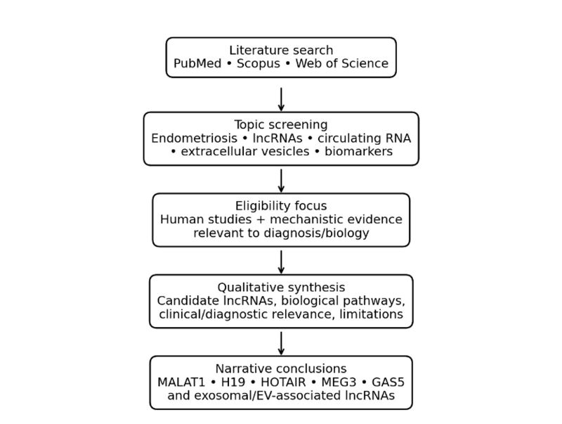
Figure 1. Literature identification and qualitative synthesis workflow used for this narrative review.
3. Biological Characteristics of lncRNAs
LncRNAs comprise a large and diverse group of non-coding RNA transcripts. Based on their genomic location and relationship to protein-coding genes, they can be broadly categorized as intergenic, intronic, antisense, sense-overlapping, or bidirectional transcripts [6,8,12]
Their functions are similarly diverse. Some lncRNAs act as molecular scaffolds, bringing proteins and chromatin-associated molecules together. Others interact with transcription factors or chromatin-modifying complexes and thereby influence gene transcription. At the post-transcriptional level, lncRNAs can affect mRNA stability and translation. Some also function as competing endogenous RNAs by binding microRNAs and altering the availability of those microRNAs to regulate their target messenger RNAs [6,8,9,12].
This range of functions provides a plausible explanation for why lncRNAs may be involved in endometriosis. The disease itself involves several overlapping processes, including inflammation, hormonal signaling, immune responses, angiogenesis, cellular proliferation, apoptosis, and fibrosis [18-21].
4. Why lncRNAs May Be Relevant to Endometriosis
4.1 Inflammation
Inflammation plays a central role in both the development and persistence of endometriotic lesions. LncRNAs can influence inflammatory signaling pathways, including nuclear factor-κB and cytokine-related networks. Changes in lncRNA expression may therefore contribute to the inflammatory environment surrounding endometriotic lesions [18,21].
4.2 Angiogenesis
For ectopic endometrial tissue to survive and expand, it must establish an adequate blood supply. Several lncRNAs have been linked to angiogenic pathways and signaling involving vascular endothelial growth factor. This raises the possibility that abnormal lncRNA activity could contribute to the vascularization and continued growth of endometriotic lesions [18,20].
4.3 Estrogen Signaling
Endometriosis is strongly dependent on estrogen. Abnormal estrogen metabolism and signaling are thought to contribute to both lesion growth and inflammation, while lncRNAs may influence estrogen receptor activity and the expression of estrogen-responsive genes [5,18].
4.4 Cellular Proliferation and Apoptosis
expression through transcriptional, post-transcriptional, epigenetic, and competing endogenous RNA mechanisms. MALAT1, H19, HOTAIR, MEG3, and GAS5 have been investigated in endometriotic tissue and, in selected studies, in serum, plasma, or extracellular vesicles. These molecules appear to participate in pathways involving inflammation, angiogenesis, cellular proliferation, apoptosis, estrogen signaling, invasion, and tissue remodeling. Their relative stability in biological fluids makes circulating lncRNAs attractive candidates for non-invasive biomarker development. However, substantial heterogeneity exists in patient selection, disease stage, menstrual-cycle phase, specimen type, RNA extraction, normalization, and analytical methodology.
Conclusion: LncRNAs represent promising molecular candidates for non-invasive diagnosis and potentially disease monitoring in endometriosis. Nevertheless, current evidence is not sufficient for routine clinical implementation. Large prospective multicenter studies using standardized pre-analytical and analytical protocols, clinically relevant control groups, and independent external validation are required before lncRNA-based biomarkers can be incorporated into clinical practice.
Keywords: Endometriosis; long non-coding RNA; lncRNA; biomarker; non-invasive diagnosis; MALAT1; H19; HOTAIR; MEG3; GAS5; extracellular vesicles.
The establishment of an endometriotic lesion depends partly on the ability of ectopic cells to survive and proliferate. Alterations in lncRNA expression have been linked to pathways involved in cell-cycle regulation, proliferation, and apoptosis [6,12,18]. These changes may allow ectopic endometrial cells to survive in an environment in which they would otherwise undergo programmed cell death.
4.5 Invasion and Tissue Remodeling
Endometriotic cells have invasive properties and interact with the surrounding extracellular matrix. Some lncRNAs have been associated with epithelial-mesenchymal transition and pathways involved in extracellular-matrix remodeling. Such effects could contribute to implantation, invasion, and progression of endometriotic lesions [18,19,23].
5. Results: Evidence for lncRNAs in Endometriosis
The literature reviewed provides consistent evidence that altered lncRNA expression is associated with several molecular processes involved in endometriosis, although the findings are not completely uniform. MALAT1, H19, HOTAIR, MEG3, and GAS5 are among the most frequently discussed candidates. Their reported expression patterns vary depending on the tissue or biological fluid examined and on the design of individual studies. For this reason, the findings were considered qualitatively rather than combined into pooled estimates of diagnostic accuracy.
5.1 Circulating and Extracellular-Vesicle-Associated lncRNAs
One of the most appealing features of lncRNAs from a clinical perspective is that some can be detected in peripheral blood. Circulating lncRNAs may be present as free RNA associated with proteins or may be transported within extracellular vesicles. Exosomes and other extracellular vesicles can protect their RNA cargo from degradation, potentially allowing these molecules to remain detectable in biological fluids [14,15].
This has led to interest in circulating and exosomal lncRNAs as possible diagnostic biomarkers for endometriosis. However, simply detecting an lncRNA in blood does not demonstrate that it is clinically useful. A diagnostic biomarker needs to reliably distinguish patients with endometriosis from appropriate control populations and should ideally demonstrate similar performance in independent cohorts
It is also important not to assume that changes observed in endometriotic tissue will necessarily be reproduced in blood. Circulating RNA is influenced by its release from different tissues, transport, degradation, and clearance. As a result, tissue expression and circulating expression may reflect different aspects of disease biology [14,15].
5.2 Candidate-Specific Findings
MALAT1: MALAT1 is one of the most extensively studied mammalian lncRNAs. In endometriosis, changes in MALAT1 expression have been associated with cellular proliferation, migration, angiogenesis, inflammation, and regulatory RNA networks. Its expression can be measured using techniques such as qRT-PCR, making it an appealing candidate for biomarker research. Nevertheless, reported diagnostic performance has varied between studies, and evidence specifically supporting its use as a circulating biomarker remains less developed than the broader evidence concerning its biological role [6,12].
H19: H19 is an imprinted lncRNA involved in developmental and regulatory processes. It has attracted interest in endometriosis because of its reported links to estrogen-related signaling and cellular proliferation. Experimental studies suggest that abnormal H19 expression may affect the behavior of endometriotic cells through interactions with microRNAs and downstream signaling pathways. However, differences in sample type and normalization methods make it difficult to compare results directly between studies.
HOTAIR: HOTAIR is a trans-acting lncRNA that can interact with chromatin-modifying complexes. In endometriosis, altered HOTAIR expression has been associated with invasion, proliferation, and epigenetic regulation.
These findings support a possible biological role for HOTAIR, but they do not yet demonstrate that it can function as an independent diagnostic biomarker [6,9].
MEG3: MEG3 has been associated with inhibition of cellular proliferation and promotion of apoptosis in several biological settings. Studies of endometriosis have reported altered MEG3 expression, while experimental findings suggest that reduced MEG3 activity could contribute to abnormal cell survival and proliferation. Whether MEG3 can be reliably measured in circulation and used diagnostically remains to be established in well-designed human studies.
GAS5: GAS5 is involved in cell growth, apoptosis, and signaling related to steroid receptors. Changes in GAS5 expression may affect the survival and apoptotic behavior of ectopic endometrial cells. Although its detection in circulation makes GAS5 an interesting biomarker candidate, current evidence is not sufficient to support its use as a standalone diagnostic test.
5.3 Exosomal and Extracellular-Vesicle-Associated lncRNAs
Extracellular vesicles contain a range of molecular components, including RNA, proteins, and lipids, and are involved in communication between cells [14,15]. Because lncRNAs contained within these vesicles may be protected from degradation, extracellular vesicles have attracted considerable interest as a possible source of minimally invasive biomarkers.
In endometriosis, RNA contained within extracellular vesicles may provide information about communication between endometriotic lesions and surrounding tissues. However, research in this area is complicated by substantial methodological differences. Various techniques are used to isolate extracellular vesicles, and the resulting preparations may differ in their composition and biological properties. Standardization of vesicle isolation, characterization, RNA extraction, and normalization is therefore essential if findings from different studies are to be compared reliably [14-16].
5.4 Overall Diagnostic Implications
At present, the available evidence does not support a single, reproducible circulating lncRNA signature with a clinically established sensitivity, specificity, or diagnostic threshold. Instead, the findings suggest that a combination of several lncRNAs may eventually be more useful than an individual transcript. Such a model could potentially be combined with clinical characteristics, imaging findings, or established biomarkers such as CA-125 to improve diagnostic discrimination.
6. Summary of Major lncRNA Candidates
Table 1. Principal lncRNA candidates discussed in the reviewed literature.
|
Candidate |
Main biological associations |
Potential biomarker role |
Key evidence gap |
|
MALAT1 |
Proliferation, migration, angiogenesis, RNA-regulatory networks |
Circulating/multimarker candidate |
Limited external validation |
|
H19 |
Estrogen-related signaling, proliferation, cellular regulation |
Potential circulating candidate |
Heterogeneous expression and methods |
|
HOTAIR |
Epigenetic regulation, invasion, proliferation |
Biological/diagnostic candidate |
Limited prospective evidence |
|
MEG3 |
Growth regulation and apoptosis |
Candidate biomarker |
Limited circulating validation |
|
GAS5 |
Growth arrest, apoptosis, steroid-related signaling |
Potential diagnostic candidate |
Small, heterogeneous studies |
|
Exosomal lncRNAs |
Intercellular communication and RNA protection |
Potentially stable non-invasive biomarkers |
Isolation and normalization not standardized |
7. Discussion
The main conclusion from this review is that lncRNAs have a strong biological rationale for involvement in endometriosis and may have potential as non-invasive biomarkers, but the evidence is not yet sufficient for routine clinical use. It is important to distinguish between demonstrating that an lncRNA is biologically involved in disease and demonstrating that it can be used reliably as a diagnostic test. MALAT1, H19, HOTAIR, MEG3, and GAS5 have all been investigated repeatedly, yet their reported expression patterns and diagnostic performance differ between tissues, blood-derived samples, and experimental platforms.
The biological rationale is consistent with the complex nature of endometriosis. The disease does not result from a single molecular abnormality. Instead, inflammatory, endocrine, immune, angiogenic, proliferative, apoptotic, and fibrotic processes interact with one another over time [18-21]. LncRNAs are capable of influencing several of these processes because individual transcripts can affect chromatin structure, transcription, post-transcriptional regulation, microRNA activity, and intracellular signaling [6,8,12]. Their ability to operate across several levels of gene regulation may therefore explain why they continue to emerge as potential candidates in endometriosis research.
7.1 Interpretation of Candidate lncRNAs
The candidate lncRNAs considered in this review appear to be associated with somewhat different aspects of endometriosis biology.
MALAT1 has been linked to proliferation, migration, angiogenesis, and regulatory RNA networks. H19 has been more closely associated with estrogen-related signaling and cellular proliferation, whereas HOTAIR has been implicated in epigenetic regulation, invasion, and proliferative behavior. MEG3 has mainly been studied in relation to growth inhibition and apoptosis, while GAS5 has been associated with growth arrest, apoptosis, and steroid-related signaling.
These differences are important because they suggest that individual lncRNAs may reflect distinct components of endometriosis rather than representing different versions of the same biological signal.
At the same time, biological plausibility should not be mistaken for diagnostic validity. An lncRNA that is clearly altered in endometriotic tissue may have an important role in disease mechanisms without being sufficiently abundant or specific in peripheral blood to serve as a useful diagnostic marker. Circulating RNA is affected by release from multiple tissues, transport, degradation, and clearance. Extracellular vesicles may provide some connection between lesion biology and the circulation by protecting their RNA cargo, but methods for isolating and characterizing these vesicles are still not sufficiently standardized [14,15].
7.2 Why Promising Biomarkers Have Not Yet Translated into Clinical Tests
Several factors may explain why promising molecular findings have not yet resulted in clinically available tests. Many published studies are exploratory and involve relatively small patient populations. In addition, control groups are often composed primarily of healthy women. This can make a biomarker appear more accurate than it would be in clinical practice, where a patient with suspected endometriosis may instead have another cause of pelvic pain or infertility.
Small discovery cohorts are also particularly vulnerable to overestimating diagnostic performance. A marker may appear highly sensitive and specific in the population in which it was initially identified but perform much less well when tested in a separate group of patients.
Pre-analytical and analytical differences add another layer of uncertainty. Menstrual-cycle phase may influence RNA expression, and serum, plasma, whole blood, tissue, and extracellular-vesicle preparations represent different biological compartments [14,15]. Differences in RNA extraction kits, quantification techniques, reference genes, and normalization procedures can also alter measured expression levels. Therefore, conflicting findings between studies do not necessarily mean that one study is biologically incorrect; methodological differences may account for at least some of the variation.
7.3 Disease Heterogeneity and Clinical Context
Endometriosis includes several clinical phenotypes, including superficial peritoneal disease, ovarian endometriosis, and deep endometriosis, and these forms may have different molecular characteristics [1,23]. If these phenotypes are grouped together without appropriate stratification, clinically relevant molecular differences may be overlooked.
Future studies should therefore record factors such as hormonal treatment, previous surgery, disease stage, phenotype, and menstrual-cycle phase. Where possible, these variables should either be standardized or incorporated into the statistical analysis.
The choice of control group is equally important. Comparing patients only with healthy women may overestimate the usefulness of a proposed biomarker. In clinical practice, patients investigated for endometriosis may have pelvic pain, infertility, adenomyosis, benign ovarian lesions, or other gynecological conditions. Future diagnostic studies should therefore include control groups that reflect the patients who would actually be encountered in clinical settings [1,23,24].
7.4 Single Biomarkers Versus Multimarker Models
The evidence reviewed here suggests that a multimarker approach may ultimately be more realistic than relying on a single lncRNA.
Given the biological heterogeneity of endometriosis, several molecular signals may provide a more stable and informative profile than one transcript alone.
A future diagnostic model could combine lncRNA expression with symptoms, imaging findings, age, reproductive history, and established biomarkers such as CA-125. Such an approach may provide more clinically useful information than measuring one molecular marker in isolation.
Diagnostic studies should also go beyond reporting the area under the receiver-operating-characteristic curve. Sensitivity, specificity, likelihood ratios, predictive values, calibration, and decision-curve analyses are important for determining whether a test would actually alter clinical decision-making. Candidate signatures should first be developed in an appropriate training population and then tested in an independent external cohort.
7.5 Clinical Translation and Research Priorities
Before an lncRNA-based test could realistically be introduced into clinical practice, several questions need to be answered. The biological association should be reproducible, diagnostic performance should be demonstrated prospectively in adequately sized populations, and the findings should be independently validated. Researchers also need to consider practical issues such as reproducibility, cost, clinical utility, and the potential consequences of false-positive and false-negative results.
Future studies should use consistent procedures for blood collection, processing, storage, RNA extraction, normalization, and reporting. Menstrual-cycle phase and hormonal exposure should also be documented. Studies involving extracellular vesicles should provide sufficient information about their isolation and characterization methods to allow other researchers to reproduce the work [14,15].
7.6 Strengths and Limitations of the Present Review
A major strength of this review is its clinically focused discussion of both the biological and diagnostic relevance of lncRNAs, particularly those detected in circulation or associated with extracellular vesicles. However, the review is narrative rather than systematic. Consequently, pooled diagnostic estimates were not calculated, and the available literature should not be interpreted as providing a quantitative measure of the diagnostic performance of any individual lncRNA.
Another limitation is the possibility of publication or selective-reporting bias. Positive molecular findings may be more likely to be published than studies reporting weak or negative associations. This possibility further supports the need for larger prospective studies and independent validation before strong clinical conclusions can be drawn.
8. Future Perspectives
Future research should move beyond small studies focused on individual candidate lncRNAs and toward standardized, multicenter biomarker-development programs. Large prospective studies should include clearly defined patient populations representing the different clinical phenotypes of endometriosis, together with control groups that reflect real-world diagnostic practice.
Blood collection should be standardized as far as possible, including the timing of collection, sample processing, storage conditions, and documentation of menstrual-cycle phase. Analytical methods should also be harmonized so that results from different centers can be compared more reliably.
LncRNAs identified during discovery studies should subsequently undergo independent validation using predefined statistical criteria. Combining lncRNAs with other molecular biomarkers, imaging findings, and clinical variables may ultimately provide better diagnostic performance than any individual marker. Machine-learning methods could also be useful for identifying molecular patterns that are difficult to detect using conventional approaches. However, such models would still require rigorous external validation before they could be considered clinically reliable.
It will also be important for future studies to distinguish between different clinical purposes.
A biomarker capable of identifying the presence of endometriosis may not necessarily be useful for determining disease severity, predicting treatment response, or identifying recurrence. These outcomes should therefore be evaluated separately rather than treated as interchangeable measures of biomarker performance.
9. Conclusion
Long non-coding RNAs have emerged as potentially important regulators of several molecular processes involved in endometriosis. MALAT1, H19, HOTAIR, MEG3, and GAS5 are among the most extensively investigated candidates and have been associated with inflammation, estrogen signaling, cellular proliferation, apoptosis, angiogenesis, invasion, and tissue remodeling [6,18-21].
The detection of selected lncRNAs in blood and extracellular vesicles provides a plausible basis for developing non-invasive biomarkers [14,15]. However, the evidence remains heterogeneous, and most candidate biomarkers have not yet undergone the level of external validation required for clinical use. At present, lncRNAs should therefore be regarded as promising research biomarkers rather than established diagnostic tests.
Large prospective multicenter studies with standardized protocols, appropriate control populations, adequate sample sizes, and independent validation are needed. In the longer term, combining lncRNA signatures with clinical assessment and imaging may offer a more practical diagnostic strategy than relying on a single molecular marker. Such an approach could potentially help reduce diagnostic delays and improve the management of women living with endometriosis.
Declarations
Conflict of Interest: The authors declare no conflict of interest.
Funding: No specific funding was received for this work.
Ethical Approval: Ethical approval was not required because this study was based entirely on previously published literature.
Author Contributions: The authors contributed to the conception of the review, literature search, interpretation of the available evidence, and preparation of the manuscript.

Open Access By Aditum Open Access Journals id licensed under Creative Commons Attribution 4.0 International License. Based On a Work at aditum.org


