
International Journal of Clinical Case Reports and Medical Cases
OPEN ACCESS | Volume 1 - Issue 1 - 2026
ISSN No: - | Journal DOI: 10.61148/IJCCRM


Mamoona Arshad1, Ayesha Malik1, Zahra Batool1, Muhammad Shaheer2, Muhammad Adil Umar1, Muhammad Aetesam Nasir1, Fatima Mazhar3*
1Department of Medicine and Surgery, Hitec-institute of medical sciences Taxila Cantt.
2Department of Medicine and Surgery, CMH Multan Institute of Medical Sciences Multan.
3Department of Microbiology, MNSUAM.
*Corresponding author: Fatima Mazhar, Department of Microbiology, MNSUAM.
Received: September 20, 2026 | Accepted: September 29, 2026 | Published: October 05, 2026
Citation: Arshad M, Malik A, Batool Z, Shaheer M, Muhammad A Umar, Muhammad A Nasir, Mazhar F. (2026) “Interleukin Networks in Breast Cancer: Mechanisms, Clinical Significance, and Therapeutic Opportunities” International Journal of Clinical Case Reports and Medical Cases, 1(2); DOI: 10.61148/IJCCRM/006.
Copyright: © 2026 Fatima Mazhar. This is an open access article distributed under the Creative Commons Attribution License, which permits unrestricted use, distribution, and reproduction in any medium, provided the original work is properly cited.
Breast cancer remains the most frequently diagnosed malignancy among women worldwide and is a leading cause of cancer-related mortality. A growing body of research indicates that immunological dysregulation and chronic inflammation are important factors in the development, spread, invasion, and metastasis of breast cancer. In the tumour microenvironment, interleukins (ILs), a broad class of cytokines involved in inflammatory responses and immune control, have become important mediators. The biological roles of key interleukins and their connection to the pathophysiology of breast cancer are outlined in this article. Through various signalling pathways, a number of interleukins, such as IL-1, IL-6, IL-8, IL-10, IL-11, IL-13, IL-17, IL-19, IL-21, IL-23, IL-32, and IL-33, contribute to tumour growth, angiogenesis, immune evasion, epithelial–mesenchymal transition, and metastatic dissemination. Aggressive tumour characteristics, a poor prognosis, treatment resistance, and lower survival rates have all been linked to elevated expression of these cytokines. On the other hand, some interleukins have anti-tumor properties through boosting immune surveillance and encouraging cytotoxic reactions against cancerous cells. Genetic variables such BRCA1, BRCA2, HER2, TP53, and vitamin D receptor polymorphisms affect breast cancer susceptibility and disease development in addition to cytokine-mediated processes. Gaining an understanding of the intricate relationships that exist between interleukins, tumour cells, and the immunological milieu may help uncover new biomarkers for diagnosis, prognosis, and targeted treatment. Future research on interleukin-based treatment approaches may help enhance clinical results and provide patients with breast cancer with more individualised care.
Breast cancer, Interleukins, Cytokines, Inflammation, Tumor microenvironment, Metastasis, Biomarkers, Immunotherapy
ILs are a type of cytokine that was first thought to only be made by white blood cells (leukocytes), but it has since been found that many other cells in the body also make them [1]. They are very important for turning on and differentiating immune cells, as well as for their growth, movement, adhesion, and multiplication. On top of that, they can both cause and stop inflammation [2].
So, interleukins' main job is to control cell growth, differentiation, and activity during immunity and inflammatory reactions. Interleukins are a big group of proteins that can make cells and tissues do a lot of different things by attaching to special spots on the outside of cells [3]. They work in both autocrine and paracrine ways. Interleukins are also used to study things that have to do with clinical medicine in animals.
Cytokines and interleukins in general:
Cytokines are proteins that are created when the body senses pathogens or other antigens. They help control and direct immune and inflammatory responses[4].
The process of making interleukin stops on its own. The messenger RNA that codes for most interleukins is fragile, which leads to a short-term production. Once these molecules are made, they are quickly released.
Cellular reactions to interleukins involve both up- and down-regulation, as well as the activation and involvement of genes that code for cytokine receptor inhibition.
Interleukins do more than one thing. IL-4, IL-5, and IL-13 are examples of B-cell growth factors that help B cells differentiate.
Cytokines cause B cells to switch between different types of antibodies, helper T cells to differentiate into Th-1 and Th-2 groups, and phagocytes to start killing microbes [1].
The production and activities of other interleukins are often affected by interleukins. In this case, IL-1 helps activate lymphocytes, which then releases IL-2.
High-affinity receptors or messages from outside the cell control and drive how cells respond to cytokines. As an example, viruses that activate B cells cause more cytokine receptors to be expressed.
Most cytokines work on the cell that makes them or on a cell nearby. For example, IL-2 made by T cells works on the T cells that made it or on a cell nearby. Moreover, cytokines can get into the bloodstream and work in places other than where they were made. For instance, IL-1 is a natural pyrogen that affects the central nervous system (CNS) and leads to fever[5].
Only small amounts of a cytokine are needed to bind to receptors and cause biological impacts.
Breast cancer is the most common type of cancer in women and makes up about 30% of all new cases of cancer in women [6]. It is also the second most common type of cancer that kills women in the United States. Breast tumor growth is still not fully understood, but over the past few years, a number of ideas have been put forward to try to explain what starts the process. In 1863, Virchow R. noticed that leukocytes were present in cancerous cells. He was the first person to talk about a possible link between inflammation and the development of cancer. Inflammation has been shown to be a key part of tumor growth and progression over the last few decades. Many of the main molecular mechanisms have now been uncovered, highlighting the important role of cytokines, especially interleukins (ILs), in the starting, spreading, and progressing of breast cancer. The study's goal is to look at the part ILs play in breast cancer [1].
Interleukins were linked to breast cancer
Cytokines are biomolecules that play a key role in infections, hematopoiesis, and homeostasis. Their multipurpose role controls the body's reaction to infectious diseases and even cancer by managing tissue repair, cellular sprouting, and growth [7]. ILs are proteins that are secreted and play a role in immunology. They are in the same family as cytokines and have complex immune activities as cytokines. In the immune system, ILs' main job is to help cells talk to each other. This includes cell movement, growth, maturation, and adhesion, all of which are very important for the inflammatory response [8]. Interleukins play a part in both short-term and long-term inflammation. When certain receptors on the cell surface are stimulated, they react by starting up a different signaling route each time. About 38 different interleukins have been discovered so far. Each one binds to a different type of receptor and has its own history, structure, and qualities. Many of them are thought to be present in breast cancer and play a part in its development and spread.
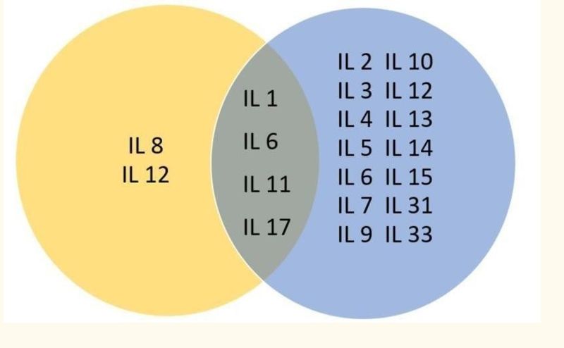
Interleukin-1
The cytokines 17–20 kilodaltons (kDa) that make up interleukin-1 (IL-1) have a lot of different biological roles. These are mainly the proinflammatory cytokines IL-1a and IL-1b, as well as the interleukin-1 receptor antagonist (IL-1ra). All of these are involved in starting and spreading inflammatory processes. IL1A and IL1B, two nearby genes found on chromosome 2, control how active they are. A lot of different kinds of cancer have high levels of IL-1, and a lot of those types of cancer have an aggressive tumor phenotype that means they don't have a good outlook. It is thought that IL-1β and the induction of the NF-kB pathway may be connected in an autocrine way [9]. Metastatic breast cancer cells release IL-1β and possibly other substances that affect mesenchymal stem cells (MSCs). With the help of chemokines, the MSCs will have a big effect on the breast cancer cells' ability to spread and invade other tissues. Within the tumor microenvironment, MSCs can either help or hurt the tumor. They do this by making chemokines that change chemotaxis and other aspects of how cells behave. Chemokines act as chemoattractants, which means they pull cells to places where there are more of the factor[10].
By making growth factors and angiogenic proteins from nearby cells, IL-1 encourages the production of metastatic genes like matrix metalloproteinases (MMP). This helps cancer grow through neovascularization and spread. In 2009, Perrier et al. suggested a model that shows how IL-1 might be able to control breast cancer [11]. According to their findings, IL-1, leptin, and adiponectin, which are found in mammary adipose cells, can affect the growth, migration, and invasion of breast cancer. These chemicals work on specific receptors in both an autocrine and a paracrine way on mammary tumor cells [12]. Controlling the production of growth factors and epithelial-derived and angiogenic proteins that help other cancer cells invade and multiply. Filippi et al. also agreed that IL-1 plays a part in breast cancer spread. They said that low oxygen levels helped the MDA-MB-231 triple negative breast cancer cell line move, along with the formation of hypoxia-inducible factor 1-alpha (HIF-1α) and an increase in chemokine (CXCR1) levels. By stopping IL-1R signaling, Holler et al. looked into how IL-1 is expressed in breast cancer. The writers said that IL1 plays a useful part in the development and spread of breast cancer to bones [13].
Interleukin 2
The 15.5 kDa glycoprotein interleukin-2 (IL-2) is found in the plasma membrane and is found in normal tissues, endothelial cells, and the intestinal epithelium. This one-of-a-kind interleukin plays a big part in the growth, sprouting, and differentiation of T and B cells, as well as non-lymphoid cells, and it helps natural cells (NK cells) break down cells [14]. The IL-2 receptor complex (IL-2R) is made up of three different parts that are coded for by genes that are not connected to each other. CD4+ T helper (Th) lymphocytes make it. It used to be called the T cell growth factor. The production of IL-2 was first linked to the development of breast cancer in women who had been treated for breast cancer. After 10 to 12 months, the return rate was 4.7% if the plasmatic level of IL-2 was normal, but it rose to 33.3% if IL-2 was low. In clinical tests, Garcia-Tuñón et al. found that IL-2 expression, including its three receptor chains, was higher in cancers that had spread into the breast compared to surgical samples that had not spread. In this case, it makes sense because we know that invasive neoplasms are more aggressive. Xiao-Bo et al. found that polymorphisms in the IL-2 gene were linked to higher rates of breast cancer and could be used as a marker for the prognosis of the disease. Meanwhile, Muraro et al. looked at patients with human epidermal growth factor receptor 2 (HER-2) that was overexpressing or negative and had locally advanced breast cancer. They found that IL-2 can affect the start and progression of cancer in HER-2 patients because they had much lower amounts of IL-2 [15].
Interleukin 4
To control allergic reactions, interleukin 4 (IL-4) works on B lymphocytes, monocytes, dendritic cells, and fibroblasts. It has also been shown to fight tumors and reduce inflammation. The Janus kinase/signal transducers and activators of transcription (JAKs and STAT) pathways handle IL-4 messages. Activated T lymphocytes, mast cells, and basophils are the only cells that can produce and make IL-4. Several studies have shown that IL-4 plays a role in the development of breast cancer and its resistance to apoptosis and local spread. The IL-4 receptor is highly expressed in breast cancer, and IL-4 must bind to it in order for it to work on cancer cells. Nagai and Toi found that IL-4 controls the enzymes that make estrogen-promoting death happen in breast cancer cells that have been grown in a lab. Gaggianesi et al. looked at what happened when they blocked IL-4 with the IL4Rα blocker IL4DM in mammary gland tumors. They found that this stopped cancer cells from multiplying, spreading, and growing by lowering the activity of the mitogen-activated protein kinase (MAPK) pathway. IL-4 and the IL-4/IL-4R signaling interleukins have been studied a lot because they can be used to treat cancer. For example, IL-4 protects tumor cells from CD95- and chemotherapy-induced apoptosis by increasing antiapoptotic proteins. IL-4R has been shown to directly promote tumor metastasis in the breast, and blocking IL-4 protects against the macrophage-mediated radioresistance of inflammatory breast cancer [9].
Interleukin 6
IL-6 is a 26 KD cytokine that is made by vascular endothelial cells and mononuclear phagocytes and fibroblasts that are found in bladder and cervical cancer. It is released when IL-1 and TNF do something, and it mostly affects liver and B cells. As the main trigger of the inflammatory reaction, interleukin-6 plays a key part in the development of cancer and its pathology. When IL-6 is present, it makes neoplastic cells more likely to break through the extracellular matrix (ECM) and make drugs less effective [16]. Cancer cells from the bladder, kidneys, cervix, and breast were tested and found to possibly produce IL-6. After that, the IL-6 receptor is found on the cell walls of the prostate, ovaries, kidneys, and breasts. In many ways, IL-6 shows that it can do more than one thing. Several types of tumors release it, and its growth is linked to an auto-paracrine way of stimulating neoplasms. IL-6 can increase the levels of any proteins that stop cells from dying or cause a grouping of cytokines that help blood vessels grow. On top of that, IL-6 makes cell lines make vascular endothelial growth factor (VEGF). A lot of cancer people have thrombocytopenia because of their treatment. In these cases, IL-6 is a strong molecule that helps blood clot. We know that IL-6 controls the discovery of VEGF in MEG-01, a human megakaryoblastic leukemia cell line. Many studies have shown that IL-6 can help blood vessels grow; it does this by controlling the amount of VEGF in platelets and encouraging endothelial cells to divide. The IL-6 amounts in the blood show that the growth process of the tumor is still going on. There is a good chance that the low mortality rate has something to do with blood IL-6. Xian-Peng found that human IL-6 and the IL-6 soluble receptor kept the MCF-7 breast cancer cell line growing when they were grown with human IL-6. Additionally, treating breast cancer cells with IL-6 stops the growth of estrogen receptor (ER) positive cells [24]. However, IL-6's main use in breast cancer is as a prognostic marker, with high amounts of IL-6 in the blood being linked to a poor outcome in many studies.
Interleukin 7
Interleukin-7 is a about 25kDa glycoprotein that is controlled by a gene on chromosome 8q12–13. In the bone marrow and the thymus, stromal cells make IL-7, which helps pre-pro B cells and hepatocyte growth factor (HGF) grow. In the thymus, IL-7 also helps the development of T cell precursors [17]. There is a cell surface receptor (IL-7R) made up of two chains: the γc chain and the IL-7Rα chain. This receptor maps to chromosome 5p13. The IL-7 can attach to an IL-7R receptor on the surface of cells. This receptor has two parts: the γc chain and the IL-7Rα chain. The γc chain maps to chromosome 5p13 [26]. IL-7 affects the growth of new lymphatic veins when it is expressed on vascular cells. In the last ten years, studies have shown that abnormal production of IL-7 and IL-7Ra polymorphism play different roles in the development of breast cancer depending on the genetic subtype. Recently, it was suggested that IL-7 may play a part in the development of mammary gland cancer by helping cancer cells survive and grow in culture and being linked to a poor outcome in human samples. A study by Al-Rawi et al. found that wortmannin-sensitive IL-7 caused breast cancer cells to grow in the lab. In 2017, Boesch et al. published a study that found IL-7-expressing cancer-associated fibroblasts helped breast tumors grow and provided important niches for keeping breast cancer stem cells alive. They also found that CXCL12 was an important niche factor in Il7-expressing cancer-associated fibroblasts (CAFs), which suggests that the stromal cell-derived factor 1/cluster of differentiation 184 CXCL12/CXCR4 pathway may be a therapeutic target for anti-cancer stem cells (anti-CSCs) [18].
Interleukin 8
IL-8, also known as neutrophil-activating protein-1, is an 8 KD protein with 72 acids that is made by macrophages and endothelial cells. It has a strong chemotactic effect on T-lymphocytes and neutrophils, and it increases the ability of leucocyte adhesion receptor CD11b/CD18 to bind. IL-8 has properties that stop the growth of viruses and immune cells. These properties can have a good or bad effect on the immune system. There is inflammation and cell movement caused by it [19]. IFN-8 is a chemokine that belongs to the CXC group. It can be made and released in both healthy and cancerous human cell lines, such as those from breast cancer, ovarian cancer, prostate cancer, thyroid cancer, and many others [20]. Nitric oxide (NO) is a key second messenger that controls the translation of the IL-8 gene in response to hypoxia and anoxia. NO is made from L-arginine by an enzyme called NO synthase with oxygen and other substances [21].
IL-8 has both an autocrine and a paracrine identity. It has a face that causes tumors and a lot of promise as a predictor and predictive marker for cancer [22]. It is found in ER- breast cancers and makes them more likely to invade and spread to other parts of the body in both ER- and ER+ breast cancer patients. When it comes to medical diseases that involve inflammation, IL-8 comes from fibroblasts, endothelium cells, and tumor cells. The IL-8RA and IL-8RB are the two receptors for IL-8 [23]. The important production of IL-8 shows how breast cancer cells spread; even higher amounts of IL-8 in the blood show that breast cancer cells are spreading [24].
A lot of different cytokines are found in breast tissue, both healthy and cancerous. In their study, Green et al. used reverse transcriptase-linked polymerase chain reaction (RT-PCR) to look into a group of messenger RNA (mRNA) transcripts for cytokines [25]. We looked at IL-1β, IL-2, IL-3, IL-4, IL-5, IL-6, IL-7, IL-8, tumor necrosis factor (TNF)-β, TNF-β, and interferon (IFN)-γ. The in-depth study only found higher levels of IL-8. What is important about this lab test is that the above-mentioned cytokines are not
present at all; these cytokines are found in immune system cells [26]. Another study looked at the mRNA profiles of 13 cytokines in 20 normal breast tissues and 73 cancer breast tissues. It was tried on the MCF-7 and MDA-435 cell lines, which can grow tumors and spread to other places. Metastasis did not happen in tumors filled by MCF-7 cells, but it did happen in about half of the cases where MDA-435 cells were present. As expected, this supports the original idea that the MDA-435 cell line, which released the most IL-8, invaded faraway body parts more actively than the MCF-7 tumors. IL-8 was found to have the highest amounts of the cytokines that were tested [14].
Interleukin 10
The molecular weight of IL-10 is about 18 kDa, and it is a pleiotropic cytokine. IL-10 is made by TH0, TH1, TH2, Treg, cytotoxic T cells, mast cells, and active monocytes. It was first named the cytokine synthesis inhibitory factor because it stops the production of some cytokines. The anti-inflammatory cytokine IL-10 has been studied the most and is the most well-known [14]. A lot of research has shown that IL-10 plays a role in the development of breast cancer by showing that breast tumor cells have high levels of IL-10 mRNA. It has been shown to play different parts in the development, metastasis, and growth of breast cancer because it has multiple functions, including immunosuppressant and antiangiogenic ones. It's also possible to tell how long someone will live with breast cancer that isn't basic, triple-negative, ER-positive, or progesterone (PR)-positive by looking at their IL-10 levels [27].
Interleukin 11
IL-11 is a 65–85 kDa protein made by fibroblast and stromal cells in the bone marrow. It is part of a big group of cytokines that also includes IL-6, IL-11, LIF, IL-31, IL-27, and CLC. Interleukin-11 works with a transmembrane receptor called IL-11Rα to encourage the growth of breast cancer cells. This growth includes both the original cancer cells and cancer cells that have spread to other parts of the body [28]. IL-11 levels in breast cancer cells are also linked to a high pathological grade and a low chance of survival. In breast cancer, Mengyao et al. found that IL-11 expression levels were linked to ER, PR, and Her-2 expression, and that IL-11Ra expression levels were linked to ER, PR, and Her-2 expression. They also found that co-expression of IL-11 and IL-11Ra was linked to microvessel density and angiogenesis in breast cancer patients [29].
Interleukin 13
Interleukin-13 (IL-13) is a 12-kDa cytokine that is mostly made by TH2 cells but can also be released by CD8+ T cells, mast cells, eosinophils, and basophils [30]. IL-13 is mostly the same as IL-4. It has a lot of the same biological effects on B cells, mononuclear phagocytic cells, endothelial cells, and epithelial cells, even though cells react less strongly to IL-13. It has been shown that breast cancer cells have too much IL-13. A lot of new studies have also shown that IL-13 plays a part in the development of breast cancer. There are CD4+ T cells inside breast cancer tumors that release IFN-γ and IL-13. Breast cancer also tells dendritic cells to activate IL-13-releasing CD4+ T cells that help the tumor grow [31]. In addition, the level of IL-13 is inversely related to ER and PR, suggesting that it may play a role in how aggressive ER- breast tumors are. Because of this, it is being looked at as a possible indicator of prognosis for breast cancer, with studies testing the outcomes of blocking it [32].
Interleukin 17
The interleukin 17 IL-17 is an inflammatory cytokine that is made by CD4 Th17 and CD8 Tc17 cells. Its production is the most complicated of all the cytokines. Th17 cells are the main ones that make IL-17. IL-17A through IL-17F are the names of the six members of the IL-17 family [33]. IL-17 causes inflammation in many types of cells by activating prostaglandins, nitric oxide, cytokines, and chemokines. IL-17 cytokines can either directly target tumor cells or indirectly affect the patient's immune response to the tumor [34] and cause changes in the microenvironment that make the disease more likely to invade and spread. In 2013, Chen et al. found a link between high numbers of IL-17-producing cells, a high morphological grade, negative ER/PR state, and triple-negative molecular subtypes that are separated by immunoprofiles. High amounts of IL-17-producing cells in the microenvironment of a mammary gland tumor are linked to a poor outlook for grading, overall survival, and disease-free survival [35].
Interleukin 19
IL-19 is a part of the IL-10 family and is released by monocytes. The IL-19 protein makes fibronectin (FN) assemble and be expressed, and it also helps breast cancer cells divide and spread [36]. Fibronectin is a protein in the stroma that helps cancer cells spread to the lungs in breast cancer. Cells and the matrix interact with each other and release cytokines that help the infection spread to other areas. It turns out that peptide and antibody inhibitors of fibronectin are good at stopping spread and could be useful tools for studying and controlling cancer [37]. IL-19 levels rise when GM-CSF, LPS, IL-6, and TNF-a are present. And transforming growth factor (TGF)-β, as well as MMP2, MMP9, and CXCR4, have their genes changed by IL-19. At the cellular level, shape is set by interactions between cells and between cells and the matrix. Both of these interactions are needed for spread to happen. IL-19 is linked to breast disease, has an autocrine effect on cells in the mammary gland, and is a major predictor of outcome for many types of tumors, including breast cancer [38]. IL-19 directly encourages cell growth and migration and, more indirectly, creates an environment that supports tumor growth. High amounts of IL-19 are linked to a worse result for breast cancer patients. It is said that IL-19 directly affects the growth, migration, and spread of breast cancer cells by increasing the production of cytokines and chemokines. Higher levels of expression are linked to later stages, higher mitotic rates, and metastasis. Researchers have found that higher levels of IL-19 are linked to worse disease-specific survival and metastasis-free survival. It is thought that IL-19 mainly acts as a local mediator in the microenvironment that affects breast cancer cells and that it acts in an autocrine way in breast cancer [14].
Interleukin 21
The gene IL21 codes for interleukin-21, which is found on chromosomes 4q26 and q27. It has a 131-amino-acid four-helix-bundle cytokine sequence that is physically similar to IL-15 and IL-2 [39]. CD4+ cells and natural killer T cells are the main places where release comes from. IL-21 and its specific receptor, IL21R, work together in many different biological and immune processes. With the help of a reverse transcriptase-polymerase chain reaction (RT-PCR), western blotting, and sequence analysis of triple-negative breast cancer cells lines (MDA-231), Li-Wang et al. showed that IL-21R levels are different in breast cancer tissues. It was shown that IL-21 helped MDA-231 cells multiply, move, and invade, which can even make tumor cells more likely to do these things [14].
Interleukin 23
IL-23 is a heterodimeric cytokine that is part of the IL-6 family. It is made up of two disulfide-linked polypeptide chains, p19 and IL-12 p40. The composition of IL-23 is very similar to that of IL-12. When naive CD4+ T cells are exposed to IL-23, they change into Th17 cells. These cells are an important therapeutic target for many long-term immune-inflammatory diseases [40]. Gangemi et al. published a study in 2012 in which they found a negative association between higher amounts of IL-23 and the overall survival of breast cancer patients. Sheng et al. found a stronger link between IL-23 and breast cancer by looking at the levels of the interleukin (IL)-23/IL-23 receptor (R) gene reporting higher results in breast cancer tissues and connecting these levels with the size, stage, and metastasis of the patients' tumors [41].
Interleukin 32
Interleukin (IL-32, NK4) is a new cytokine that was first found in T cells that had been triggered. IL-32 is a complex cytokine that plays a part in autoimmune diseases, infections, and cancer. It causes inflammation around tumors and often stops viruses from multiplying, and it is also very important for endothelial functions and angiogenesis [42]. The production of IL-32 is linked to breast tumors spreading and growing faster, and it also changes how fast breast cancer cells grow and survive [43]. Researchers Wang et al. did experiments in the lab and focused on how IL-32 can help cancerous breast cells divide and multiply. They found that IL-32 stops cancer cells from apoptotic [44].
Interleukin 33
Interleukin-33 (IL-33) is a cytokine in the IL-1 family that makes helper T cells, mast cells, eosinophils, and basophils make type 2 cytokines. Through the IL-33/ST-2 pathway [45], IL-33 and its target ST-2 work together to make it both pro-inflammatory and protective. There are two types of ST-2 receptors: the full-length membrane type (ST-2L) and the soluble subtype (sST2). Liu et al. confirmed that breast cancer samples had higher levels of IL-33 compared to healthy tissues. They also confirmed that HER-2 was overexpressed and that significant lymph nodes were found, along with a positive medical record for mammary gland cancer[46]. A study by Jafarzadeh et al. looked at IL-32 levels in the blood and found that IL-33 levels and the IL-33/IL-12 ratio were significantly higher in patients with stage IV breast cancer compared to other stages and controls (P<0.0001 and P<0.001, respectively). This showed that an imbalance in helper T (Th) cell responses Th1/Th2 played a role in the progression of breast cancer. [47].
In Egypt around 1600 BC, breast cancer was first written about. In 1860, the Edwin Smith papyrus found in an old Egyptian tomb talked about eight cases of breast tumors or sores [48]. Breast cancer is when breast tissue keeps growing. It can be caused by an excess in hormones, a genetic change, or something in the environment. Women are more likely than men to get breast cancer, which makes up 23% of all cancers identified in women [49]. Every year, 1.4 million new cases of breast cancer are found around the world. Breast cancer is the most common type of cancer, and 6.6% of women younger than 40 are identified with it. There are about 459000 deaths. It happens a lot more often in growing countries than in underdeveloped countries. It is higher in Asian people (not including Israelis) than in other places in the world [50]. For example, in Karachi, Pakistan,
51.7 out of every 100,000 people get breast cancer every year. In Asia as a whole, the risk of breast cancer is less than 40 per 100,000 per year, except in Manila, Philippines, where it is 47.7 per 100,000 per year. In India, the rate of breast cancer is three times higher in cities than in rural places[51]. Indian Council of Medical Research says that the number of cases of breast cancer will go up from 106,124 in 2014 to 123,634 in 2020 [52]. In Pakistan, 60% to 76% of marriages are between relatives, and 75% of Pakistanis living in the United States are married to relatives [53].
Women who get this disease are usually young and have severe need disease [54]. It is known that inbreeding raises the risk of getting sick from having two copies of harmful recessive genes. In Pakistani children, parental consanguinity has been linked to 60% of deaths and serious illnesses. In Britain, there was also an increase in childhood cancers among children of relatives who were married [55]. Not much is known about the possible role of negative genes in adult cancer. One study from Pakistan found a link between being related to someone and having a higher risk of getting breast cancer [56].
In Pakistan, 24.4% of all cancers in women are breast cancer [57]. This makes it the most common cancer in women. Nine out of ten women have breast cancer [58].. A study from the Shaukat Khanum Memorial Cancer Hospital in Pakistan found that breast cancer made up 21.5% of all cancers in the general population and 45.9% of all cancers in women [59]. Between 1995 and 1997, 53.1% of people in Karachi were diagnosed with breast cancer. From 1998 to 2002, that number rose to 69.1%. Some of the reason for this could be the lack of early screening programs that can find cancers before they get too big. There are more cases of breast cancer in people who are already in an advanced state.
The largest group of breast cancer patients in KPK, Pakistan, came from the Bannu area, making up 78 of the 120 cases (65%). Other places are Karak (19/120), which is 15.833%, and Lakki Marwat (17/120), which is 14.166 %. Other places that made the list were Kohat (0.83%), North Waziristan Agency (NWA) (2.5%), and South Waziristan Agency (SWA) (1.66%). When it comes to age, the most cases are between 15 and 30 years old and between 31 and 60 years old, with 55 out of 120 cases (45.83%). The fewest cases are between 61 and 90 years old [60].
Studies from Pakistan and India were interesting because they found that people who had more negative scores for estrogen receptors (ER) and progesterone receptors (PR) were more likely to get breast cancer. Eighty to ninety percent of breast cancer cells express ER, and seventy to eighty percent express PR [61]. The reason is that most cases come in at a very late point. Our work shows how important it is to find ALN early to stop spread. The most important factor in predicting disease-free mortality and the total survival rate for breast cancer is the status of the axillary lymph node (ALN). Different types of breast cancer with different prognoses were found by looking at the estrogen receptor, the progesterone receptor, the Her2/Neu status, and how these things were linked to ALN spread in a group of women from Northern Pakistan[62]. Studies have shown that differences in culture can cause a disease to act differently in different groups of patients. More of these kinds of studies on different racial groups have been done in the US, on black, Hispanic, white, and other groups. Triple-negative breast cancers spread faster and worse than other types of breast cancer [63].
Around 5–10% of breast cancer cases are estimated caused by germline mutations in breast cancer genes [64]. Two major breast cancer susceptibility genes, BRCA1 and BRCA2 have been identified on chromosomes 17q and 13q respectively [65]. People who carry certain different versions of the genes BRCA1 and BRCA2 have a high chance of getting breast cancer. This is because these genes are called "high penetrance." The proteins that these genes make are thought to help stop tumors from growing. They are also involved in keeping the purity of the genome. Through homologous recombination, they help fix DNA damage and are also thought to control transcription. It shows that people who carry the BRCA1 and BRCA2 at-risk genes get breast cancer earlier than people who do not carry them [66]. The total risk of getting breast cancer in the BRCA1 and BRCA2 families was thought to be 87% and 84%, respectively, by age 70. However, studies among Ashkenazi Jews have suggested that the estimated BRCA1/2-associated breast cancer risks are lower (56% by age of 70 years) [67]. Furthermore, risk estimates are lower among unselected patients for family history; 65% and 45% cumulative risk by age 70 years, for BRCA1 and BRCA2 variant allele carriers, respectively [68]. In Finland, there are unique founder mutations in BRCA1 and BRCA2 not reported in other populations [69]. Also, carriers of the BRCA1 and BRCA2 variant genes are said to make up only 21% of Finnish breast cancer families, which is a much smaller percentage than in other groups. The condition of Li-Fraumeni and Cowden disease is linked to a higher risk of getting cancers, including in breast tissue. These genetic conditions that make people more likely to get cancer are caused by changes in the tumor suppressor genes p53 and PTEN. On the other hand, these genes only explain about 1% of all inherited breast cancers. In breast cancer, The Genome Atlas found that increasing the levels of RAC1 and VASP was helpful. They are linked to fewer cancer cells differentiating and stopping cancer cells from moving [70].
There are four main types of breast cancer that have been found. Two of the subtypes come from tumors that don't have ER, and the other two come from tumors that do have ER [71]. .Triple-negative breast cancers have a more aggressive clinical course than other forms of breast cancer.ER is more express 80- 90% in breast cancer while PR express 70-80% [72]. Furthermore, one polymorphism known as variable length poly (A) sequence is located in the 3’UTR. The alleles are divided according to length of poly(A) sequence to short (S, A13–A17) or long (L, A18–A24) [73]. This polymorphism has been shown to be linked with the BsmI, ApaI and TaqI polymorphisms in Caucasians, resulting in two common haplotypes baTL and BAtS[74] .Furthermore, one polymorphism in the promoter region of VDRwas found later. This cdx2 polymorphism creates a nucleotide change from G to A, which is located in a binding site for an intestinal-specific transcription factor (Cdx2). It was found to be independent on FokI and BsmI polymorphism [75]. Cdx2 polymorphism was recently reported among Caucasians, albeit at a low frequency [76].
VDR POLYMORPHISM
In many cases, breast cancer’s growth is due to the estrogen receptor and vitamin D Receptor.The observed associations between VDR genotypes and breast cancer risk have varied in different ethnic groups [77]. The VDR TaqI polymorphism has major risk effect on the breast cancer development, particularly in Caucasians [78]. Moreover, no associations between TaqI polymorphism and breast cancer have been found in Swedish, Turkish, Taiwanese. The investigations on functional effects of VDR polymorphisms, the BAt haplotype compared to baT haplotype exhibited a slightly higher tendency for increased levels of VDR mRNAexpression, although contrasting results also exist. In contrast, FokI and Cdx2 polymorphisms would seem to possess functional effects on binding efficiency to transcription factors TFIIB and Cdx2, respectively [75].
Breast Anatomy
The female breast is mostly made up of fat cells called adipose tissue. This tissue extends from the collar-bone down to the underarm and across to the middle of the ribcage. Healthy female breast is made up of 12–20 sections called lobes. These lobes are made up of many smaller lobules, the gland that produces milk in nursing women. Both the lobes and lobules are connected by milk ducts, they act as tubes to carry the milk to the nipple. Within the adipose tissue is a network of ligaments, fibrous connective tissue, nerves, lymph vessels, lymph nodes, and blood vessels[79].
The lymph system is a part of the immune system, a network of lymph vessels and lymph nodes running throughout the entire body. Similar to how the blood circulatory system distributes elements throughout the body, the lymph system transports disease-fighting cells and fluids [80].
The type of breast cancer is generally determined by the origin of the growth of cancer nearby lymph nodes, it helps doctors to identify just how far cancer has spread. If the nearest nodes contain cancer, additional nodes are usually examined for the presence or absence of cancer cells to understand how far the disease has progressed [80].
1.1.2 Breast Function
In women, the main job of their breasts is to make milk to feed their babies. This is called breastfeeding. The breasts take water and nutrients from the bloodstream and turn them into milk. The lobules hold the milk until the hormone oxytocin tells the lobules' tiny muscles to tense up. This forces the milk through the tubes. This process is called let-down reflex or the milk-ejection reflex [81].
Breast cell lines
Human breast cell lines exhibits a cellular hierarchy that is characteristics of primary breast tumors, in which small population of cells such as CD44+CD24+ESA+ for initiatingcells that have self-renewable property in vitro, target to these breast cell tumor lines CD24+ESA+CD44+ to identify that therapies that prevent self-renewal and force depletiontumor original breast cancer stem cell lines [82]. Symptoms:
Some signs of breast cancer are a lump in the breast or armpit, a bloody nipple, breast pain or sore nipple discharge, skin that feels like orange peel or has dimpling, a twisted nipple, swollen lymph nodes, and changes in the size and shape of the breasts [83]..
Cholesterol Effect
Fatty acid decomposition can come from triglycerides alone.There isn't a clear link between triglycerides before a breast cancer diagnosis and the disease getting worse by molecular group , HDL cholesterol may have different effects on breast cancer outcome depending on the type of breast cancer[84].
P53:
P53 is a transcriptional factor activated by genotoxic stress, depending upon the level of DNA damage. P53 can trigger cell cycle, apoptosis, and DNA repair. If p53 inducible proteins that promote repair and inhibit apoptosis are up-regulated. Tumors can become resistant to many types of treatment, multidrug resistance leads to treatment failure and death in breast cancer patient .those patient relies solely on chemotherapy because they don’t express hormone receptors [85].
Hormonal effect
All aspects of breast development and function are influenced by hormones.Before puberty, there is little difference between the male and female breast. As a girl enters puberty, the hormones estrogen and progesterone cause the breasts to undergo significant changes, eventually leading to the development of mature female breasts [86]. ER is more express 80-90% in breast cancer while PR express 70-80% [87].
Risk Factor:
One study from Pakistan has described an association between consanguinity and the risk of breast cancer [88]. Race, genetics, culture, and environmental stressors are the main things that make the frequency of breast cancer different in different parts of the world. [89]. Reproductive factor like parity,the age of first childbirth and lactation also affectbreast cancer [90]. The risk of breast cancer is higher among women have relatives with this disease. Women diagnosed with benign breast conditions have ahigh risk of breast cancer, include hyperplasia: a condition in which abnormal development of cells but no cancer development. White women have ahigh risk of breast cancer than African women. First childbirth after the age of 30 increase the breast cancer risk. Breastfeeding one and ahalf to two years lower the risk of breast cancer [91]. Breast cancer risk factor mutation in gene sequence, breastfeeding durationless than 1.15--2 years , 1st child after the age of thirty ,diet , Physical activity, smoking, chemicals, carcinogenic elements that expose in environment, Hormonal imbalance,obesity, alcohol drinking, dense breast tissue increase ,ROS [92].
Diagnose:
Breast cancer diagnoses by physical exam, by self-examination of the breast,mammography, biopsy test, ultrasound test and by molecular analysis. HER2/neuis a breast cancer protein human epidermal growth factor receptor II. The molecular analysis of breast cancer help in early detection on the basis of genes BRCA1 and BRCA2.48 differentially expressed genes inthe tumor, [93] showed that 3 differentially expressed genesIGHG3, CDK6 and RPS9 in tumors were suggestedto play a novel role in breast cancer
GENES
Breast cancer is highly identified in BRCA1, BRCA2, PTEN, and TP53, also have some other genes that are involved in DNA repairing like RAD59C, PALBL, ATM, CHEK2, ATM, BRTP1 also affiliated with modest breast cancer [94]. Genetic mutation BRCA1 and BRCA2 are the major cause of breast cancer. There are 48 differentially expressed genes in the tumor,
[95] from which 3 differentially expressed genes IGHG3, CDK6,and RPS9 were suggested to playa novel role in breast cancer
BRCA1:
DNA repair, genome stability, and checkpoints in the cell cycle are all important jobs that BRCA1 does. When BRCA1 binds to different adaptor proteins, it makes several complexes. Each complex forms in a way that prevents the formation of the others [96]. It is on chromosome 17q
21.13 and is called BRCA1. Miki et al. (1994) say that the BRCA1 gene has 22 exons and 110 kb of DNA. BRCA1 is a tumor suppressor gene that codes for nuclear Phospho-proteins help keep the genome stable, and gene products connect to DNA-polymerase II through the C-terminal region and the histone deacetylase complex. 40% chance of being passed down after a gene change.
BRCA2:
In families where breast cancer was linked to chromosome 13q12, Wooster et al. (1995) found 6 different hereditary mutations in the BRCA2 gene (see, for example, 600185.0001). Each of these caused major problems with the open reading frame of the transcriptional unit. If you have a variant in BRCA1 or this gene, BRCA2, you are more likely to get breast or ovarian cancer in your lifetime. BRCA1 and BRCA2 both play a part in keeping the genome stable, especially in the homologous recombination process for fixing double-strand DNA. One of the parts of the BRCA2 protein is the BRC motif, which is 70 amino acids long[97]. This motif helps the BRCA2 protein connect to the RAD51 recombinase, an enzyme that fixes DNA. It is thought that BRCA2 is a gene that stops tumors from growing because tumors that have BRCA2 mutations usually lose the wild-type allele [98].
Human Epidermal Growth Factor Receptor 2
The HER2 oncogenic protein is a transmembrane glycoprotein, member of the HER family encodes by ERBB2 [99]. HER2 express at a low level in several epithelia, including the breast. HER2 overexpressionoccurs approximately 15% to 20% [100]. HER2 status can be found in formalin-fixed, paraffin-embedded (FFPF) sections by looking at the protein expression on the tumor cells' membranes with IHC.. In situ hybridization methods include fluorescence in situ hybridization, chromogenic in situ hybridization, dual in situ hybridization, and silver-enhanced in situ hybridization. Some assay use single probes [101]
Spk2
"F-box" protein S-phase kinase protein-2, or skp2, is a key player in the development of breast cancer. The ubiquitin-proteasome system, which Skp2 is part of, is an important part of many biological processes because it makes sure that proteins are properly recycled[102]. The S-phase kinase-associated protein-2 is a specific part of the SCFskp2 E3 ligase that helps the cell cycle move forward by changing the target it binds to. P27 binds to Skp2, and its low amount is caused by Skp2 being overexpressed, which is linked to cancer in people. Skp2 is a predictive sign and a key player in breast cancer cell growth, invasion, apoptosis, and spread [103].
Treatment
Propranolol, a beta-blocker, stops breast cells from migrating when adrenaline is present. Treatment of breast cancer depends on the type of cancer and itsstages (0-4) and may involve surgery, radiation or chemotherapy. Chemotherapies are things like doxorubicin and anthracyclines[104]. Doxorubicin stops topoisomerase from working and DNA synthesis from happening. This creates free radicals and cell damage. Doxorubicin is more similar structure to Daunorubicin. It is more abundant, a natural product produced by wild types strains of Streptomyces [105].
Neupogen ( Filgrastim) Neutrophils are a type of white blood cell that help the body make more of them. Nuculocytecolony stimulating factors do this. Neupogen is used to lower the chance of getting an illness while on chemotherapy [106].
Aromasin (Exemestane) Hormonal treatment with an aromatase inhibitor. After menopause, aromatase inhibitors lower the amount of estrogen in women. Aromasin is used to treat women who have gone through menopause and to lower the chance that early-stage, hormone-receptor-positive breast cancer will come back after surgery and other treatments[107].
Xeloda(Capecitabine) is an Antimetabolite chemotherapy. Antimetabolites kill tumors by putting fake building blocks into the genes of cancer cells. This makes the cancer cells die. Xeloda is often taken with other drugs that fight cancer. It is used to treat breast cancer that has spread and is no longer responsive to Taxol, Taxotere, or Adriamycin. Xeloda is taken by mouth as a pill [108].
Conclusion
Interleukins are important immunological and inflammatory response regulators that are crucial to the development, spread, and metastasis of breast cancer. Numerous interleukins, such as IL-1, IL-6, IL-8, IL-10, IL-17, IL-19, IL-21, IL-23, IL-32, and IL-33, have been shown to affect tumour cell proliferation, angiogenesis, invasion, immune evasion, and resistance to therapy through intricate signalling pathways within the tumour microenvironment. Aggressive tumour behaviour, advanced disease stage, poor prognosis, and decreased survival outcomes are often linked to their dysregulated expression.
Apart from their function in the advancement of cancer, interleukins have great promise as biomarkers for diagnosis and prognosis.Furthermore, novel opportunities for targeted treatments and immunotherapeutic strategies focused at modifying the inflammatory tumour microenvironment have been made possible by developments in our understanding of cytokine-mediated signalling.

Open Access By Aditum Open Access Journals id licensed under Creative Commons Attribution 4.0 International License. Based On a Work at aditum.org


