
International Journal of Clinical Case Reports and Medical Cases
OPEN ACCESS | Volume 1 - Issue 1 - 2026
ISSN No: - | Journal DOI: 10.61148/IJCCRM


Pankaj Goyal1, Kishan Kumawat2, Manisha Chouhan3
1Apollo E.N.T. Hospital, pal road, Jodhpur, Rajasthan, India.
2Apollo ENT Hospital, Pal road, Jodhpur, Rajasthan, India.
3JIET medical College, Jodhpur, Rajasthan, India.
*Corresponding author: Pankaj Goyal, Apollo E.N.T. Hospital, pal road, Jodhpur, Rajasthan, India.
Received: September 10, 2026 | Accepted: September 26, 2026 | Published: September 28, 2026
Citation: Yonas K Gebre. (2026) “Endoscopic Transcanal Management of Restricted Attic Cholesteatoma: A Retrospective Clinical Study” International Journal of Clinical Case Reports and Medical Cases, 1(1); DOI: 10.61148/IJCCRM/001.
Copyright: © 2026 Pankaj Goyal. This is an open access article distributed under the Creative Commons Attribution License, which permits unrestricted use, distribution, and reproduction in any medium, provided the original work is properly cited.
Background: A minimally invasive exclusive endoscopic transcanal technique, which offers wide-angled imaging of hidden epitympanic recesses, may be a successful treatment for restricted attic (epitympanic) cholesteatoma.
Objective: To assess the results of endoscopic transcanal treatment for attic cholesteatoma limited to the epitympanum.
Methods: 45 patients (ages 15 to 37) with cholesteatoma limited to the epitympanum underwent exclusive endoscopic transcanal surgery in the absence of canaloplasty. Demographics, ossicular status, audiology, surgical findings, complications, and recurrence at one-year follow-up were among the information gathered.
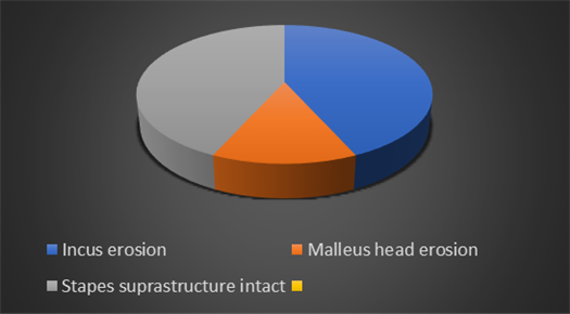
Results: There were 23 men and 22 women in the cohort, with 27 right ears and 18 left. Every patient exhibited erosion of the incus, and 14 additionally had erosion of the malleus head. The average preoperative air-bone gap (ABG) was between 35 and 40 dB. ABG improved to 5–10 dB after surgery. There were no issues. One patient experienced a recurrence after 1-year otoendoscopic follow-up, which was successfully treated using an endoscopic transcanal technique.
Conclusion: For attic cholesteatoma limited to the epitympanum, comprehensive endoscopic transcanal surgery is safe and successful, with good vision, low morbidity, steady improvement in hearing, and no recurrences in this series.
endoscopic ear surgery, attic cholesteatoma, epitympanum, minimally invasive, transcanal approach
Attic cholesteatoma, which often develops from a pars flaccida retraction in Prussak's space, is noted for its ability to erode the ossicles early, particularly the incus and malleus heads, even when the disease is limited to the epitympanum. Traditional microscopic techniques frequently necessitate canal wall-up or canal wall-down mastoidectomy to see critical recesses such the anterior epitympanum, lateral attic, and aditus ad antrum. Despite excellent microscopic access, some concealed locations may remain difficult to evaluate due to the straight-line view of the microscope and the anatomic restrictions of the external auditory canal, frequently needing more extensive mastoid surgery even for modest attic disease. (1-3)
With the progress of endoscopic ear surgery (EES), the treatment of epitympanic cholesteatoma has evolved significantly. The endoscope provides wide-angled, panoramic views of the attic and its subsites, providing direct access to areas that are difficult to see with the microscope. Tarabichi was among the first to demonstrate the feasibility and benefits of transcanal endoscopic removal of limited attic cholesteatoma, which resulted in improved visualization and a reduced requirement for mastoidectomy. (4) Marchioni and colleagues' subsequent anatomical and clinical research provided extensive mapping of the epitympanum and confirmed the use of rigid endoscopes to identify important regions such as the anterior epitympanic recess, lateral epitympanic space, and supratubal recess. (5-7) These efforts helped to establish EES as a potent tool for safe and effective attic disease management. Multiple investigations have since demonstrated that endoscopic transcanal methods can completely remove cholesteatoma in early or limited attic disease while conserving normal tissues, minimizing external incisions, and lowering morbidity. (8-10) Endoscopic techniques have produced comparable or better results than microscopic surgery in terms of disease clearance, postoperative hearing results, and recurrence rates. (9,11) Endoscopic approaches have also been linked to better identification of residual matrix, fewer blind patches, and lower incidence of second-look procedures. (8-11)
Given these benefits, endoscopic transcanal surgery has emerged as the recommended treatment for carefully chosen instances of attic cholesteatoma confined to the epitympanum. The current case series seeks to assess the outcomes of this minimally invasive method in 45 consecutive patients, focusing on surgical efficacy, safety, hearing improvement, and recurrence over a one-year follow-up period.
Methods
Study Design
Retrospective case series of 45 consecutive patients treated at a tertiary otology center.
Inclusion criteria
Exclusion criteria
Preoperative Evaluation
Surgical Technique
Results
Demographics
|
Parameters |
Value |
|
Sample size |
45 |
|
Age range |
15-37years |
|
Sex |
23 males / 22 females |
|
Side |
27 right / 18 left |
|
Extent |
Epitympanum only |
Ossicular Status
Audiological Outcomes
|
Outcome |
Value |
|
Pre-op Air bone gap |
35-40db |
|
Post-op air bone gap |
5-10db |
|
Hearing improvement |
25-30db |
|
|
|
Recurrence
• One patient experienced a recurrence after 1-year otoendoscopic follow-up, which was successfully treated using an endoscopic transcanal technique.
Discussion:
Exclusive endoscopic transcanal surgery is a very successful treatment for attic cholesteatoma confined to the epitympanum, as the current case series shows. At a one-year follow-up, the 45-patient group had only one recurrence, no problems, and continuous hearing improvement. These results are consistent with the increasing amount of evidence that endoscopic ear surgery (EES) for restricted cholesteatoma is beneficial. Surgeons can examine hidden recesses such the anterior epitympanic space, lateral epitympanum, and supratubal recess thanks to rigid endoscopes' improved visibility. These recesses are often missed by microscopic procedures because of their straight-line optical restrictions. (1-3) The remarkable clearance rates seen in endoscopic surgery are probably a result of this enhanced access. By showing that angled endoscopes permitted full view of Prussak's space and the epitympanum without the requirement for mastoidectomy, Tarabichi's seminal work first demonstrated the viability of endoscopic removal of attic cholesteatoma.(4) Marchioni and associates built on these discoveries by providing thorough endoscopic mapping of the epitympanum and stressing the significance of examining the anterior attic and supratubal recess, regions that are frequently in charge of residual disease when treated with conventional microscopy. (5-7)
The study's hearing results were good, with postoperative ABG closure to 5–10 dB. Similar gains have been documented in a number of EES series, where improved disease clearance and middle ear structural preservation lead to improved functional results.(8,11,12) The endoscopic technique allowed for the atraumatic dissection of matrix and ossicular chain, allowing for the best repair when necessary, despite the fact that all patients showed incus erosion and one-third had malleus head erosion. Strong postoperative audiometric outcomes were probably also influenced by the preservation of the stapes superstructure in all instances. The minimally intrusive nature of the endoscopic approach is one of its key advantages. No patient in this series needed postauricular access or canaloplasty. This is in line with research by Ayache and others that support the transcanal endoscopic technique as a tissue-sparing substitute that improves cosmetic results and lowers postoperative morbidity.(9,13) The safety profile of EES for limited attic disease is further supported by the lack of sequelae such tympanic membrane reperforation, facial nerve damage, and postoperative infection. Compared to microscopic surgery, endoscopic cholesteatoma surgery often has similar or lower recurrence rates. In a systematic review, Hunter et al. shown that endoscopic methods lessen residual disease by enabling examination of previously unreachable areas. (10) In a similar vein, Kuo and Shiao highlighted the importance of EES in spotting tiny residual pearls that could otherwise go unnoticed. (14) This series' primary drawback is its one-year follow-up. Longer-term observation, preferably with diffusion-weighted MRI, is recommended since cholesteatoma recurrence may appear later. A comparison microscopic arm, which would confirm the endoscopic method's superiority, is also absent from the study. However, with 45 consecutive patients and consistent surgical technique, the results provide compelling evidence that endoscopic transcanal surgery is the best treatment option for confined attic cholesteatoma.
Our results support the safety, efficacy, and low invasiveness of exclusive endoscopic transcanal treatment of attic cholesteatoma limited to the epitympanum. Excellent visibility is made possible, total disease removal is made easier, middle ear structures are preserved, and dependable hearing results with minimal morbidity and recurrence are produced. EES is the best approach for early attic cholesteatoma when used on carefully chosen patients.
Conclusion:
Exclusive endoscopic transcanal surgery is an excellent treatment option for attic cholesteatoma limited to the epitympanum. It provides minimal morbidity, wide vision, low recurrence, and satisfactory hearing outcomes. For some patients, it might be the suggested first course of action.
Compliance with Ethical Standards:
The procedure performed in this case report was in accordance with the ethical standards of the institutional and/or national research committee and with the 1964 Helsinki declaration and its later amendments or comparable ethical standards.”
Funding:
This study is not funded by any resources.
Conflict of Interest:
The author(s) declare no potential conflicts of interest with respect to the research, authorship, and/or publication of this paper.
Ethical Approval:
The study was published with written consent of the patient.
Compliance with Ethical Standards:
The procedure performed in this case report was in accordance with the ethical standards of the institutional and/or national research committee and with the 1964 Helsinki declaration and its later amendments or comparable ethical standards.”
Funding:
This study is not funded by any resources.
Conflict of Interest:
The author(s) declare no potential conflicts of interest with respect to the research, authorship, and/or publication of this paper.
Ethical Approval:
The study was published with written consent of the patient.

Open Access By Aditum Open Access Journals id licensed under Creative Commons Attribution 4.0 International License. Based On a Work at aditum.org


