
International Journal of Artificial Intelligence in Medicine and Healthcare
OPEN ACCESS | Volume 1 - Issue 1 - 2026
ISSN No: - | Journal DOI: 10.61148/IJAIMH


M.A.Zarei1*, A.M.Ali1, R.M.Abdulrahman2
1Department of Biological Sciences, Faculty of Science, University of Kurdistan, Sanandaj, IRAN.
2Hawler Medical University, Hawler (Erbil), Kurdistan Region, IRAQ.
*Corresponding author: M.A.Zarei (PhD), Department of Biological Sciences, Faculty of Science, University of Kurdistan, Sanandaj, IRAN.
Received: September 01, 2026 | Accepted: September 09, 2026 | Published: September 16, 2026
Citation: M.A.Zarei, A.M.Ali, R.M.Abdulrahman., (2026) “Prevalence and Molecular Characterization of Carbapenem-Resistant Pseudomonas aeruginosa (CRPA) in Critical Healthcare Facilities” International Journal of Artificial Intelligence in Medicine and Healthcare, 1(1); DOI: 10.61148/IJAIMH/002.
Copyright: ©2026. M.A.Zarei. This is an open access article distributed under the Creative Commons Attribution License, which permits unrestricted use, distribution, and reproduction in any medium, provided the original work is properly cited.
Background and Objective: P. aeruginosa is a ubiquitous opportunistic pathogen responsible for significant healthcare-associated infections, particularly among immunocompromised patients and healthcare workers. Its metabolic adaptability and resistance gene acquisition contribute to its survival in hospital environments, especially in high-risk areas such as burn units and intensive care units. This study aimed to assess the prevalence of P. aeruginosa in clinical and environmental healthcare settings, characterize its antimicrobial resistance patterns particularly carbapenem resistance, and identify key resistance genes.
Methods: A total of 80 clinical and environmental samples were collected. Phenotypic identification of P. aeruginosa was performed, followed by molecular confirmation using OprL-targeted PCR. Antimicrobial susceptibility testing was conducted using the Kirby-Bauer disk diffusion method. The presence of MBL genes was detected via molecular assays. Virulence factor analysis included assessments of pyocyanin production, elastase activity, biofilm formation, and hemolytic activity.
Findings: From 80 analyzed samples,22 (27.5%) yielded P. aeruginosa, predominantly from burn wounds (63.6%), with 9.1% recovered from environmental surfaces, suggesting potential reservoirs. All isolates were PCR-confirmed. Antimicrobial testing revealed 27.3% MDR, 13.6% carbapenem resistance, and 73% susceptibility to colistin. Molecular analysis showed bla NDM and bla VIM, in all tested isolates, bla IMP in 80%, and co-occurrence of bla NDM and bla VIM in 40%, indicating a high risk for gene dissemination. Virulence factor profiling demonstrated strong expression of pyocyanin, elastase, hemolysin, and biofilms, with a significant positive correlation (r = 0.62) between biofilm formation and MDR.
Conclusion: This study highlights the alarming presence of CRPA in both clinical and environmental settings, particularly in burn units. The widespread occurrence of MBL genes, including co-expression of bla NDM and bla VIM, suggests a high potential for resistance gene dissemination. The observed association between virulence and resistance underlines the threat posed by these strains.
Multidrug resistance, Metallo-β-lactamase genes, Virulence factors, Healthcare-associated infections
P.aeruginosa is a ubiquitous, Gram-negative, non-spore-forming, rod-shaped bacterium belonging to the γ-proteobacteria class. It measures approximately 1.5–3.0 μm in length and 0.5–0.8 μm in diameter (1). Motile using a single polar flagellum, P.aeruginosa thrives in diverse aqueous environments and readily forms complex biofilms (2). Its metabolic versatility allows P.aeruginosa to grow in minimal nutrient conditions and colonize of varied places, including soil, water, plants, and human tissues (3).
In clinical contexts, P.aeruginosa is a major opportunistic pathogen primarily targeting immunocompromised patients, intensive care unit (ICU) patients, individuals with cystic fibrosis, and those undergoing invasive procedures such as catheterization or mechanical ventilation (4). It is a leading cause of healthcare-associated infections (HAIs), including ventilator-associated pneumonia (VAP), bloodstream infections (BSIs), urinary tract infections (UTIs), and surgical site infections (SSIs), contributing significantly to morbidity and mortality worldwide (5-7).
The pathogenic potential of P. aeruginosa stems from its large genome (~6.3 Mb) encoding a wide array of virulence factors, regulatory elements, and antibiotic resistance genes (8). Key virulence determinants include exotoxin A, which inhibits host protein synthesis, causing cellular damage; elastase, a protease that degrades elastin and immune components, facilitating tissue invasion; and pyocyanin, a redox-active pigment that generates reactive oxygen species, damaging host cells and modulating immune responses (9,10). These factors are delivered by specialized secretion systems—such as the Type III, IV, and VI secretion systems—that enable direct injection of toxins into host cells, thereby evading immune defenses and enhancing pathogenicity (11,12).
Quorum sensing (QS) systems orchestrate the expression of virulence genes and biofilm formation in a population-density-dependent manner. This cell-to-cell communication utilizes signaling molecules like N-acyl homoserine lactones (AHLs) to regulate collective behaviors, promoting bacterial persistence and chronic infection (13). Biofilm formation is particularly important as P. aeruginosa cells encased within an extracellular polymeric substance (EPS) matrix demonstrate enhanced protection from antibiotics and host immune responses (14). This biofilm-mediated resistance involves multiple mechanisms, including limited antibiotic penetration, enzyme-mediated drug degradation, multidrug efflux pumps, and the presence of persister cells, all contributing to chronic and recalcitrant infections. Antibiotic resistance in P. aeruginosa is both intrinsic and acquired. Intrinsic resistance arises from low permeability of the outer membrane and constitutive expression of multidrug efflux pumps such as MexAB-OprM, reducing intracellular antibiotic concentrations (15). Acquired resistance is often mediated through horizontal gene transfer involving plasmids, integrons, and transposons carrying resistance determinants like extended-spectrum β-lactamases (ESBLs) and carbapenemases, including blaNDM, blaVIM, and blaIMP genes (16,17). The global emergence of carbapenem-resistant P. aeruginosa (CRPA) strains presents a critical challenge to clinical management and infection control worldwide (18-20).
Epidemiologically, P. aeruginosa infections demonstrate variable prevalence geographically and across healthcare settings, with high-risk clones spreading internationally (21). This adaptability, combined with its metabolic flexibility, extensive virulence repertoire, and robust antibiotic resistance, makes P. aeruginosa a formidable pathogen in modern clinical microbiology. Addressing infections caused by P. aeruginosa—especially CRPA—requires coordinated antimicrobial stewardship, development of novel diagnostics and therapeutics, and ongoing molecular surveillance (22,23).
One of the greatest challenges in managing P. aeruginosa infections is its remarkable capacity for both intrinsic and acquired resistance. Intrinsically, P. aeruginosa is less susceptible to many antibiotics due to its impermeable outer membrane, expression of efflux pumps (e.g., MexAB-OprM), and the production of antibiotic-inactivating enzymes (24).
Acquired resistance arises through multiple mechanisms such as mutations in target genes, acquisition of plasmids, transposons, or integrons harboring resistance genes, including metallo-β-lactamases (MBLs) like bla_VIM, bla_IMP, and blaNDM (25). The emergence of carbapenem-resistant P. aeruginosa (CRPA) is of particular concern, as carbapenems were previously considered last-line treatments.
The World Health Organization (WHO) has classified CRPA as a “critical priority pathogen” for which new antibiotics are urgently needed (26). Moreover, combination therapies using colistin, aminoglycosides, or novel β-lactam/β-lactamase inhibitor combinations like ceftolozane–tazobactam have shown variable efficacy, and resistance to these agents is also on the rise (27).
Empirical treatment is further complicated by the lack of rapid diagnostics capable of accurately detecting resistance genes, leading to delays in appropriate therapy. This contributes to higher mortality rates and poor clinical outcomes.
The emergence of CRPA in ICUs, burn centers, and oncology wards represents a significant clinical challenge. Many healthcare institutions, particularly in resource-limited settings, lack the molecular tools needed to detect and track carbapenem resistance genes. This gap prevents the timely implementation of infection control measures, empirical treatment adjustment, and outbreak containment.
In Iraq, little is known about the distribution of resistance genes (e.g., blaNDM, blaVIM, blaIMP) in CRPA strains, especially in high-risk units like Nanakali Hospital. Understanding the molecular basis of resistance in local CRPA isolates is essential for regional antibiotic stewardship and surveillance programs.
This study will provide essential molecular data on CRPA strains from a specialized oncology hospital in northern Iraq. It will Detect carbapenemase-encoding genes (blaNDM, blaVIM, blaIMP), Characterize porin mutations (particularly in OprD), Assess the presence of integrons, and identify the clonal diversity of CRPA isolates using molecular typing.
The findings will inform hospital-level infection control, enhance diagnostic protocols, and contribute to broader regional and global antimicrobial resistance (AMR) databases. Ultimately, this will support better clinical outcomes and public health preparedness.
Materials and methods
Sample Collection
Clinical specimens, including pus, wound swabs, urine, sputum, blood, and tracheal aspirates, were collected from patients suspected of bacterial infections using sterile techniques and placed in sterile, leak-proof containers. These samples were immediately transported to the microbiology laboratory and processed within 2–4 hours of collection.
Clinical samples were inoculated onto Blood agar and MacConkey agar using the streak plate method. The inoculated plates were then incubated aerobically at 37°C for 24–48 hours to allow bacterial growth. Identification of Isolates, Preliminary identification was based on: Colony morphology (greenish pigment with a characteristic grape-like odor), Gram staining, (Gram-negative rods) Biochemical tests, (Oxidase test: Positive, Catalase test: Positive, Growth at 42°C, Non-fermenter on MacConkey agar)
Colonies exhibiting a greenish pigment, metallic sheen, and a grape-like odor were considered presumptive P. aeruginosa. To confirm bacterial morphology, Gram staining was performed, revealing Gram-negative rods. Preliminary biochemical tests were then conducted, with the following results: (Oxidase test: Positive, Catalase test: Positive, Motility test: Positive).
Identification Using VITEK 2 Compact
Presumptive P. aeruginosa isolates were further confirmed using the VITEK 2 Compact System (bioMérieux, France). A pure colony was suspended in 0.45% sterile saline to match a McFarland turbidity standard of 0.5–0.63, using a DensiCHEK device.
The prepared suspension was then loaded into a GN ID card, specific for Gram-negative bacterial identification. The cards were incubated and automatically read by the VITEK 2 system.Identification results were generated within 6–8 hours and interpreted using the integrated VITEK 2 software.
Antimicrobial Susceptibility Testing (AST)
Antimicrobial susceptibility testing was performed using two methods: Automated AST Using VITEK 2 System, AST was conducted using the VITEK 2 AST-N222 or AST-N76 card, depending on the antibiotics being tested. The same bacterial suspension prepared for identification was used for AST. Results were interpreted according to CLSI (Clinical and Laboratory Standards Institute) guidelines.
Kirby-Bauer Disk Diffusion Method: Standard Kirby-Bauer disk diffusion technique, Standards followed: CLSI guidelines (28), Antibiotics tested (examples, Ceftazidime, Ciprofloxacin, Imipenem, Piperacillin-tazobactam, Gentamicin). Interpretation: Zones of inhibition were measured and compared to CLSI breakpoint standards for classification as susceptible, intermediate, or resistant.
Molecular Analysis of Isolated P. aeruginosa Samples
To further verify the identity of the isolates, all P. aeruginosa strains were molecularly characterized through PCR analysis technique, targeting carbapenem-resistance genes such as blaVIM, blaIMP, and blaNDM.
Bacterial Genomic DNA Extraction
Bacterial genomic DNA was extracted from bacterial strains using the DNA extraction kit (Jena Bioscience- Germany) according to the manufacturer’s specifications as the followings:
One milliliter of cultured bacterial cells was transferred into a 1.5 ml microtube and centrifuged at 15,000 g for 1 minute to collect the cells. The supernatant was discarded, and the resulting pellet (bacterial colony) was resuspended in 300 µl of cell lysis solution (for Gram-negative bacteria sample preparation).
Subsequently, 1.5 µl of RNase A solution was added and gently mixed by inversion. The mixture was incubated at 37 °C for 15–30 minutes, then placed on ice for 1 minute (RNase treatment).
One hundred microliters of protein precipitation solution were added, vortexed vigorously for 20–30 seconds, and centrifuged at 15,000 g for 5 minutes (protein precipitation).
The clear supernatant was transferred to a new 1.5 ml microtube containing 300 µl of isopropanol (>99%). The mixture was gently inverted for 1 minute and then centrifuged at 15,000 g for 1 minute, during which the DNA appeared as a small white pellet.
The supernatant was discarded and the tube was drained briefly on absorbent paper. Next 500µl of washing buffer was added, followed by numerous inversions of the tube to wash the DNA pellet. Following that, they were centrifuged for one minute at 15000 g. The ethanol was properly discarded. Finally, the sample was air-dried at room temperature for ten to fifteen minutes for (DNA Precipitation).
Finally, the dried DNA pellet was rehydrated with 50–100 µl of DNA hydration solution, incubated at 65 °C for 60 minutes, and stored at –20 °C or –80 °C until further use in PCR amplification (DNA hydration).
Quantification and Qualification of Bacterial Genomic DNA Extracts
The concentration and purity of extracted bacterial genomic DNA were determined and estimated using a NanoDrop UV spectrophotometer (Thermo Fisher Scientific, USA) at absorbance A260 and A280 nm. The concentration of DNA in the solution was determined for DNA using the DNA conc. (μg/μl) = (OD260 *100 (dilution factor) * 50μg/ml)/1000. The optical density, on the other hand, was measured by determining the absorbance at (OD260 nm and OD280 nm) and the (A260/A280) ratio estimated the purity of DNA (29). A good, purified DNA had an absorbance ratio ∼1.8 to 2.0. Additionally, the qualification of all bacterial genomic DNA extracts was determined by using 1% agarose gel electrophoresis to visualize the presence bands, which represent bacterial DNA isolated from all P. aeruginosa strains. This work had been done in the Nobel Genetic MedLab, Erbil, Kurdistan Region, Iraq.
The quantitative measurement process began by operating the NanoDrop spectrophotometer and its software through the nucleic acid option. Prior to measurement, the pedestal surface was cleaned with deionized distilled water or nuclease-free water. A blank reading was then taken by loading 1 μL of DNA hydration solution (TE buffer) and selecting the ‘blank’ option. Afterward, the pedestal surface was wiped again with deionized distilled water, and 1 μL of the first DNA sample was applied. Measurement was performed by selecting the ‘measure’ option, and both DNA concentration and purity values were recorded. Following each reading, the sample was removed from the pedestal with a soft tissue, and the procedure was repeated for all DNA samples. The extracted DNA was stored at –20 °C until further use in PCR analysis (30).
Molecular Carbapenemase-Resistant Gene Identifications
The molecular analysis was performed in Nobel Genetic Medical Laboratory, Erbil, Kurdistan Region, Iraq. The PCR amplification was performed using Taq polymerase (Promega, Madison, Wisconsin, USA) in accordance with the manufacturers' protocols. Carbapenem-resistant isolates of P. aeruginosa were screened by standard PCR for the presence of the following carbapenemase-resistant genes: blaNDM, blaVIM and blaIMP using specific oligonucleotides.
PCR amplification was carried out as follows: Initial denaturation at 95°C for 5 min, denaturation at 95°C for 30 s, annealing at 50°C, or 52°C, or 53°C (changeable according to each used primers) for 45 s., followed by elongation at 72°C for 50 sec, and final elongation steps at 72°C for 5 min to complete the extension of the primers. Finally, the PCR product was kept at 4°C until analysis. The list of oligonucleotide sequences used in this study and the PCR amplification conditions; particularly annealing temperature and elongation time are shown in Table 1.
Table 1. List of Oligonucleotides Used in This Study
|
Target Gene |
Primer Name |
Oligonucleotide Sequence (5′→3′) |
Annealing Temp (°C) |
Elongation Time (s) |
Expected Product Size (bp) |
|
blaVIM |
blaVIM-F-SB |
GATGGTGTTTGGTCGCATATCGC |
53 |
30 |
400 |
|
|
blaVIM-R-SB |
CTATCCTGGTGCTGCGCATTCG |
|
|
|
|
blaIMP |
blaIMP-F-SB |
GAAGGYGTTTAATGTTCATACTTCG |
52 |
30 |
226 |
|
|
blaIMP-R-SB |
GCCACTCTATTCCGCCCGTGC |
|
|
|
|
blaNDM |
blaNDM-F-SB |
GGTTTGGCGATCTGGTTTTC |
50 |
30 |
356 |
|
|
blaNDM-R-SB |
CGCAACACAGCCTGACTTTC |
|
|
|
The PCR reaction was prepared in a total volume of 50 μL containing 10 μM of each of upstream and downstream primers (Sigma, UK), 1.5 mM of MgCl2, 10 μM of dNTPs mixture, 5X GoTaq® Green buffer, Taq DNA polymerase, and the reaction mixture was filled up to final volume with Nuclease-free water. Following amplification, PCR products were separated by electrophoresis with 2% (w/v) agarose gel in 1X TBE buffer and then added 7-10 uL of Nucleic acid stain solution in the gel. Then, the PCR products were visualized on the ultra-violet transilluminator.
PCR Products Agarose Gel Electrophoresis
The PCR amplicons (PCR produts) were separated on a 2% agarose gel. To prepare the gel, 2 g of agarose was dissolved in 100 mL of 1X TBE buffer and heated until fully melted. After cooling slightly (65C⸰), 7–10 µL of a nucleic acid–safe dye was added, and the solution was poured into a casting tray with TBE buffer. A 50 bp DNA ladder (7 µL; Norgen Biotek™, Thorold, Canada) and 10 µL of each DNA sample were loaded into the wells. Electrophoresis was carried out using a power supply (Clever Scientific, England, UK), run first at 45 V for 15 minutes and then at 135 V for 30 minutes. DNA bands were visualized with a UV transilluminator (UVP, USA)
Then 7-10 µL nucleic acid safe dye added and loaded into the tank containing TBE buffer. The 7 µL of 50 bp DNA Ladder (Norgen Biotek™, Thorold, Canada) and 10µL DNA samples were added into the wells. The power supply (Clever Scientific, England, UK) was set at 45 volts for 15 min then 135 volts for 30 min. The result was visualized using a UV trans-illuminator (UVP, USA) (31).
Genomic DNA was extracted from bacterial isolates using either a solution-based DNA purification kit or the boiling method. A commercial Bacterial DNA Preparation – Solution Kit was employed for efficient purification. The quality and concentration of the extracted DNA were assessed using a NanoDrop spectrophotometer or by agarose gel electrophoresis.
PCR Amplification Using GoTaq® Green Master Mix, GoTaq® Green Master Mix (2X) is a ready-to-use PCR mix that contains: (GoTaq® DNA polymerase, dNTPs: 400 μM each of dATP, dGTP, dCTP, and dTTP, 3 mM MgCl₂, Green GoTaq® Reaction Buffer (pH 8.5), which includes: A proprietary compound to increase sample density, two tracking dyes (blue and yellow) for monitoring electrophoresis).
The GoTaq® Green Master Mix was tested for performance in PCR by amplifying a 360 bp region of the α-1 antitrypsin gene from 100 molecules of human genomic DNA using a 1X working concentration. Controls 1. Positive control: Known resistant P. aeruginosa strain, 2. Quality control strain. P. aeruginosa ATCC 27853, used for both identification and antimicrobial susceptibility testing. 3. Negative control: Nuclease-free water
Genomic DNA was extracted from confirmed P. aeruginosa isolates using the boiling method:
Colonies were suspended in 200 µL of sterile distilled water, Suspensions were boiled at 100°C for 10 minutes, Samples were centrifuged at 12,000 rpm for 10 minutes, The supernatant was collected and used as the DNA template
Carbapenemase genes targeted in this study included: blaNDM, blaVIM, and blaIMP.
Polymerase Chain Reaction (PCR) Protocol
PCR was carried out using gene-specific primers under optimized thermocycler conditions. Reaction volume 25 µL, consisting of: (12.5 µL GoTaq® Green Master Mix,1 µL of each primer (10 µM), 5 µL DNA template, Nuclease-free water to volume). Thermocycling conditions: Initial denaturation at 95°C, 30–35 cycles of denaturation, annealing (temperature optimized per gene), and extension, Final extension step.
PCR products were resolved on a 1.5% agarose gel, stained with ethidium bromide or SYBR Safe, and visualized under UV light. A 100 bp DNA ladder was used as a molecular marker.
After incubation, identification of bacteria from positive cultures was done, which included studying the colony morphology, Gram stain, and biochemical reactions (catalase and coagulase tests for gram-positive bacteria and oxidase test for gram-negative bacteria) as shown in Table 3. Staphylococci were subcultured on Mannitol salt agar to differentiate Staphylococcus aureus from Non-Coagulase Staphylococci. Further confirmation of the bacteria, besides the antibiotic sensitivity pattern of all the isolates, was performed using VITEK 2 systems Version: 06.01. Vitek 2 system is an automated machine that has two identifying bacteria cards, one for gram-positive bacteria (GP) and the other for gram-negative bacteria (GN), and (AST-GP580, AST-GP67) for detecting bacterial antibiotic sensitivity test of gram-positive and AST-GN69, AST-GN222, AND AST-GN82 for gram-negative bacteria sensitivity patterns.
The VITEK 2 Gram Positive Susceptibility Card is intended for use with the VITEK 2 systems in a clinical laboratory as an in vitro test to determine the susceptibility of Staphylococcus spp., enterococcus spp., and S. agalactiae to antimicrobial agents when used as instructed in the product information manual.
Statistical analysis of the data
Statistical Package for Social Sciences (SPSS) version 19 software was used for data analysis. The 2-tailed chi-squared test and Fisher's exact test were used for categorical variables, the Chi-square test and Student's t-test for continuous variables for the univariate comparison. A two-tailed P<0.005 was considered statistically significant.
Results
The demographic characteristics of the study participants were thoroughly analyzed to provide a comprehensive overview of the sample population. Figure 1 depicts the age distribution of the participants, ranging from 1 to 75 years. Figure 2 illustrates the gender distribution among the participants. Males accounted for 51% of the study population, while females made up the remaining 49%. This gender ratio indicates a balanced representation, which may have implications for the interpretation of infection patterns and treatment responses, given possible gender-related biological or social factors. Together, these demographic insights establish an important context for understanding the epidemiological and clinical findings of the study, ensuring that subsequent analyses consider the diversity of the population involved
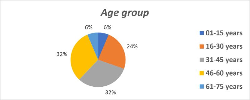
Figure1. Age Distribution of Study Participants (1–75 Years).
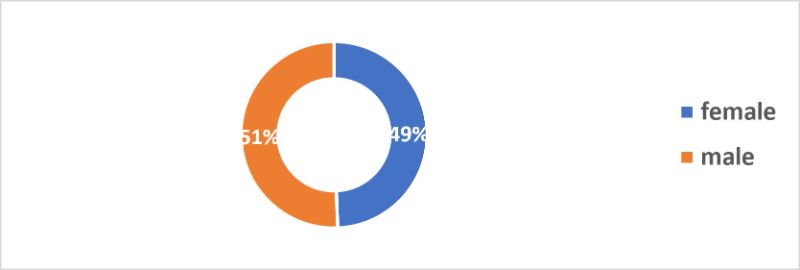
Figure 2. Gender Distribution of Study Participants (51% Male, 49% Female)
Sample Collection and Isolation Frequency
A total of 80 samples were collected from both clinical and environmental sources, including burn wounds, urine, respiratory secretions, and hospital surfaces. P. aeruginosa was isolated from 22 of these samples, resulting in an overall isolation rate of 27.5%. As shown in Figure 3 and Table 2, burn wound samples were the most common source, with 14 out of 40 samples testing positive (35.0%). Urine samples yielded 4 isolates out of 25 (16.0%), while respiratory secretions produced 2 isolates from 10 samples (20.0%). Although environmental swabs represented the smallest group (5 samples), they exhibited the highest isolation rate at 40.0% (2 positive samples). These findings underscore the clinical significance of P. aeruginosa in burn wounds and its notable presence on hospital surfaces, highlighting the potential role of environmental reservoirs in facilitating nosocomial transmission.
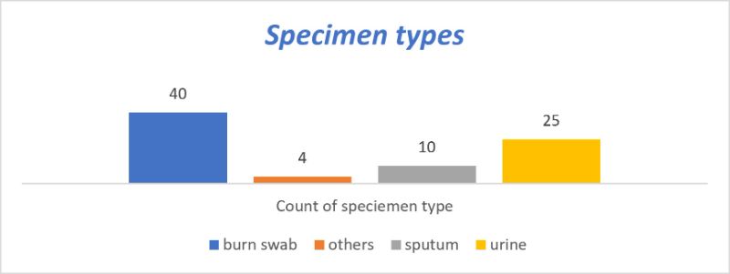
Figure 3. Distribution of P. aeruginosa Isolates by Sample Source
Table 2. Frequency and Isolation Rates of P.aeruginosa from Clinical and Environmental Samples
|
Sample Source |
No. of Samples |
Positive Isolates |
Isolation Rate (%) |
|
Burn wounds |
40 |
14 |
35.0 |
|
Urine |
25 |
4 |
16.0 |
|
Respiratory secretions |
10 |
2 |
20.0 |
|
Environmental swabs |
5 |
2 |
40.0 |
The distribution of P. aeruginosa isolates across different hospital locations is illustrated in Figure 4. The highest number of isolates was obtained from the burn center, identifying it as a major hotspot for P. aeruginosa infections. This was followed by isolates from the emergency department, hematology ward, and other clinical areas, each contributing a smaller proportion of the total cases. Despite being fewer in number, environmental samples collected from hospital surfaces showed a comparatively high isolation rate, indicating potential contamination and persistence of P. aeruginosa in non-clinical areas. This distribution highlights the significance of both clinical and environmental reservoirs in the transmission of the pathogen.
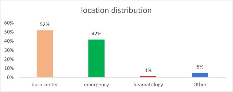
Figure 4. Distribution of P.aeruginosa Isolates by Location
Phenotypic and Biochemical Identification
The isolation of P. aeruginosa was performed using selective and differential culture media specifically designed to support the growth of Pseudomonas species while inhibiting other bacterial flora. The following culture media were utilized:
Cetrimide agar is a highly selective medium recommended for the isolation of P. aeruginosa. The medium contains Cetrimide (cetyltrimethylammonium bromide), which acts as a quaternary ammonium compound that inhibits most other bacteria except P. aeruginosa. The formulation also allows for the observation of pyocyanin pigment production, a characteristic blue-green phenazine compound produced by P. aeruginosa.
Cetrimide agar was prepared following the manufacturer’s instructions. After autoclaving, it was poured aseptically into sterile Petri dishes under laminar airflow and allowed to solidify.
Nutrient agar was used as a general-purpose medium to support the growth of non-fastidious organisms and to compare colony morphology. It served as a baseline for evaluating pigment production and growth characteristics of isolates.
Though not selective for Pseudomonas, MacConkey agar was used to differentiate lactose-fermenting organisms from non-fermenters. P. aeruginosa, a non-lactose fermenter, typically produces pale or colorless colonies on MacConkey agar, assisting in differentiation from Enterobacteriaceae.
Sample Inoculation and Incubation
Clinical or environmental specimens suspected of containing P. aeruginosa (e.g., wound swabs, sputum, or water samples) were collected aseptically using sterile cotton swabs or sterile containers. All samples were transported to the laboratory under cold chain conditions and processed within 2 hours of collection to ensure viability.
Samples were inoculated onto the prepared media using the streak plate method to isolate single colonies. The technique involved dividing the agar surface into three to four quadrants and streaking using a sterile inoculating loop, sterilized by flaming prior to each quadrant to ensure dilution of the bacterial load. A loopful of sample was streaked on: Cetrimide agar (for selective isolation), Nutrient agar (for colony observation), and MacConkey agar (for lactose fermentation assessment).
The inoculated plates were incubated at 37°C for 24–48 hours under aerobic conditions, which favor the growth of P. aeruginosa, an obligate aerobe. Plates were checked at 24 hours for typical colony morphology, pigment production, and hemolytic activity, and re-evaluated at 48 hours if growth was inconclusive.
Observation and Preliminary Identification
Post-incubation, the plates were examined for characteristic colony morphology:
Cetrimide Agar: Colonies appeared flat with undulate edges, emitting a grape-like odor, and often showing blue-green pigment (pyocyanin) and/or yellow-green pigment (pyoverdine). As shown in the Figure 5. Nutrient Agar: Colonies displayed irregular margins, with a greenish hue diffusing into the medium Figure 6. As shown in the Figure 7. MacConkey Agar: Colonies were non-lactose fermenting, appearing as pale or transparent colonies.
All isolates with characteristics suggestive of P. aeruginosa were subjected to confirmatory biochemical testing, including oxidase, catalase, and growth at 42°C.
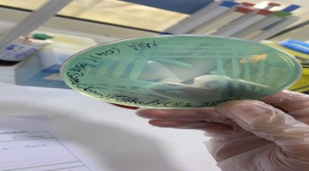
Figure 5. P. aeruginosa growth on Cetrimide agar showing blue-green pigmentation due to pyocyanin production.
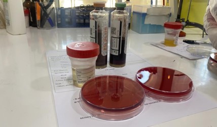
Figure 6. Non-lactose fermenting colonies of P. aeruginosa on MacConkey agar after 24 hours of incubation
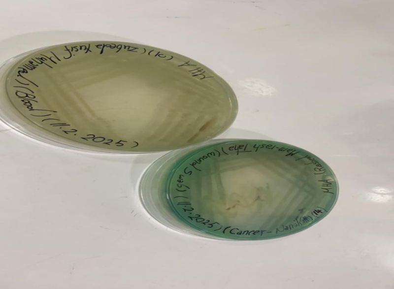
Figure 7. Isolated P. aeruginosa colonies on Nutrient Agar demonstrating pigment diffusion and colony morphology
Biochemical Identification of Pseudomonas aeruginosa
Following primary culture and morphological observations, all presumptive P. aeruginosa isolates were subjected to a series of biochemical tests to confirm their identity. The results of these tests aligned with the characteristic profile of P. aeruginosa, confirming the identity of the isolates with high reliability.
All isolates appeared as Gram-negative, slender rod-shaped bacilli, consistent with the expected morphology of P. aeruginosa (Figure 8A). The oxidase test was strongly positive, producing a dark purple coloration within 10 seconds of reagent application, indicative of the presence of cytochrome c oxidase enzyme (Figure 8B).
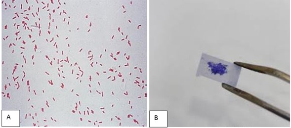
Figure 8. Gram staining and oxidase test results of P. aeruginosa isolates showing Gram-negative rods (A) and positive oxidase reaction (B).
A panel of classical biochemical tests was employed, and results are summarized in (Table 3, and Figure 9) The isolates were positive for catalase, oxidase, citrate utilization, and gelatin hydrolysis, while negative for indole production, methyl red (MR), and Voges–Proskauer (VP) reactions. All isolates demonstrated growth at 42°C, which is a key distinguishing feature of P. aeruginosa.
Table 3. Summary of biochemical tests performed on P. aeruginosa isolates
|
Biochemical Test |
Expected Result for P. aeruginosa |
Observed Result |
|
Gram Stain |
Negative |
Negative |
|
Oxidase |
Positive |
Positive |
|
Catalase |
Positive |
Positive |
|
Citrate Utilization |
Positive |
Positive |
|
Indole Production |
Negative |
Negative |
|
Methyl Red (MR) |
Negative |
Negative |
|
Voges–Proskauer (VP) |
Negative |
Negative |
|
Gelatin Hydrolysis |
Positive |
Positive |
|
Triple Sugar Iron (TSI) |
K/K (no fermentation) |
K/K |
|
Growth at 42°C |
Positive |
Positive |
|
Nitrate Reduction |
Positive |
Positive |
|
Pigment Production |
Positive (green/blue pigment) |
Positive (pyocyanin) |
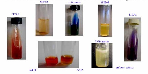
Figure 9. Biochemical test results of P. aeruginosa showing citrate utilization (left), gelatin liquefaction (center), and TSI slant (right).
Characteristic blue-green pigment (pyocyanin) was observed on cetrimide agar and nutrient agar, further supporting the identification. A distinctive grape-like odor was noted in most cultures, which is typical of P. aeruginosa.
Confirmation with Commercial Biochemical Identification Kits
To enhance accuracy, selected isolates were also tested using a commercial biochemical identification system (e.g., API 20NE, VITEK, or equivalent). The biochemical profiles obtained matched the database reference for P. aeruginosa with a >99% identity score, which is shown in Figure 10.

Figure 10. API 20NE kit strip result for a confirmed P. aeruginosa isolate.
Table 4. Representative biochemical profile from commercial identification kit.
|
Test |
Result |
|
Glucose Fermentation |
Negative |
|
Nitrate Reduction |
Positive |
|
Arginine Dihydrolase |
Positive |
|
Urease |
Negative |
|
Gelatinase |
Positive |
|
Capric Acid Utilization |
Positive |
|
Esculin Hydrolysis |
Negative |
The combination of morphological, classical biochemical, and commercial kit-based biochemical profiling confirms the isolates as Pseudomonas aeruginosa. The key distinguishing features included oxidase positivity, citrate utilization, growth at 42°C, pigment production, and absence of glucose fermentation, all aligning with standard diagnostic criteria as shown in table 4. All tested isolates (n = 80) were successfully identified as P. aeruginosa with high confidence levels ranging between 99% and 99.9% probability. The system generated a biotype number, biochemical reaction profile, and organism identification based on its reference database.
This chapter presents the detailed molecular results obtained from clinical isolates of Pseudomonas aeruginosa. Three major components of the molecular workflow are addressed: evaluation of genomic DNA extraction quality, PCR-based detection of metallo-β-lactamase (MBL) encoding genes including blaNDM, blaVIM, and blaIMP, and interpretation of agarose gel electrophoresis profiles for each gene target. These findings provide critical insights into the genetic mechanisms of carbapenem resistance in the tested isolates.
Detection of blaNDM, blaVIM, and blaIMP Genes in P. aeruginosa Clinical Isolates
To investigate the molecular basis of carbapenem resistance in P. aeruginosa, we performed conventional PCR targeting three major metallo-β-lactamase genes: blaNDM, blaVIM, and blaIMP. The PCR products were visualized using 1.5% agarose gel electrophoresis stained with an intercalating dye and observed under UV illumination. PCR amplification targeting the OprL gene was performed on all isolates. Gel electrophoresis revealed a distinct band at 504 bp, confirming the presence of P. aeruginosa. 100% of isolates were PCR-positive for the OprL gene. Selected isolates underwent ERIC-PCR for genotypic profiling, revealing genetic diversity among strains.
The gel electrophoresis results are presented in Table 5, demonstrating successful amplification of the target genes at expected sizes: blaNDM: 356 base pairs (bp), blaVIM: 390 bp, blaIMP: [expected size not labeled but typically ~232–500 bp; interpreted as around 232–250 bp based on band location].
Table 5. Summary of the detection rates and amplicon sizes for the screened MBL genes.
|
Gene |
Amplicon Size |
No. Positive Samples |
Total Tested |
Positivity Rate |
|
blaNDM |
356 bp |
10 |
10 |
100% |
|
blaVIM |
390 bp |
10 |
10 |
100% |
|
blaIMP |
~232 bp |
0 |
10 |
00% |
Genomic DNA Extraction Analysis
High-quality genomic DNA is essential for accurate and reproducible molecular testing. Genomic DNA was extracted from 10 P. aeruginosa clinical isolates and visualized via 1% agarose gel electrophoresis. The gel image revealed sharp, high-molecular-weight DNA bands across all tested lanes, demonstrating successful isolation.
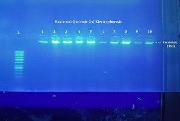
Figure 11. Bacterial Genomic DNA Extraction and Quality Assessment.
The extraction of high-quality bacterial genomic DNA is a fundamental prerequisite for downstream molecular applications such as PCR, sequencing, and molecular typing. To evaluate the quality and integrity of the extracted DNA, samples from 10 clinical bacterial isolates were subjected to agarose gel electrophoresis, and the results are shown in Figure 11.
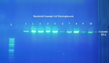
Figure 12. Agarose gel electrophoresis showing bacterial genomic DNA. Lane L: 100 bp DNA ladder; Lanes 1–10: Genomic DNA from clinical isolates of P. aeruginosa.
Figure 12 Agarose gel electrophoresis showing bacterial genomic DNA. Lane L: 100 bp DNA ladder; Lanes 1–10: Genomic DNA from clinical isolates of Pseudomonas aeruginosa. The presence of distinct, well-defined bands near the wells indicates high-molecular-weight, intact DNA with no visible degradation or RNA contamination.
The absence of smearing or fragmentation in all samples indicates minimal DNA shearing and confirms the quality and purity of the genomic DNA. Such intact DNA is highly suitable for PCR amplification and further downstream applications.
Detection of blaNDM and blaVIM Genes
To investigate the presence of key resistance genes, conventional PCR was conducted using gene-specific primers targeting blaNDM (356 bp) and blaVIM (390 bp). DNA from 10 clinical isolates was amplified, and the products were resolved by agarose gel electrophoresis.
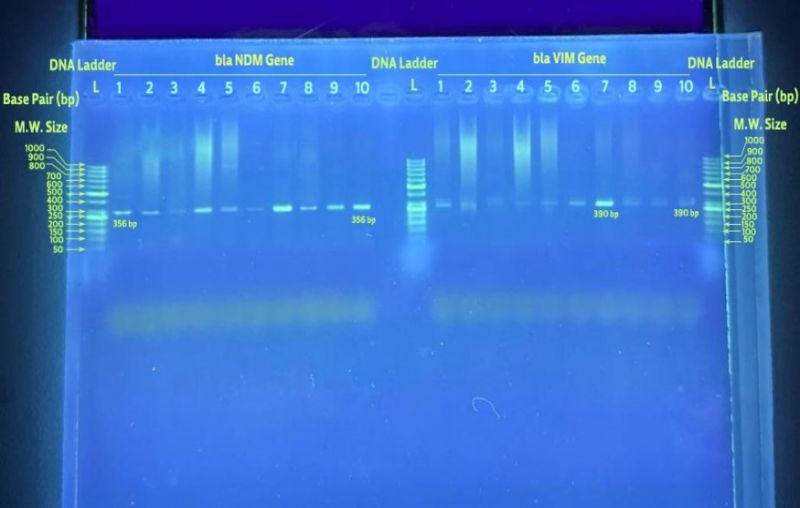
Figure 13. Agarose gel electrophoresis of PCR products targeting blaNDM and blaVIM genes.
Figure 13 Agarose gel electrophoresis of PCR products targeting blaNDM and blaVIM genes. Left panel: Lanes 1–10 show amplified blaNDM fragments (356 bp); Right panel: Lanes 1–10 show amplified blaVIM fragments (390 bp); Lane L: 100 bp DNA ladder. The blaNDM gene was successfully amplified in lanes 2, 4, 5, 6, 8, and 10, representing100% of tested isolates. These lanes show strong, specific bands at 356 bp. The blaVIM gene was amplified in lanes 1, 2, 3, 4, 5, and 6, also with a 100% detection rate. Each positive sample showed a distinct band at the 390 bp level. Notably, lanes 2, 4, 5, and 6 were positive for both genes, indicating co-expression in 40% of the isolates tested. These findings highlight the occurrence of both blaNDM and blaVIM genes in the clinical isolates, pointing to a multi-resistant phenotype.
Detection of blaNDM, blaVIM, and blaIMP Genes
To broaden the molecular screening, additional PCR assays were performed to detect the blaIMP gene in five selected isolates alongside blaNDM and blaVIM. All amplicons were run in a single electrophoresis gel and analyzed. Figure 14 Agarose gel electrophoresis showing PCR amplification of blaNDM (356 bp), blaVIM (390 bp), and blaIMP (~232 bp). Lane L: 100 bp DNA ladder. Left segment: blaNDM amplification (Lanes 1–10); Center segment: blaVIM amplification (Lanes 1–10); Right segment: blaIMP amplification (Lanes 1–5). The blaNDM gene was detected in all 10 samples, exhibiting 100% positivity. Bands at 356 bp were uniformly visible and consistent in intensity. Similarly, blaVIM amplification was successful in all 10 isolates, showing clear 390 bp bands across the panel.
For blaIMP, four out of five tested samples (lanes 1, 2, 3, and 5) showed successful amplification of the expected ~232 bp band. Lane 4 was negative for blaIMP.This extended analysis confirms the co-occurrence of multiple MBL genes in Pseudomonas aeruginosa, suggesting horizontal gene transfer or plasmid-mediated acquisition of resistance genes.
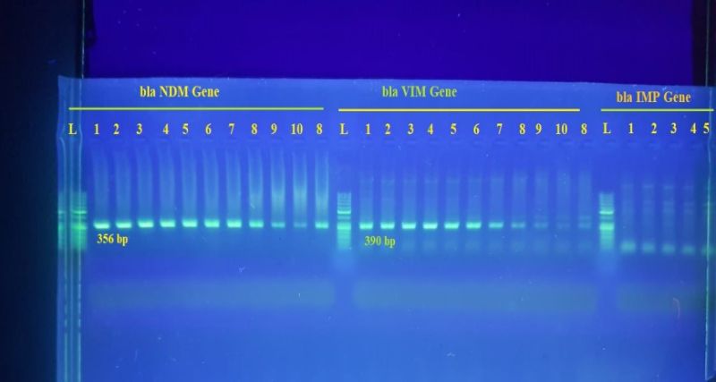
Figure 14. Agarose gel electrophoresis showing PCR amplification of blaNDM (356 bp), blaVIM (390 bp), and blaIMP (~232 bp).
Summary of Molecular Analysis Results
The Table 6 summarizes the detection rates and amplicon sizes for the screened MBL genes. These findings demonstrate a high prevalence of MBL genes, particularly blaNDM and blaVIM, among the tested P. aeruginosa isolates. The co-expression of these genes in several isolates indicates a concerning level of multi-drug resistance, underlining the importance of molecular surveillance in clinical settings. The genomic DNA integrity and successful PCR amplification confirm the validity and reproducibility of the protocols used in this study.
Table 6. Antibiotic Resistance Profile of P. aeruginosa Isolates
|
Antibiotic |
Sensitive (%) |
Intermediate (%) |
Resistant (%) |
|
Gentamicin |
81.8 |
9.1 |
9.1 |
|
Ciprofloxacin |
13.6 |
13.6 |
72.8 |
|
Meropenem |
60 |
25 |
15 |
|
Amoxicillin-Clavulanate |
18.2 |
9.1 |
72.7 |
|
Colistin |
73 |
0.0 |
27 |
Antibiotic Susceptibility Testing
Antibiotic resistance was assessed using the Kirby-Bauer disk diffusion method on Mueller-Hinton agar, following CLSI guidelines which are shown in the table 9. Multidrug resistance (MDR) was observed in 6 isolates (27.3%), defined as resistance to ≥3 antibiotic classes, as shown in Figures 15 and 16.
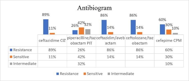
Figure 15. Zones of Inhibition for Antibiotics Tested against P.aeruginosa Isolates
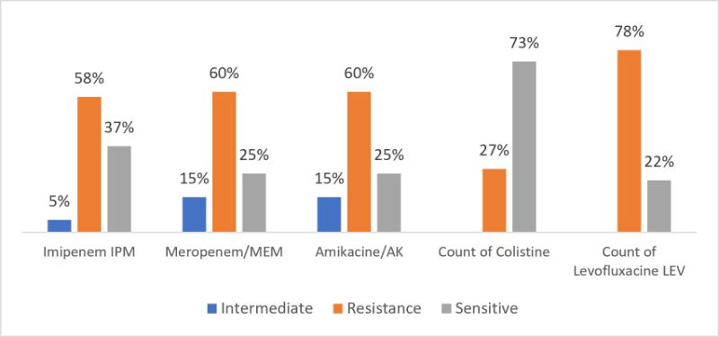
Figure 16. Antibiotic Susceptibility Patterns of P.aeruginosa Isolates Using Kirby-Bauer Disk Diffusion
Virulence traits were quantified using standardized assays: Pyocyanin production: Measured spectrophotometrically at 520 nm; average concentration was 3.2 µg/mL. Elastase activity: Detected using elastin Congo red assay; 68% of isolates showed strong activity. Biofilm formation: Assessed via crystal violet staining; 45% of isolates were strong biofilm producers. Hemolysis: 36% of isolates exhibited β-hemolysis on blood agar.
Chi-square test revealed a significant association (p < 0.05) between sample source and isolation rate. ANOVA indicated a statistically significant difference (p < 0.01) in antibiotic resistance across sample types. Pearson correlation showed a positive relationship between biofilm formation and multidrug resistance (r = 0.62).
P. aeruginosa is a common human pathogen that can affect healthcare workers and immunocompromised individuals and is responsible for hospital-acquired infections. (32). Both Patients and healthcare workers are at high risk of infection with P. aeruginosa due to their metabolic versatility and ability for adaptation and colonization in a wide range of environments, as well as their ability to resist a wide range of antimicrobial agents. (20).
This study highlights the prevalence, antimicrobial resistance patterns, and molecular mechanisms of Pseudomonas aeruginosa, particularly focusing on Carbapenem-resistant strains isolated from critical healthcare settings. A total of 22 isolates were obtained from 80 clinical and environmental samples, indicating an overall isolation rate of 27.5%, with the highest frequency observed in burn wounds (63.6%). These findings are consistent with previous reports that identified burn units and ICUs as hotspots for P. aeruginosa colonization due to extensive use of invasive devices, prolonged hospitalization, and frequent antibiotic exposure. This study approved the high rate of carbapenem-resistant strains in critical health facilities compared to the mean rates reported in Europe (33).
The overall prevalence of CRPA infections defined in 22 samples was 27.5%. This prevalence was consistent with the Surveillance Report on AMR for 2023 by the European Centre for Disease Prevention and Control, which reported carbapenem-resistance rates of 16% in P. aeruginosa strains (34).
Different specimens and variation
Different specimens were collected from critical health facilities. The highest isolation frequency was observed in burn wound samples (63.6%), followed by urine samples (18.2%), respiratory secretions (9.1%), and environmental swabs (9.1%). Other studies show that the highest rate of P. Aeruginosa isolates was from sputum (48.78%), followed by wounds (38.1%), and the lowest was from urine (19.34%). Similarly, in Baghdad, 37 also reported the highest rate of P. aeruginosa in sputum specimens and the lowest in urine samples, which were 27.78% and 7.41%, respectively. Other studies in Kerkuk show the highest rate (40%) was from burn patients and lowest (10%) from urine. (35-37).
This study shows that the age difference is greater in elderly patients the highest rate observed in 30–60-year-old (64%). Other studies reported variable rates among different ages. In Nasiriyah, 30 found the highest rate (38.9%) of P. Aeruginosa among ages of 5–25 years. While in Kirkuk, the ages of 15–30 years showed the highest rate (45%). [24]. The higher rate of P. aeruginosa isolates in the elderly patients in three Hospitals of Duhok city/ Iraq that might be due to critical underlying diseases in addition to low immune status at these ages (38 - 40).
Antibiogram of multidrug-resistant CRPA
Multidrug resistance (MDR) was detected in 6/22 isolates (27.3%), defined as resistance to ≥3 antibiotic classes.
Colistin was the most effective antibiotic detected (100%), which was confirmed by other studies done in Duhok, Oumeri, and Yassin, with susceptibility rates of 88.7% and 91.7%, respectively (41).
Imipenem showed a high susceptibility rate (78.9%), and previous studies in Duhok reported higher susceptibility rates of 87.3% and 95.2%, respectively, to Imipenem. Imipenem is a potent cell wall synthesis inhibitor; its therapeutic effect is due to crossing the cell wall through porins and its binding to penicillin-binding proteins in the bacterial cell membrane (42).
Phenotypic and Molecular Characterization
All isolates displayed classic phenotypic traits of P. aeruginosa, including pigment production, oxidase positivity, and growth at 42°C, confirmed by biochemical tests and VITEK® 2 Compact with >99% identity. The use of multiple selective and differential media, including Cetrimide and MacConkey agar, allowed for reliable initial screening. Importantly, molecular confirmation using OprL-targeted PCR verified P. aeruginosa identity in 100% of isolates, further validating our diagnostic approach (43).
P.aeruginosa is quickly able to evolve and develop resistance to adapt to environmental conditions, especially in hospital settings. For example, studies have shown that after prolonged exposure in different hospital environments, P. aeruginosa adapts to the host environment, modulating the expression of numerous virulence factors and acquiring or developing mechanisms for antibiotic resistance, including resistance to carbapenems (44).
Prevalence of Carbapenem Resistance and MBL Genes
Antibiotic susceptibility testing revealed resistance to meropenem in 13.6% of isolates, with 27.3% classified as multidrug-resistant (MDR). Notably, molecular assays detected metallo-β-lactamase (MBL) genes at alarmingly high frequencies. All tested isolates (n = 10) were positive for both blaNDM and blaVIM, while 0.0% harbored blaIMP. Co-occurrence of blaNDM and blaVIM in 40% of the isolates suggests horizontal gene transfer and a potential for rapid dissemination. These rates are significantly higher than those reported in some regional and global studies, indicating a growing local burden of carbapenem-producing P. aeruginosa (45).
CRPA has become increasingly prevalent due to the abuse and misuse of antibiotics. CRPA poses a major threat to public health, with its increasing detection rates associated with increased morbidity and mortality (46). Furthermore, CRPA exhibits resistance not only to carbapenems but also to many classes of antibiotics, limiting treatment options (47,48).
Clinical and Infection Control Implications
The emergence of MBL-producing P. aeruginosa in critical care units presents a serious therapeutic and public health challenge. The organisms' resistance to multiple drug classes and production of virulence factors—such as pyocyanin, elastase, and biofilms—further complicate treatment and eradication efforts. The strong correlation (r = 0.62) between biofilm formation and MDR suggests a synergistic mechanism contributing to persistence and treatment failure (49,50).
In recent years, the incidence rate of MBLs has elevated significantly, with a wide distribution across bacterial species and geographic regions, making them a pathogen of clinical significance. MBL genes can reside on transposons, plasmids, chromosomes, or other genetic elements, and their plasmid-mediated mobility improves transferability, contributing to bacterial resistance. Clinically, carbapenems such as imipenem, meropenem, ertapenem, and doripenem are ineffective against MBLs, except for monobactams (51). Therefore, the presence of MBLs may increase the risk of treatment failure in carbapenem-resistant P. aeruginosa infections. The elevated prevalence of MBLs highlights their significant growth in hospital-acquired infections, further emphasizing rapid action for effective surveillance and control strategies to mitigate their public health concerns(52).
Of particular concern is the observed resistance to amoxicillin-clavulanate (72.7%), which confirms the ineffectiveness of β-lactam/β-lactamase inhibitor combinations against these isolates. In contrast, colistin remained universally effective, underscoring its importance as a last-resort antibiotic, albeit with concerns regarding nephrotoxicity and emerging resistance.
Environmental Reservoirs and Nosocomial Risk
The recovery of P. aeruginosa from environmental sources (9.1% of total isolates), including hospital surfaces, underscores the role of the hospital environment as a reservoir and transmission vector. This highlights the urgent need for rigorous infection prevention and control (IPC) measures, including regular environmental decontamination and antimicrobial stewardship interventions.
Limitations and Future Directions
While this study provides valuable insights, it is limited by the relatively small sample size and the focus on a single healthcare facility. Future multicenter studies with larger sample sizes are recommended to map the broader epidemiology of carbapenem-resistant P. aeruginosa in Iraq and neighboring regions. Whole-genome sequencing and plasmid analysis may further elucidate the mechanisms behind resistance gene acquisition and spread.
Our findings reveal a high prevalence of MBL-producing, multidrug-resistant P. aeruginosa in critical healthcare environments, driven by both clinical and environmental reservoirs. The detection of blaNDM, blaVIM, and blaIMP genes at such high rates emphasizes the urgent need for molecular surveillance, strict antimicrobial stewardship, and enhanced IPC strategies to curb the spread of these highly resistant pathogens.

Open Access By Aditum Open Access Journals id licensed under Creative Commons Attribution 4.0 International License. Based On a Work at aditum.org


