Dental Science and Innovative Research
OPEN ACCESS | Volume 5 - Issue 1 - 2026
ISSN No: 3065-7008 | Journal DOI: 10.61148/DSIR


Nagaveni NB
Professor, Consultant Pediatric Dentist “Garike Dental Care” Davangere, Karnataka, India.
Corresponding author: Nagaveni NB, Consultant Pedodontist, “Garike Dental Care”, Davangere, Karnataka, India.
Received: February 16, 2024
Accepted: February 20, 2024
Published: February 29, 2024
Citation: Nagaveni NB. (2024) ““Mirror Image Canines” in Association with Multiple Dental Anomalies – Report of A Unique Case”. Dental Science and Innovative Research, 3(1); DOI: 10.61148/DSIR/004.
Copyright: © 2024 Nagaveni NB. This is an open access article distributed under the Creative Commons Attribution License, which permits unrestricted use, distribution, and reproduction in any medium, provided the original work is properly cited.
“Mirror Image Canines” or “Kissing Canines” or Transmigration of bilateral permanent maxillary canines is an extremely rare dental eruption phenomenon showing scarcity in publications. The aim of the present manuscript, is to show the occurrence of such an uncommon dental variation in association with other dental anomalies such as congenital agenesis of mandibular both right and left first premolars and severe taurodontism (hypertaurodont) affecting mandibular second molars which all occurred together in Indian female patient.
Introduction
Permanent canines are the most commonly disputed teeth in the literature pertaining to its position, eruption pattern, impaction, congenital agenesis and associated pathology. ‘Canine transmigration’ is an uncommon dental abnormality referring to ‘intra-osseous migration of impacted teeth across the dental midline to the opposite side of the dental arch’ [1]. The teeth most frequently disturbed by this variation are the permanent mandibular canines [2]. These may see unilateral or bilateral with no gender predilection. However, some prevalence studies reported more in females than males. Two canines getting impacted at the same time, located in the same eruption pattern and moving at the same rate in same direction is really a strange occurrence in the dental anomalies’ literature. Such presentations are termed as ‘bilateral transmigration’ or ‘mirror image canines’ and literature shows common occurrence of bilateral transmigration well reported in the mandibular arch compared to maxilla [3,4]. Although impaction of maxillary canines in the maxillary arch is more common, transmigration is rarely reported. In addition to this, bilateral impaction along with transmigration is still an extremely rare entity.
The term “Mirror Image Canines” also referred as “Kissing Canines” are suggested by an author Idris Faiq Qaradaghi who defined it as ‘the migration of both canines at the same rate and on the same horizontal axis parallel to each other and meeting each other at the midline [5]. In this unusual dental scenario, most of the time, transmigrated canines remain impacted. Very rarely it can erupt either labially or palatally or remain in mirror image pattern in association with contralateral canine. Extensive review of literature revealed scanty publications showing only one case report on ‘kissing canines’ or ‘mirror image canines’ [6] and is elaborated in Table 1. The exact etiology and pathogenesis behind this ‘impaction love’ or kissing canines phenomenon is not well understood. The present article is the second article showing ‘mirror image canines’ in the maxillary arch which occurred in female patient from Indian ethnicity.
Table 1: Published Research Data on ‘Mirror Image Canines’ or ‘Kissing Canines’ confined to Maxillary Arch
|
Case No. |
Author/Year |
Ethnicity |
Patient age (in years)/Gender |
Retained primary teeth |
Associated Anomalies
|
|
|
Present case |
India |
38/Female |
No |
Bilateral Congenital agenesis of mandibular first premolars + Bilateral Taurodontism in mandibular second molars (Hypertaurodont) |
|
|
Kiyumi et al/2021 [5] |
Oman |
28/Male |
Right canine |
- |
Case Report
A 38-year-old female patient reported to a private dental clinic complaining of pain in the upper left tooth from past 15 days. On physical examination, patient appeared healthy and there were no signs and symptoms of particular disorder. On intraoral examination, all permanent teeth were erupted including third molars. Permanent maxillary first molar was totally destroyed with no crown structure remaining due to dental caries. Clinically, in the maxillary arch permanent right and left canines appeared missing and also in the mandibular arch, permanent first premolars were missing on both right and left side. Small gap was evident distal to the lateral incisors on both right and left side. Patient was subjected to radiographic evaluation to rule agenesis of these missing teeth using orthopantomograph. On radiographic examination, surprisingly permanent maxillary both right and left canines were impacted in horizontal position (Figure 1). The left canine was transmigrated with its full crown component crossing the mid-palatal suture and was seen in the opposite arch above the root apex of the right central incisor. On right side, the canine was located above the root apex of right lateral incisor with the crown facing mesial direction with root facing distal direction. Both right and left canines were appeared mirror image of one another or looked like they are kissing or touching each other in the same angulated position. Radiograph also showed presence of severe taurodontism affecting mandibular second molars and congenital agenesis of both right and left first premolars (Figure 1). There was no any associated pathology observed with these two teeth. A final diagnosis of transmigration of left canine and horizontal impaction of right canine was made. Patient was explained about the varied dental phenomenon and kept under observation as patient did not have any symptoms associated with it and extraction of the upper left first molar was advised.
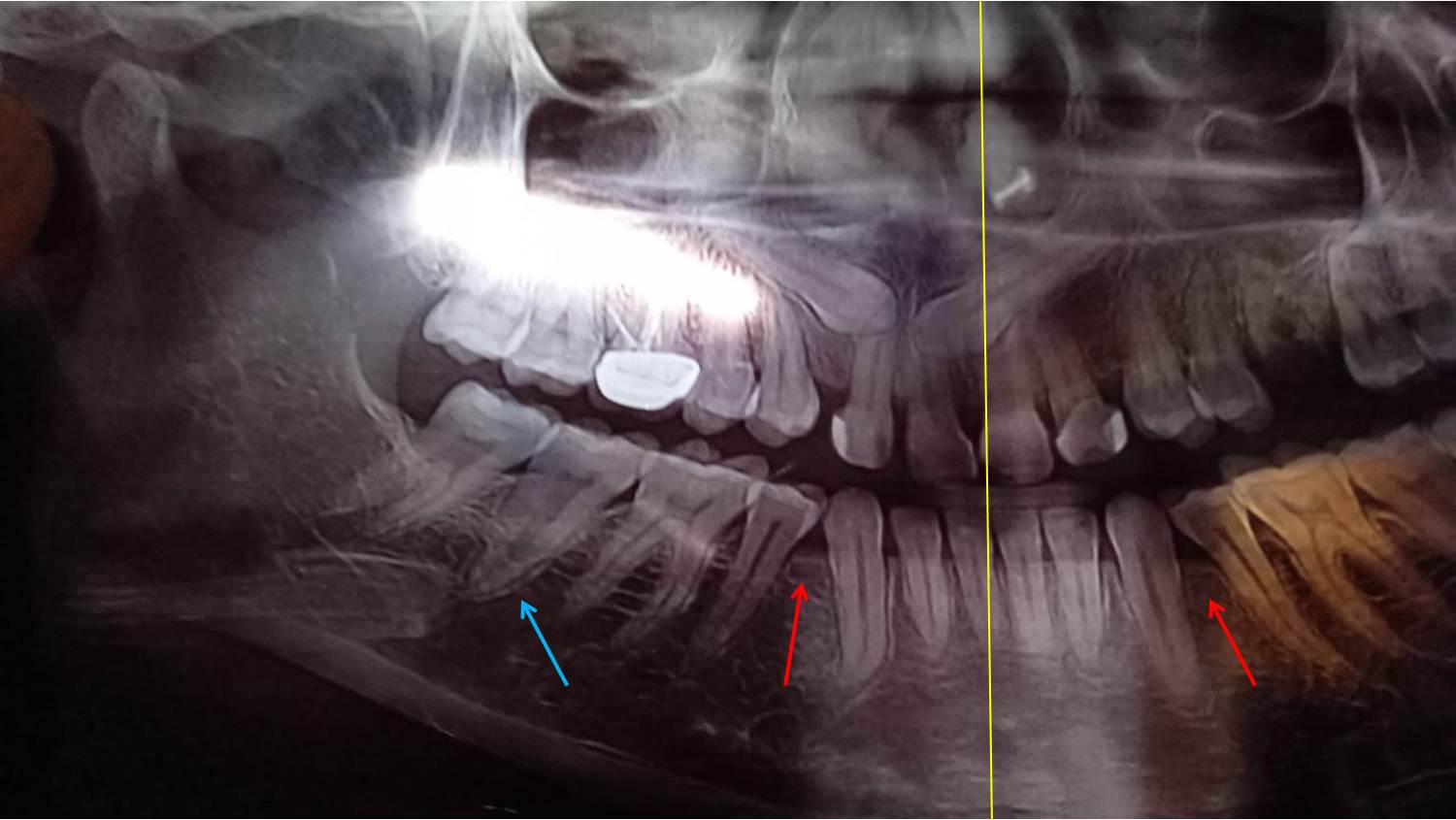
Figure 1: Orthopantomograph radiograph showing “Mirror Image Canines” or “Kissing Canines” in the maxillary arch. Intraosseous transmigration of permanent maxillary left canine (two fourth portion of the crown – yellow line) crossing the mid-palatal suture to the opposite side of the arch is evident. Right canine is impacted in mesioangular position similar to left canine. Congenital agenesis of bilateral mandibular first premolars (red arrows) and hypertaurodontism in mandibular second molar (blue arrow) is also present.
Discussion
There is no classification system for categorizing the maxillary canines which are in transmigration. This is due to low prevalence of transmigrated maxillary canines from that it is difficult to frame classification system. Moreover, the whole tooth crossing the mid-palatal suture is also not found or identified till date. In the present case, full crown portion was crossed the mid-palatal suture and seen opposite side of the arch above the root apex of the right central incisor. Transmigration of permanent maxillary central incisor is also reported by the current author which is also rarely reported [7].
As transmigrated teeth are always impacted, most of the time they are not diagnosed from routine daily examination unless a radiographic examination is carried out. Therefore, radiographic examination is mandatory in diagnosis of transmigrated canines. This indicates necessity of future studies including large sample size in different population across the globe. There are only few studies showing impaction of maxillary canines. One study carried out in 2011, reported only six cases on transmigrated maxillary canines out of 4500 patients examined [8]. Prior to this in 2010, Celikoglu et al [9] investigated panoramic radiographs of 2,215 patients of Turkish origin from an orthodontic department consisting of 940 males and 1,275 females. The maxillary transmigrated canine was associated with impacted mesiodens. The percentage of maxillary canine impaction was more compared to transmigrated canines.
Different researchers investigated impaction of maxillary canines and formulated various classifications. In 2003, one searcher (Yamamoto) classified impacted maxillary canines based on the orientation of the long axis of the impacted tooth with the occlusal plane into seven types grading from type I to type VII [10]. The present case was classified as type IV according to this classification as the tooth was impacted above the roots of central and lateral incisors with crown facing mesially (towards midline) and root distally. Using the same classification, a recent study evaluated the pattern and prevalence of maxillary canines by studying 5000 orthopantomograph radiographs among Saudi population. They discovered a prevalence of 46% of type I cases and 37% of type II [11]. Similar studies are highly warranted including different ethnic groups across the globe. Regarding treatment of ‘mirror image canines’ or ‘kissing canines,’ various modalities have been suggested such as periodic observation, surgical exposure followed by orthodontic alignment, transplantation and surgical removal [12,13]. Impacted canines are most of the time associated with retained primary teeth due to delayed root resorption process. But in this case such incidence was not observed.
Congenital agenesis of tooth most commonly involves permanent maxillary lateral incisor followed by mandibular second premolars. Second premolar agenesis is more common in the mandible (4.4%) than in the maxilla (1.7%). But agenesis of first premolar is still rarer with a documented prevalence of 0.1% in the mandibular arch compared to maxillary arch (0.2%) [14,15]. In the case reported here, exhibited congenital agenesis of both right and left first premolars making this case a rarest entity.
Taurodontism also termed as ‘Bull Tooth’ is another dental anomaly rarely seen during clinical practice. It is characterized by an apically displaced pulp chamber, a shortened root and an enlarged pulp chamber and apical displacement of the bifurcation or trifurcation of the roots in the affected tooth [16,17]. Shaw et al in 1928 [18], classified these teeth into three types as hypo, meso and hypertaurodont based on apical displacement of the root. Based on this classification the present case was classified as hypertaurodont as the pulp chamber was displaced apically nearly reaching the apex giving prismatic or cylindrical forms.
Conclusion
The current case report is one more addition to the occurrence of ‘mirror image canines’ which is extremely rare encountered dental phenomenon in the maxillary arch in association with other dental anomalies which are also rarer. Author wishes to welcome future publications involving prospective studies of large sample size to come up with more research output.


Open Access By Aditum Open Access Journals id licensed under Creative Commons Attribution 4.0 International License. Based On a Work at aditum.org