Dental Science and Innovative Research
OPEN ACCESS | Volume 4 - Issue 2 - 2025
ISSN No: 3065-7008 | Journal DOI: 10.61148/DSIR
Hanali Abu Shilbayeh
Department of Pediatric Dentistry, Al-Quds University, Jerusalem, Palestine.
Corresponding author: Hanali Abu Shilbayeh, Department of Pediatric Dentistry, Al-Quds University, Jerusalem, Palestine.
Received: June 10, 2025 |Accepted: July 20, 2025 |Published: July 28, 2025
Citation: Hanali Abu Shilbayeh., (2025) “The Hall Technique in Pediatric Dentistry” Dental Science and Innovative Research, 4(1); DOI: 10.61148/3065-7008/DSIR/053.
Copyright: ©2025. Hanali Abu Shilbayeh. This is an open access article distributed under the Creative Commons Attribution License, which permits unrestricted use, distribution, and reproduction in any medium, provided the original work is properly cited.
The Hall technique is a novel method of managing carious primary molars by cementing preformed metal crowns, also known as stainless steel crowns, over them without local anaesthesia, caries removal or tooth preparation of any kind. Clinical trials have shown the technique to be effective, and acceptable to the majority of children, their parents and clinicians. The Hall technique is NOT, however, an easy, quick fix solution to the problem of the carious primary molar. For success, the Hall technique requires careful case selection, a high level of clinical skill, and excellent patient management. In addition, it must always beprovided with a full and effective caries preventive programme. The aim of this article is to investigate the opportunity for treatment of the approximal dentin caries/ asymptomatic closed pulpitis with primary metal crowns (PMCs) using the Hall technique.
Crowns Hall Technique; Minimal intervention dentistry; stainless steel crowns; Deciduous
Introduction:
The modern approach in caries management focuses on arresting the lesions and disturbing or modifying the plaque [1-9]. Restorations have finite success, and the repeat restoration cycle often reduces the survival of a tooth [6-13]]. The rationale for complete caries removal is debated in the existing literature and considered as “potentially damaging even to attempt to remove all infected dentin in a symptomless, vital tooth” .[9-14] It may not be possible or essential to remove all caries, and moreover, a properly placed restoration that seals the cavity, can stop the progress of lesion without jeopardizing pulpal vitality. Once the cariogenic bacteria are isolated from the nutrient source, i.e., the oral environment, it can bring about successful treatment outcomes .[8-15]
Hall technique of stainless steel crown placement has proved to be a viable restorative option for carious primary molars resulting in its successful exfoliation. The rising evidence based on pragmatic clinical trials exhibits successful outcomes in terms of clinical performance and acceptability to Hall technique crown. It involves no local anesthesia, no removal of caries, no preparation of tooth to receive crown (only separation of teeth at the contacts, if necessary). [16] A randomised trial found that the HT and non-restorative caries treatment (NRCT) have greater acceptability than the conventional restorations (CR). [17] A retrospective analysis reported that sealing caries using Hall technique significantly outperformed standard restorations over 10 years, both clinically and statistically [18].
The Hall Technique (HT) presents an alternative for treating primary molars by seating stainless steel crowns without tooth preparation, caries removal, or local anesthesia, effectively halting cavity progression . Introduced by Dr. Norna Hall in Scotland , HT is recognized for treating early to moderately advance active caries lesions in primary molars with evidence of effectiveness and acceptability.[18]
In a biological approach, most restorations involve stainless steel crowns through HT (95%), followed by selective removal to firm dentin (5%) . While awareness of HT is high globally (92.32%), its utilization varies widely among countries (50.6%) .[19]
The Hall Technique (HT) presents dentists with a swift and definitive treatment, effectively minimizing patient anxiety . By eliminating the need for local anesthesia, HT aims to enhance child compliance and operator comfort. Beyond cavity sealing, it anticipates providing a less traumatic dental experience early in a child's life, fostering a likelihood of their return for more complex treatments in the future.
The use of preformed metal crowns through HT in managing dentinal caries in primary teeth reduces the risk of pain and restoration failure. HT has demonstrated a reduction in discomfort, earning preference from both patients and parents . Notably, HT boasts a shorter treatment duration, enhanced cost-effectiveness, and higher acceptability among parents compared to conventional techniques.
Children treated with HT over whel mingly reported positive experiences immediately after treatment, with nearly 90% expressing enjoyment. A retrospective study in the United States demonstrated similar success rates in clinical and radiological outcomes for stainless steel crowns used in restoring primary molars with carious lesions, comparing the conventional technique and HT . HT is well-tolerated bychildren, acceptable to parents, and associated with minimal adverse effects . When applied alongside silver diamine fluoride and atraumatic restorative treatment (ART), HT gained high acceptance from parents/caregivers . Its restoration survival rate was nearly three times higher than ART (93.4% compared to 32.7%) for occluso-proximal dentin lesions in primary molars after three years [24].
HT demonstrates cost-effectiveness, with significantly lower total cumulative costs compared to the conventional technique. Long-term practice-based trials affirm HT's superior cost-effectiveness, as it maintains longer with fewer complications at lower costs . Positioned as a cost-effective approach, HT contributes to anxiety reduction in dental caries treatment.[20-22]
The Hall Technique (HT) is not without drawbacks, with its chief adverse consequences encompassing an unsightly appearance and increased plaque accumulation when optimal adaptation is lacking . Major failures, such as irreversible pulpitis or dental abscess, are infrequent but significant . Cases involving the loss of restoration or crown, rendering the tooth unrestorable, are rare . Both major failures (irreversible pulpitis, dental abscess, periradicular radiolucency, and loss of crown with non-restorable tooth) and minor failures (loss of crown and restorable tooth, crown perforation, secondary/marginal caries, and reversible pulpitis) have been documented . Additionally, cases of the ectopic first permanent molar adjacent to the crowned tooth have been reported [30], and in rare instances, internal root resorption can occur .[23,24,25]
Stress distribution in tooth-supporting tissues is noted to be greater with HT within the initial 2 weeks compared to the conventional crown placement technique, where settling occurs within 2 days. Concerns about increased occlusal vertical dimensions with HT are addressed by evidence showing occlusion equilibration after 30 days without longterm problems .Parents or legal guardians in clinical practice commonly express apprehension about unremoved caries with HT . Another potential adverse outcome is the perforation of the occlusal surface.[24,26]
The Hall Technique (HT) necessitates meticulous case selection and evaluation of pulp status. Determining pulp status involves a comprehensive approach, including medical history, clinical examination, mechanical tests (probing, blowing air), test cavities, percussion, and radiography. In situations where children cannot cooperate or obtain diagnostic images, the extent of decay and pulp vitality (vital or non-vital) becomes crucial for deciding on a treatment modality . Excellence in diagnosis, treatment planning, and follow-up is pivotal for success.[13,27]
HT is not a "fit and forget" technique. Teeth should exhibit no symptoms of pulpal pathology, such as irreversible pulpitis, when considering Hall crowns. If a Hall crown is inadvertently placed due to diagnostic error or reaches the pulp causing irreversible pulp disease, prompt detection during reviews is essential . Hall crowns should be placed only when clinical examination and radiographic investigation indicate a very low risk of irreversible pulpal pathology .[15,28]
Recommended applications include occlusal caries if fissure sealant, partial caries removal, or conventional restoration is not accepted. Proximal cavitated or non-cavitated caries are suitable if the patient rejects partial caries removal or conventional restoration . Asymptomatic proximal primary molars with multisurface caries, asymptomatic occlusal lesions, hypoplastic primary molars, and asymptomatic carious lesions in primary molars (active or inactive) are also candidates for HT [36]. It is applicable to children and adults with intellectual/physical disabilities.[17,29]
Proper diagnosis is paramount for creating an appropriate treatment plan, considering pulp status. Emphasizing the importance of post-treatment follow-up to parents/caregivers is crucial, as ongoing monitoring is necessary to detect any failures promptly.[27]
HT is contraindicated when signs or symptoms of irreversible pulpitis, dental abscess/fistula, radiological signs of pulp involvement, or periradicular pathology are present. It is not recommended when there is no cooperation due to the risk of corona aspiration or swallowing. Contraindications also include patients at risk of infective endocarditis, immunocompromised children, severely destroyed crowns with non-restorable cavities, very young children unable to understand or tolerate the procedure without local anesthesia, allergy or vulnerability to nickel, a temporary tooth close to exfoliation, x-ray evidence of more than half tooth root resorption, pulp exposure during treatment, excessive tooth mobility, pulp necrosis, or dental abscess .[30]
Hall crowns require careful follow-up after placement, with prompt treatment of pulp pathology if it develops. While HT is not a universal solution for providing oral health care to disadvantaged or underserved populations , the patient's medical history should always be analyzed to consider any medical alerts contraindicating the treatment. In young children, analyzing pulpal behavior can be challenging, underscoring the importance of adequate post-treatment follow-up and clear communication with parents/caregivers about expectations.[31,32]
The aim of this article is to investigate the opportunity for treatment of the approximal dentin caries/ asymptomatic closed pulpitis with primary metal crowns (PMCs) using the Hall technique.
The Hall Technique in Five Steps;
The Hall Technique is a technically less complex procedure to perform in terms of the clinician’s dexterity, and it can be performed successfully in five steps;

Fig.1;Crown restoration with the Hall technique. (a) Pretreatment condition: The interproximal contacts are very tight. (b) Separating ring placed with the aid of rubber dam forceps. (c) The separating ring was removed after 2 days. Note the interproximal space gained for restoration.
Step 1: Assess Crown Morphology, Contact Areas, and Occlusion
The assessment of the crown morphology is decisive before performing a Hall crown. Anomalies of tooth size and form can make placing a Hall crown more challenging. This can also be problematic when there is a proximal carious lesion with marginal ridge breakdown in one of the primary molars, causing migration of the adjacent molar into the cavitated area as this leaves less mesio-distal space to place the crown. The margin of the crown can be adjusted to accommodate the intruding margin of the adjacent tooth.
In addition to crown morphology, the contact areas of the molar to be treated should be carefully assessed. Absence of interdental spaces can make it difficult to place a Hall crown. In contrast, Hall crowns can be easily fitted in Type I arches, where the marginal ridge is still intact. For closed contacts, it can be necessary to create a small amount of space for the crown by using orthodontic separators. These should be placed 2–3 days before fitting the Hall crown and removed immediately prior to crown placement.
Step 2: Size the Hall Crown
The smallest crown size, which will seat over, and cover the whole tooth, should be selected. The clinician should have the sensation of a slight feeling of “spring back” when trying on the crown (Fig. 13.6). Do not fully seat the crown through the contact points before cementation; it can be very hard to remove. As described above, orthodontic separators may be placed in the interproximal spaces 2–3 days before fitting the Hall crown to facilitate the crown placement.
Step 3: Clean the Tooth and Fill the Crown
Before placing the Hall crown, clean the tooth and dry the crown. To remove the residual bacterial plaque covering the tooth, use a rotary bristle brush or a toothbrush.
Use the triple syringe or cotton wool rolls to clean and dry the crown, and then place glass ionomer luting cement into the crown to fill approximately twothirds of the crown ensuring that all inner surfaces are covered (Fig. 13.7). Be careful to avoid air blows and gaps.
Step 4: Fit and Seat the Crown
To seat the crown, it is placed on top of the tooth and pushed straight down (Fig 13.6b).
The clinician should seat the crown by finger pressure ensuring the crown seats evenly over the tooth and between the contact points. As the crown is seated over the tooth, excess cement will flow out from the margins (Fig. 13.8). As soon as the crown is seated, and before the cement sets, the patient should be asked to open their mouth to allow the crown position to be checked and to remove the excess cement. If the crown fails to fully seat, it should be removed rapidly, using a spoon excavator.
There are then two options to further seat the crown: The child is asked to bite down on the crown, or the clinician pushes the crown down with finger pressure.
Usually a combination between these two options is used.
If the crown is in the ideal position, the child should be instructed to bite down firmly again on the crown (or cotton wool) for 2–3 min until the cement has set Alternatively, the clinician should hold down the crown with firm finger pressure to prevent the crown from springing back up as this would compromise the seal and unnecessarily increase the degree of occlusal opening.
Step 5: Remove Excess Cement and Check Occlusion
The final step is to clean around the teeth with a hand excavator to remove any excess cement and floss between the contacts (Fig. 13.9). It is important to remind the parents that the child will notice the crown being high in the bite (Fig. 13.10) and that this should resolve in a few days to a week, with full occlusion equilibration within a few weeks. Many children even do not consider the increase in the OVD as something uncomfortable or even notice it. The primary dentition has great ability to adjust to a slightly opened bite over a few days, with full reestablishment within a few weeks with no evidence of adverse effects .

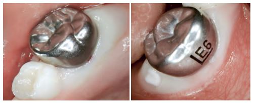
Fig. 2;(d and e) Immediately after placement of the steel crown. Circumferential ischemia is clearly visible.
DISCUSSION
The Hall Technique emerges as a significant advancement in pediatric dentistry, offering a non-invasive alternative to traditional restorative methods for managing carious lesions, particularly in primary molars. Its innovation lies in its ability to encapsulate carious lesions within stainless steel crowns, thus halting the progression of decay without the need for local anesthesia, dental drills, or the removal of carious tissue.
The Hall Technique demands a clear understanding and communication with the patient's caregivers, ensuring they are informed about the nature of the technique, its benefits, and limitations. Despite
its merits, the Hall Technique is not without challenges. Its reliance on the sealing properties of stainless steel crowns means that any breakdown in the marginal seal could lead to treatment failure. Hence, the role of regular dental check-ups ascends in importance, ensuring the integrity of the crowns and the absence of secondary caries.

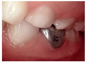
Fig.3;a) Temporary increase in vertical occlusal height after placement of a steel crown by the Hall
The Hall Technique should be considered a valuable addition to the pediatric dentist’s arsenal, one that aligns with the contemporary goals of minimally invasive dentistry and supports the psychological well-being of the child, paving the way for a lifetime of positive dental experiences.
In a study from 2007, acceptance of the technique among dentists, parents, and children and the success rate were tested versus conventional composite restorations. The results speak for themselves and have been confirmed in more recent studies from 2014. In their study, Innes et al55 restored a total of 128 teeth using the Hall technique and examined the restorations after 23 months; 89% of the teeth exhibited only very mild or no unpleasant sensations (versus 78% of teeth with composite restorations), and only 1.5% caused unacceptable discomfort (4.5% for composite fillings). Also, 77% of the children, 83% of the parents, and 81% of the dentists preferred the Hall technique to composite fillings as a better restorative technique. At follow-up of the restorations after 23 months, the teeth restored by the Hall technique performed better than teeth with composite fillings: Irreversible pulpitis was found in 2% of the Hall teeth and 15% of the composite restorations; progressive caries or loss of retention affected 5% of the Hall teeth and 46% (!!!) of those restored with composite; and pain occurred with 2% of the Hall teeth and 11% of the control restorations with composite. Equally good results were achieved by Santamaria et al in a clinical trial from 2014 with a follow-up period of 1 year.[3,25]
The Hall technique can therefore be recommended as a simple and promising method for restoring primary teeth.[25] Even for specialist colleagues, this technique is entirely recommendable for selected children and may be considered a viable option.[15,26]

Fig.4; Control radiograph 1 year after the treatment. The reaction of the pulp is clearly visible. Reactive dentin is formed in change of pulp chamber volume
The use of preformed metal crowns in minimally invasive methods of treatment like the Hall technique, provides the option for these constructions to be used in daily outpatient practice in children with a small number/single carious lesions and negative attitude, who otherwise must be treated with pharmacological control of the behavior .[16,27-30] Out of all restoration materials they are the most durable in time in regard to the mechanical resistance and must be an option of choice for the restorations of deciduous dentition, especially in children with high caries risk .[28-32]
Hall crowns are a useful technique for paediatric patients who are dentally anxious and not cooperative for conventional restorations on teeth which do not have pulpal caries.[31-35] Separator placement for Hall crowns may be difficult to complete, particularly when there are tight contacts, abnormal anatomy, or reduced cooperation from the patient. Rubber dam clamp forceps can be used effectively for separator placement and provide several advantages compared to the other techniques (especially floss) reported in the literature. However, the technique requires careful patient selection and may not be suitable for all patients.[35]
Conclusions:
The Hall Technique is a biologically based, effective management option to treat asymptomatic carious primary molars. This technique not only reduces the possibility of pulp exposure or irritation during carious lesion excavation but also
offers the benefit of full coronal coverage reducing the risk of future carious lesion development on another surface of the tooth. The Hall Technique has a high rate of success, is durable,and provides a cost-effective option for primary molars.
Compliance with ethical standards
Disclosure of conflict of interest
The author declares no conflict of interest