Dental Science and Innovative Research
OPEN ACCESS | Volume 5 - Issue 1 - 2026
ISSN No: 3065-7008 | Journal DOI: 10.61148/DSIR


Dione Samuel Silveira 1*, Rubens Kaique Queiroz Farias 1, Sandy Oliveira Simões 1,
Daniel Bastos Dos Santos Filho ¹, Stephanie Quadros Tonelli 2
1 Academics of the Graduation Course in Dentistry - Favenorte;
2 Master in Dentistry; Professor of the Undergraduate Course in Dentistry –Favenorte.
*Corresponding author: Dione Samuel Silveira, Av. José Alves Miranda, 500 - Alto São João, Mato Verde - MG, 39527-000.
Received: February 20, 2021
Accepted: February 28, 2021
Published: March 04, 2021
Citation: Dione S Silveira, Queiroz Farias R K, Sandy O Simões, Santos Filho D B D, Stephanie QTonelli. “ Glossite Migratória Benigna: Relato de Caso benign Migratory Glossitis: Case Report”. J Dentistry, Oral Maxillofacial Research, 1(1) ; DOI: http;//doi.org/03.2021/1.1001.
Copyright: © 2021 Dione Samuel Silveira. This is an open access article distributed under the Creative Commons Attribution License, which permits unrestricted use, distribution, and reproduction in any medium, provided the original work is properly cited.
Objective: Benign migratory glossitis (GMB) is a disorder that most commonly affects the back of the tongue, it is one of the most frequent pathologies of the oral cavity. In this sense, this study aimed to report a case on migratory erythema.
Case report: A 20-year-old male patient attended the Favenorte Dental Clinic, Mato Verde, MG, complaining of reddish plaques on the back of his tongue. He mentioned that the plaques persisted for days or weeks and disappeared from one place on the tongue, reappearing in another. During the anamnesis, the patient did not report any health problems or use of medications. On physical examination, normal periodontal condition and good oral hygiene were found. In the tongue, lesions were found characterized by reddish circular or elliptical erosions, with disappearance of the filiform papillae and maintenance of the fungiform ones, with well-defined and whitish edges located mainly on the back and on the lateral edges of the tongue. Due to its clinical and historical aspect, the diagnostic hypothesis of GMB was considered. After the evaluation by the academics responsible for the case, in consensus with the supervising professors, the conclusive diagnosis for the referred condition was defined.
Conclusion: The diagnosis of GMB is established eminently by clinical examination, since the aspect of the epatognomonic lesion. The patient was instructed on the disease, its benign course with spontaneous disappearance, he was instructed on brushing techniques, flossing, hygiene of the tongue and avoid eating very hot, acidic or spicy foods.
Introduction
Benign migratory glossitis (GMB), also called geographic language, wandering tongue eruption, exfoliative glossitis areata and migratory erythema are a disorder that most commonly affects the back of the tongue. This alteration is characterized by the loss of the filiform papillae that are surrounded by whitish borders on the surface of the tongue. The lesions vary in appearance and time, ranging from a few hours to several days (CARVALHO et al., 2010, SILVA et al., 2019).
Although the etiology of GMB is unknown, some factors influence the formation of this alteration, such as juvenile diabetes, genetic factors, fissures in the tongue, gastrointestinal disorders related to anemia and reactive arthritis (CALDAS et al., 2018).
Its incidence can also be related to emotional changes (stress, anxiety), irritants (hot or spicy foods), and skin allergies (atopic dermatitis). The incidence is higher in childhood and puberty, being more frequent in women than in men (PEIXOTO et al., 2018).
The diagnosis is made based on the clinical history accompanied by a previous history of the disease, compatible with chronic migratory and macroscopic lesions located on the surface of the back of the tongue, which present changes in color, shape and size. Laboratory tests, such as complete blood count and serum biochemistry are normal, except when the patient has a previous disease, such as diabetes mellitus. The differential diagnosis includes candidiasis, Reiter's Syndrome, lichen planus, leukoplakia, systemic lupus erythematosus and herpes simplex virus infection (ERRANTE et al., 2016).
Benign migratory glossitis, when asymptomatic, requires no treatment. Periodic follow-up to confirm the diagnosis is necessary in the case of a first visit and when the history is not clear. It is essential to reassure the patient about the benign and self-limiting nature of the injury (CALDAS et al., 2018). Symptomatic individuals are commonly treated with topical anesthetics and corticosteroids, whose side effects are negligible compared to systemic drugs (RIBEIRO et al., 2016). In this sense, the objective of this work is to report a case on migratory erythema.
Case Report
This study is supported by the Research Project entitled “Oral Conditions of the population of the municipality of Mato Verde, MG: epidemiological survey and importance of socioeconomic factors”, approved by the Research Ethics Committee under opinion number 2.536.216. This clinical case was conducted in accordance with the recommendations of the Resolution of the National Health Council 466/2012, with the signature of the free and informed consent form (ICF) and the authorization term for publication having been previously collected.
A 21-year-old male patient attended the Dental Clinic of Faculdade Verde Norte - Favenorte, complaining of reddish plaques on the back of his tongue. He mentioned that the plaques when they appear, persist for days or weeks, disappear from one place on the tongue and reappear in another. The patient also reported that these lesions have symptoms such as burning and irritation when he ingests citrus products and that they disappear spontaneously. During the anamnesis, the patient reported not having systemic changes or using medications.
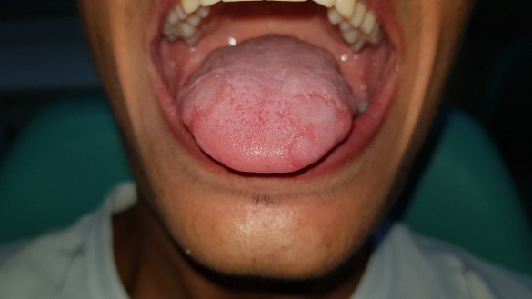
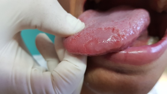
Figure 1: Clinical aspect of the lesion on the dorsum of the tongue, characteristic of benign migratory glossitis.
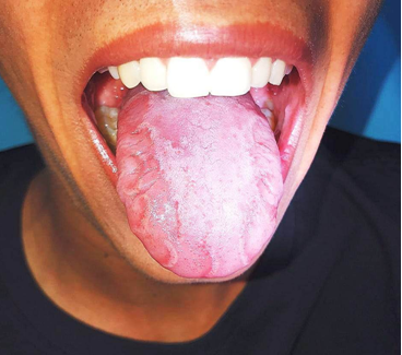
Figure 2: Areas of well-defined papillary atrophy at the lateral edges of the tongue.
On extraoral physical examination, changes in the cervical lymph nodes, facial asymmetries or signs of temporomandibular dysfunction were not identified. In the intraoral clinical examination, normal periodontal condition and good oral hygiene were found. In the tongue, lesions were found characterized by reddish circular or elliptical erosions, with consequent disappearance of the filiform papillae and maintenance of the fungiform ones (Fig. 1). Well-defined, whitish and slightly prominent edges can be seen, located mainly on the back and on the lateral edges of the tongue (Fig.2). After the evaluation by the academics responsible for the case, in agreement with the supervising professors of the Clinic, the diagnosis was defined by its clinical aspect, of GMB.
The patient was instructed on the disease, its benign course with spontaneous disappearance and the need for no interventions. Brushing techniques, flossing and tongue cleaning were also carried out, and it was also recommended to avoid eating very hot, acidic or spicy foods
Discussion
GMB or geographical tongue is a benign inflammatory disease of the tongue, which presents a variable and recurrent course, with spontaneous cure (ERRANTE et al., 2016). This type of lesion is characterized by a period of irritation and remission, during which the lesions heal without residual scarring. When the lesions return, they tend to appear in new locations and in different formats, single or multiple, thus producing the migratory effect (CALDAS et al., 2018, SANTOS et al., 2018).
To detect anomalies such as GMB, knowledge of the morphology of the language is necessary. This type of injury can vary over time and has a chronic and acute inflammatory condition (CARVALHO et al., 2010). This condition often presents without symptoms, but some patients may show symptoms of burning, sensitivity and pain, especially when eating citrus, spicy or hot foods (PILZ et al., 2015).
Several sources of information contribute to the assessment of the patient. The frequency and location of the lesions, as well as the patient's age and sex are data that contribute to the diagnosis (VOLKWEIS et al., 2016). The lesional clinical pattern is extremely suggestive, with rare situations where other diagnostic approaches are necessary, such as incisional biopsies (RIBEIRO et al., 2016). The diagnosis is commonly clinical and based on the characteristic history of migration, on the characteristics of the lesion, circumnavigated appearance and the absence or presence of significant pain as opposed to burning as a subjective complaint (SANTOS et al., 2018).
Some investigators suggest that GMB occurs more frequently in patients with psoriasis (including pustular psoriasis) or atopic dermatitis and in individuals with cleft tongue. Researchers have also indicated an association of GMB with hormonal disorders, allergies, Down syndrome, nutritional deficiencies and even a genetic predisposition (PILZ et al., 2015). Although most patients are asymptomatic, they commonly develop severe anxiety and fear of cancer. The disease is characterized by a period of irritation and remission, during which the lesions heal without residual scarring (CARVALHO et al., 2010).
In the treatment of symptomatic patients, some authors claim that the use of topical corticosteroids, oral creams with anesthetic is recommended for a better result (REGEZI et al., 2008). According to Ribeiro et al., (2016) as an example of topical medications used for the treatment of symptomatic lesions, we can mention the betamethasone elixir, clobetasol propionate and triamcinolone acetonide. Topical immunosuppressants can optionally also be used, such as tacrolimus. In the present case, due to the patient being asymptomatic, no palliative medication was prescribed.
According to Marcucci and Fonseca the treatment is empirical and according to Assimakopoulos et al., (2010) the treatment methods are disapproved, with the need to control stress. Only in some cases, can local measures be used. Most authors assure that avoiding hot and spicy foods in areas sensitive to injury reduces the burning, burning or sensitivity sensation of the patient, giving a better lifestyle in the treatment.
In the reported case, the patient presented lesions located mainly on the back and on the lateral edges of the tongue, recurrent and spontaneous healing, consistent with the literature in all aspects. The diagnosis was made only with the clinical examination, based on the characteristics of the lesion, migration and absence of significant pain, with no need for histopathological examination. The patient was instructed about GMB and all guidelines and recommendations for controlling and preventing remission were passed on to the patient.
Final Considerations
GMB is a benign, chronic and recurrent disease, associated with genetic, hereditary and environmental factors. It usually disappears from one place on the tongue and reappears in another until it disappears spontaneously, and it usually does not need treatment. It is essential that the dental surgeon has knowledge and mastery of oral changes and is prepared to diagnose these changes in advance and consequently obtain success in his clinical conduct.


Open Access By Aditum Open Access Journals id licensed under Creative Commons Attribution 4.0 International License. Based On a Work at aditum.org