
Clinical Research and Clinical Case Reports
OPEN ACCESS | Volume 7 - Issue 1 - 2026
ISSN No: 2836-2667 | Journal DOI: 10.61148/2836-2667/CRCCR


Oswaldo Núñez-Almache a,b,c,d,g*, Rómulo Lu de Lama a,c,e,f, Eliana Chávez Tejada a,c,d, Oscar Espinoza Robles a,c,d, Paola Pinto Ibarcena a,c,d, Martha Calagua Quispe a,c,d, Miguel De los Santos La Torre, Pamela Azabache Tafur
Institutional affiliations
1. Instituto Nacional de Salud del Niño (National Institute of Children's Health).
2. Universidad Nacional Federico Villarreal (National University Federico Villarreal).
Profession, speciality and academic degree
1. Physician.
2. Paediatric endocrinologist.
3. Endocrinologist.
4. Master's degree.
5. Doctorate
City, country, year: Lima, Peru, 2025
Purpose: Scientific — national/international publication.
*Corresponding author: Oswaldo Núñez-Almache, Instituto Nacional de Salud del Niño (National Institute of Children's Health).
Received: September 15, 2026 | Accepted: September 29, 2026 | Published: October 02, 2026
Citation: Núñez-Almache O, Lu-De lama R, Chávez-Tejada E, Espinoza-Robles O, Pinto-Ibárcena P, Calagua-Quispe M, De Los santos-La Torre Miguel, Azabache-Tafur, P., (2026). “Rhino-Orbital-Cerebral Mucormycosis in an Adolescent Girl with Type 1 Diabetes Mellitus at the National Institute of Children’s Health: A Case Report”. Clinical Research and Clinical Case Reports, 7(1); DOI: 10.61148/2836-2667/CRCCR/101.
Copyright: © 2026 Oswaldo Núñez-Almache. This is an open access article distributed under the Creative Commons Attribution License, which permits unrestricted use, distribution, and reproduction in any medium, provided the original work is properly cited.
Rhinoorbitocerebral mucormycosis (ROCM) is an angio-invasive mycosis with high mortality and is the most frequent form of presentation in diabetic patients. We report the case of a 17-year-old adolescent girl with type 1 diabetes mellitus (T1DM), vitiligo and autoimmune thyroiditis who developed ROCM; this is the first such case among the diabetic patients attended at the Instituto Nacional de Salud del Niño (INSN). The case is characterised by poor adherence to T1DM treatment, adverse epidemiological conditions and an initial presentation resembling an ocular conjunctival process and bacterial cellulitis, with subsequent cerebral extension, which required several surgical procedures and antifungal management. The outcome has been favourable within the severity of the condition, and medical and surgical management will be prolonged. The presentation of this case seeks to raise awareness of the need for adequate adherence to treatment in T1DM, particularly in those with concomitant autoimmune diseases, and of the early suspicion of ROCM in patients with ocular involvement.
Rhinoorbitocerebral mucormycosis (ROCM), type 1 diabetes mellitus (T1DM), diabetic ketoacidosis (DKA), vitiligo, autoimmune thyroiditis
T1DM is a disease characterised by autoimmune destruction of the pancreatic beta cells, which triggers hyperglycaemia; in women it tends to be associated preferentially with other autoimmune diseases, among them vitiligo, and this association often affects treatment and prognosis.1
ROCM is a severe angio-invasive mycosis caused by ubiquitous filamentous fungi of the order Mucorales, class Zygomycetes. It progresses rapidly and carries high morbidity and mortality, and it occurs almost exclusively in patients with underlying metabolic disorders such as type 1 diabetes mellitus or in immunocompromised patients, in whom the species Rhizopus oryzae is the organism most frequently isolated, in up to 70% of cases.2
The forms of presentation are diverse and, depending on their location, may be rhinocerebral (the most common form of mucormycosis in patients with diabetes mellitus),3 pulmonary, renal, cutaneous, gastrointestinal or systemic.2 The fungus enters the human host through the nasopharynx and usually spreads to the paranasal sinuses, the orbit and the brain; it may reach the lungs via the laryngotracheobronchial route, or disseminate to the remaining tissues. Regardless of the tissue affected, the histopathological findings include angio-invasion with subsequent infarction and tissue necrosis.4
T1DM, particularly with DKA, is a risk factor for mucormycosis, because hyperglycaemia and acidosis reduce the phagocytic capacity and adherence of neutrophils.5 However, the HbA1c level would not be the main predisposing factor, the greater risk factors being inadequate treatment, risk behaviours and the frequency of hospital admission.6
Antifungal management with liposomal amphotericin B and surgery are the pillars of treatment, on a foundation of adequate metabolic control.5
Case Report
A 17-year-and-7-month-old adolescent girl from the city of Ica, diagnosed with T1DM and vitiligo in her locality (2021), on irregular treatment until her admission to Hospital Regional de Ica in July 2024 for DKA and a facial lesion with left ocular involvement, preceded by 7 days of hyperglycaemia and an ocular lesion treated topically. The subsequent course was torpid, with an extensive soft-tissue collection (abscess) that also extended to the nasal fossa and the ipsilateral orbital region (pansinusitis); she then developed a cerebral abscess with vasogenic oedema and thrombosis of the left internal carotid artery, which prompted three surgical procedures for enucleation of the left eye, drainage of the cerebral abscess and excision of the ipsilateral hemipalate. Biopsy of the left pterygomaxillary fossa showed necrotic tissue with the presence of septate hyphae, positive PAS staining and a culture positive for mucormycosis.
She was admitted to the INSN on 15 September 2024 for specialized medical and surgical treatment, presenting: patient in good general condition, awake and responsive, with tracheostomy tube and central venous catheter in place, stable vital signs, craniotomy scars, left hemiface with dry gauze dressings, underweight, pubertal stage 5, hypochromic macules on the hands and anogenital region, and a pressure ulcer on the left heel. She remained in protective (reverse) isolation for 9 days; she subsequently transferred to Intermediate Care on 25 September 2024 and on 6 October 2024 to the Internal Medicine service to continue antifungal treatment and insulin therapy.
She is the product of a third pregnancy without complications and a normal vaginal delivery; birth weight of 4,600 g; psychomotor development apparently normal; immunizations complete (maternal verbal report). Parents and siblings apparently healthy, with a paternal grandfather diagnosed with type 2 diabetes mellitus. An epidemiological point of importance is that the patient attended irrigation canals in the rural area of her locality.
Laboratory results on admission showed negative antigens and antibodies for HIV, HBV and HCV; coagulation profile with a slight elevation of plasma fibrinogen (566.6 mg/dl; RV = 168–529); complete blood count with neutrophilia and lymphopenia, Hb 10.6 g/dl with normal corpuscular constants; total and fractionated proteins; total and fractionated bilirubins; urea and creatinine; calcium and phosphorus; and IgA, IgG and IgM within normal ranges. The course of the laboratory tests and diagnostic imaging is presented in Table 1.
Table 1. Laboratory and imaging values on admission and during the course of a patient with T1DM and ROCM.
|
Examinations and imaging |
Unit |
On admission |
During treatment |
|
Fasting blood glucose |
mg/dl |
336 |
98 |
|
HbA1c |
% |
5.6 |
6.0 |
|
ALT |
U/L |
10 |
23 |
|
AST |
U/L |
10 |
16 |
|
Total proteins / albumin / globulin |
g/dl |
6.4 / 3.7 / 2.7 |
6.4 / 3.4 / 3.0 |
|
Total cholesterol / HDL-C / LDL-C / triglycerides |
mg/dl |
— |
84 / 28 / 20 / 182 |
|
TSH |
µIU/ml |
— |
0.87 |
|
Free T4 |
pmol/l |
— |
10.2 |
|
Anti-thyroid peroxidase |
IU/ml |
— |
90 |
|
Anti-thyroglobulin |
IU/ml |
— |
20 |
|
Blood culture |
|
Negative |
Negative |
|
Urinalysis |
|
Glucose 4+; leukocyte esterase + |
— |
|
Urine culture |
|
Proteus mirabilis |
— |
|
Culture of periorbital wound secretion |
|
— |
Negative |
|
C-peptide |
ng/ml |
0.2 |
— |
|
Uric acid |
mg/dl |
1.6 |
— |
|
Ferritin |
ng/ml (6–67) |
295 |
— |
|
Serum iron |
µg/dl (50–120) |
46 |
— |
|
Transferrin |
mg/dl (215–380) |
162 |
— |
|
CRP |
mg/dl |
1.3 |
1.2 |
|
Potassium |
mmol/l |
3.4 |
3.8 |
|
Magnesium |
mg/dl |
1.5 |
2.14 |
|
CT of the facial skeleton |
|
Findings compatible with a left orbitonasal infectious process |
— |
|
Brain CT |
|
Bone defect in the anterior and posterior wall of the left maxillary sinus, excision of the ocular globe. Craniectomy and excision of the cranial vault at the left parietotemporal level, associated with slight herniation of the cerebral parenchyma; small hypodense images at the adjacent intraparenchymal level (abscesses to be ruled out). Palate preserved. |
— |
|
Carotid Doppler ultrasound |
|
— |
Right common, external and internal carotid arteries with preserved flow. Left common artery with preserved flow and absence of flow in the internal carotid. |
Notes: HbA1c = glycated haemoglobin; ALT = alanine aminotransferase; AST = aspartate aminotransferase; TSH = thyrotropin; CRP = C-reactive protein; CT = computed tomography. Reference values are given in parentheses. — = not performed / not available.
The values recorded in the table correspond to the features shown; the units and reference values follow the institution's laboratory reporting.
The patient has progressed favorably on broad-spectrum antibiotic therapy, antifungal therapy with liposomal amphotericin B and currently the deoxycholate formulation, insulin therapy (glargine/glulisine) at a dose of 0.5 U/kg/day, enteral nutrition initially and later via gastrostomy, and surgical debridement of the rhinoorbital area with postoperative wound care, up to the time of this report.
Discussion
The INSN is a referral center of the Ministry of Health for the management of T1DM in children and adolescents; in our 35 years of operation of the Comprehensive Care Programme for the Diabetic Child and Adolescent (PAINDI), this is the first case of mucormycosis, including during the COVID-19 pandemic — a period described as more dangerous for the development of mucormycosis7,8 — although our patient debuted with T1DM during the pandemic, which may have hindered her adherence to treatment. Similar to what has been reported by several studies, our patient presented extension of the rhinocerebral localization, the most frequent form of presentation of mucormycosis in children and adolescents.2,3,6,9-12 In paediatrics, mucormycosis is a rarely presenting disease but one of high lethality, and it has T1DM as an important risk factor; in this regard, the frequency varies in the literature: Pana et al.13, in an observation in European and non-European patients, found 5%, whereas Corzo-León et al.5 in Mexico found 68%, and even case reports by Simmons et al.12 in the USA, Bhadada et al.8 and Ganesh et al.10 in India indicate frequencies between 70% and 80%; however, epidemiological data in paediatrics are very limited for providing trends.
We must take into account the association of T1DM, vitiligo and autoimmune thyroiditis, a concurrence of autoimmunity that tends to cause psychological alterations such as anxiety and depression, favoring states of immunosuppression and consequently a greater risk of infectious diseases. As noted by Popoviciu et al.1, hyperglycaemia damages a multitude of cell types, among them keratinocytes and fibroblasts, producing lesions such as vitiligo, which in one third of diabetic patients appear close to the diagnosis, as occurred in the present case.
The association between mucormycosis and T1DM results from multifactorial effects of ketoacidosis and hyperglycaemia. On the one hand, acidosis causes the dissociation of iron from transferrin in plasma, favouring fungal growth through the utilization and production of keto-reductase; and, on the other hand, hyperglycaemia contributes through mechanisms such as hyperglycation of iron-sequestering proteins, up-regulation of the cellular receptor GRP78, which facilitates tissue penetration by the fungus, and defects in phagocytic function.14 In our case, the progression from hyperglycaemia to DKA during the course of the disease facilitated the rapid progression of the mycotic process. We do not know the state of glycaemic control prior to the onset of ROCM; however, authors such as Moye et al.6 state that glycaemic control is not the main factor predisposing to this infection and relate it more to poor treatment adherence, a situation objectified in the present case.
ROCM is the most frequent clinical presentation of mucormycosis in diabetic children and adolescents; the classic form is uncontrolled diabetes, unilateral blindness, ophthalmoplegia and proptosis. However, as in this case, the initial presentation may resemble an ocular conjunctival process and bacterial cellulitis, especially at an early stage; and at a later stage the cerebral extension may manifest as altered consciousness, unsteady gait and/or seizures.9,11 Moreover, the pathophysiological process has followed the classic 3-stage course described for ROCM: a first stage with the inhalation of spores that reached the paranasal sinuses, the turbinates and the hard palate; a second stage with extension to the orbital region, producing pain, proptosis, periorbital cellulitis, ophthalmoplegia and visual loss; and a third stage with intracranial involvement and carotid artery thrombosis.12 Figure 1 shows images from the multislice CT scans after the surgical interventions.
Figure
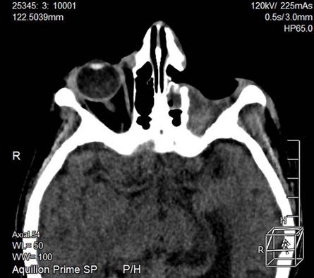
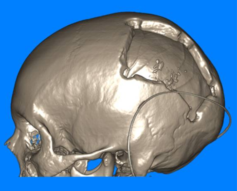
Figure 1. Post-surgical CT images of a patient with T1DM and ROCM.
Notes: CT = computed tomography; T1DM = type 1 diabetes mellitus; ROCM = rhinoorbitocerebral mucormycosis. The images correspond to the multislice CT studies performed after the surgical interventions and show the bone defect of the facial skeleton and the changes described in Table 1.
Similar to what is recommended by Cornely et al.15, management included liposomal amphotericin B at a dose of 5 mg/kg/day as the antifungal treatment of choice; combined insulin therapy (glargine and glulisine) for glycaemic control; imaging monitoring for timely surgical decisions; nutritional management and psychological support. The antifungal response was complete after 5 months of treatment (56% achieve a response within three months of starting treatment); surgical management was successful and glycaemic control has led to metabolic stability; reparative surgery of the excision area is pending, and the prognosis will depend on the course of metabolic control, the patient's immune status and psycho-emotional support.
We consider that this report will raise awareness of the need for good adherence to treatment in patients with T1DM, particularly in those with concomitant autoimmune diseases and probable psycho-emotional alterations, as well as of the early suspicion of ROCM for timely treatment and to avoid the high rate of morbidity and mortality.
Ethical criteria
The authors declare that informed consent was obtained from the patient's legal guardian for the reporting of the case, and that her privacy was respected.
Data availability
The data are recorded in the institutional clinical record and are available for review at the request of the Ethics Committee.
Conflicts of interest
The authors declare that they have no conflicts of interest.
Funding statement
The authors declare that the present report was self-funded.



Open Access By Aditum Open Access Journals id licensed under Creative Commons Attribution 4.0 International License. Based On a Work at aditum.org


