Clinical Psychology and Mental Health Care
OPEN ACCESS | Volume 7 - Issue 1 - 2025
ISSN No: 2994-0184 | Journal DOI: 10.61148/2994-0184/CPMHC


Castejón OJ1*, Carrero Gonzalez CM2, Lastre G2, Galindez P3, Salones de Castejón M3, Sierra Carrero L4
1Instituto de Investigaciones Biológicas “Drs. Orlando Castejón and Haydee Viloria de Castejón”
2Facultad de Ciencias de la Salud. Universidad Simón Bolivar. Barranquilla. Colombia.
3Instituto de Neurociencias Clínicas, Fundación Castejón, Hogar Clínica San Rafael. Maracaibo. Venezuela.
4Programa de Medicina, Universidad del Norte, Barranquilla. Colombia.
*Corresponding Author: Orlando J. Castejón, Instituto de Investigaciones Biológicas “Drs. Orlando Castejón and Haydee Viloria de Castejón”. Facultad de Medicina. Universidad del Zulia. Apdo. 526. Maracaibo. Venezuela.
Received: August 17, 2021
Accepted: September 10, 2021
Published: September 15, 2021
Citation: Castejón OJ, Carrero Gonzalez CM, Lastre G, Galindez P, Salones de Castejón M et al “Paper for Psychology and Mind health. Validating the hypothesis of blood platelets as model of serotoninergic neurons in schizophrenia and depression. A electron microscopic study”. Clinical Psychology and Mental Health Care, 3(3); DOI: http;//doi.org/03.2021/1.10048.
Copyright: © 2021 Orlando J. Castejón. This is an open access article distributed under the Creative Commons Attribution License, which permits unrestricted use, distribution, and reproduction in any medium, provided the original work is properly Cited.
The blood platelet fractions of schizophrenic and depressed patients were examined by means of electron microscopy to validate the hypothesis of blood platelets as model of serotoninergic neurons. Seventeen patients diagnosed with schizophrenia and depression according to DMS IV protocol were clinically examined. Blood platelet fractions were separated by Ingermann et al. (1978) technique and processed for transmission electron microscopy. Platelet fractions in schizophrenic patients showed activated blood platelets characterized by presence of high electron dense 5TH granules. moderately electron dense alpha granules, open and dilated canalicular system containing electron dense substance, dense and clear mitochondria, small glycogenosomes, free dispersed beta-glycogen granules, free ribosomes, pseudopods, outer surface glycocalix layer, shallow and deep invagination of limiting plasma membrane, phagocytic vacuoles, and clear endocytic and endoplasmic vesicles. On the contrary, activated and phagocytic platelets in depressed patients were found exhibiting absence of 5HT granules, disassembly of microtubules and actin filaments, depleted alpha granules, and increased membrane surface activity featured by formation of phagocytic vacuoles and endocytic vesicles. The absence of platelets 5HT granules is in agreement with clinical depressive phase of patients under study and the low concentration of biogenic amines in plasma of depressed patients. The blood platelet fraction examined from schizophrenic patients showed preservation of 5HT granules in agreement with the concentration of biogenic amine metabolites in plasma of schizophrenic patents, which validate the hypothesis of blood platelet as a good model of central serotoninergic neurons.
Introduction
Several investigators have earlier postulated the blood platelets as models of serotoninergic neurons. Detailed comparisons of the kinetic, biochemical and pharmacologic characteristics of 5-HT transport in platelets and brain support the notion that the platelet can serve as a model for 5-HT transport by central nervous system neurons [1-4]. In addition, clinical evidence of serotonin dysfunction has been sought using the blood platelet as putative model of central serotonergic neurons. The hypothesis that central biogenic amines may play a role in the pathophysiology of schizophrenia was originally based upon the fact that hallucinogenic and antipsychotic drugs have profound effects on central transmitter pathways where dopamine, noradrenaline and serotonin are involved [5].
Kinetic and pharmacologic properties of uptake of serotonin and dopamine by normal human platelets have been investigated to test whether platelets can be employed as a model system for the reuptake of serotonin and dopamine in brain [6].
Palmar et al. [7] earlier described the preliminary information about the alterations of the fine structure of blood platelet in depression to validate the hypothesis of blood platelet as model of central serotoninergic neurons using transmission electron microscopy of isolated blood platelet fraction. More recently, Castejón (2017a,b) [8,9] described in details the contribution of electron microscopy to the diagnosis of schizophrenia and bipolar disorders.
After a systematic world bibliographic review process about the contribution of electron microscopy on the morphology of blood platelets in schizophrenia and depression, we have not found additional publications in this specific subject.
In the present paper we have reexamined by means of transmission electron microscopy the blood platelet fractions of schizophrenic and depressed patients to revalidate the hypothesis of blood platelet as model of serotoninergic neurons as postulated by Da Prada et al., 1988, and Pletscher,1988. [10,11].
Material and Methods
Seventeen patients diagnosed with schizophrenia and depression according to DMS IV protocol were examined. The psychiatric study was done according with the ethical principles of Helsinki declaration for research in human beings, the Biological Institute Ethical Committee and the patient relative consent. Five millimeters of blood were obtained by venipuncture and anticoagulated on ethylenediaminetetraacetic acid (EDTA). The blood platelet fraction was immediately separated by Ingerman et al. technique [12] and primarily fixed in O.1M glutaraldehyde-buffer phosphate solution by two hours, and secondarily fixed by 1 hour in similarly buffered 1% osmium tetroxide solution. The samples were dehydrated and embedded in Epon. Ultrathin sections were obtained in MT-2B Sorvall ultramicrotome, and stained with uranyl salts and lead hydroxide. Observations were made in JEOL 100B at 80 kV. Fifty electron micrographs at intermediate magnification (20.000 to 60.000) were obtained of each case. A total 350 electron micrographs were examined, and compared with control samples as described in a previous study dealing with bipolar syndrome.
Results
Platelet submicroscopic morphology in schizophrenic patients
In schizophrenic patients the platelet fractions showed activated oval blood platelets characterized by presence of high electron dense 5TH granules and moderately electron dense alpha granules, open and closed clear dilated canalicular system, dense and clear mitochondria, glycogenosomes, free dispersed beta glycogen granules, pseudopods, outer surface glycocalix layer, shallow and deep invagination of limiting plasma membrane, phagocytic vacuoles, and clear endocytic and endoplasmic vesicles (Fig. 1).

Fig. 1: Platelet fraction from schizophrenic patient showing activated platelets characterized by emission of pseudopods (P), 5HT granules (5HT), clear and dilated open canalicular system (CS, alpha dense granules (AG), dense mitochondria (M), glycogenosomes (GG), a tick pseudopod (D )and phagocytic vacuoles (PV). The platelet microtubular ring and actin filaments are not observed suggesting cytoskeletal disassembly during centrifugation procedure.
The open canalicular system was observed as long cisterns containing a moderately electron dense substance, and partially depleted alpha granules exhibited. In addition, the dense canalicular system was observed featured by flattened and narrow cisterns. Clustered glycogen granules within membrane compartments termed glycogenosomes were observed (Fig. 2).

Fig. 2: Platelet fraction from schizophrenic patient exhibiting 5Ht granules (5HT) a discoidal platelet and a long pseudopod, dense canalicular system (S), partially depleted secretory granules (GC), clear dilated canaliculat system (CS), and glycogenosomes (GG).
Some activated and round blood platelet exhibited long and thick pseudopods and 5HT granules characterized by a clear and round vesicle containing a high electron dense nucleoid.
The phagocytic platelets also contain 5HT granules, dense mitochondria, fragments of actin filaments, phagocytic vacuoles containing dense granular substance and membrane fragments, endocytic vesicles and deep invaginations of plasma membrane (Fig. 3).
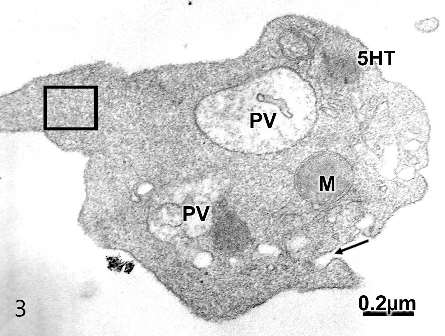
Fig. 3: Activated platelet from schizophrenic patient showing 5HT granule, disassembly of actin filaments (square), phagocytic vacuole (PV), dense mitochondrion (M), and deep invagination of limiting plasma membrane (arrow).
Phagocytic platelet exhibiting bifurcated pseudopodia engulfing plasma substance. The platelet outer surface showed released glycogenosomes and released low density glycogen granules toward the blood plasma. Large phagocytic vacuoles containing granular and fine blood plasma particles were found (Fig. 4).

Fig. 4: Phagocytic platelet exhibiting a bifurcated pseudopodia (long arrow) engulfing plasma substance and membrane fractions. The platelet outer surface shows low density glycogen granules (short arrow) released toward the blood plasma. Large phagocytic vacuoles (PV) containing granular and fine blood plasma particles are seen. Note the depleted alpha granules (AG), and low electron dense and membrane-bound glycogen granules (GG).
Degenerated blood platelet were found characterized by low electron density of cytoplasmic matrix, still remaining 5HT granules, partially depleted alpha granules, phagocytic vacuoles, and discontinuities of limiting plasma membrane. Some phagocytic leucocytes were observed engulfing the blood platelets (Fig. 5).

Fig. 5: Degenerated phagocytic platelet depicting a low electron dense cytoplasmic matrix, discontinuities of limiting plasma membrane, dense mitochondria (M), 5HT granules (5HT), phagocytic vacuole (PV), and an apparently depleted calcium body (CB).
Submicroscopic morphology of blood platelets in depressed patients
In the isolated platelet fractions of depressed patients the activated surface of platelets exhibited a number of indentations or deep invaginations that are thought to be formation of phagocytic vacuoles and endocytic vesicles. The dense canalicular system was observed as one or two flattened and narrow cisterns surrounding the mitochondria and opened to at the level of platelet outer surface. The activated and phagocytic platelets change their discoid shape and exhibit dendritic filopodia and displayed a moderate electron dense matrix and numerous pseudopodia engulfing the granular content of blood plasma. The platelets limiting plasma membrane exhibits increased surface activity revealed by the shallow and deep invaginations, corresponding to the formation of endocytic vesicles and phagocytic vacuoles (Fig. 6).
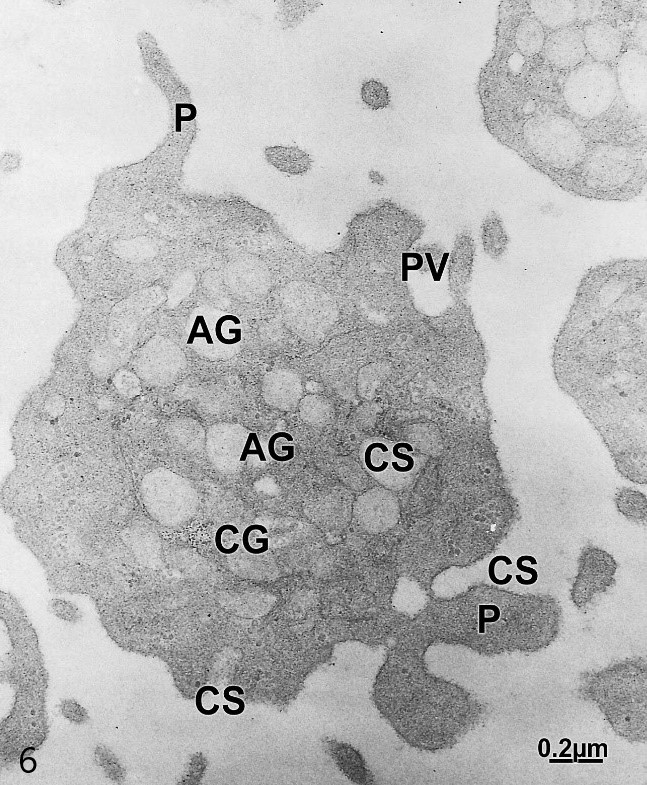
Fig. 6: Platelet fraction from depressed patient. Activated platelets are characterized by emission of dendritic and bifurcated dendritic pseudopodia (P), shallow and deep invagination of limiting plasma membrane, dilated canalicular system (CS) closed or open to the blood plasma, formation of phagocytic vacuoles (PV). Note the absence of 5HT granules and the low density alpha granules (AG) suggesting their released content.
Some highly activated blood platelets displayed numerous pseudopodic expansions phagocyting platelets fragments and undifferentiated cell organelles, and containing dense calcium granules (Figs. 7).
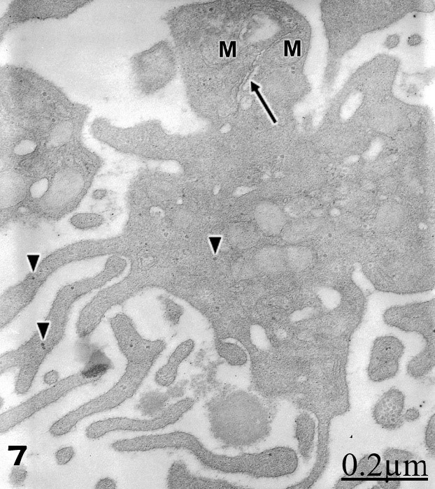
Fig. 7: Platelet form a depressed patient showimg phagocytic activity of undifferentiated processes, clear mitochondrion (M), high density tiny particles corresponding to calcium granules (triangles), segment of a clear canalicular system (arrow), mitochondria (M) and low dense alpha granules.
The dense canalicular system was observed as one or two flattened and narrow cisterns surrounding the mitochondria. In ultrathin sections the dilated canalicular system appeared in most electron micrographs as closed, round or elongated smooth surface cisterns. This canalicular system appears as a serpentine, dilated and electron lucid channel system, containing a fine granular substance and communicating the blood platelet cytoplasmic matrix and the serum blood plasma.
Most phagocytic platelets observed showed absence of 5T granules and release of glycogen granules (Fig. 8).
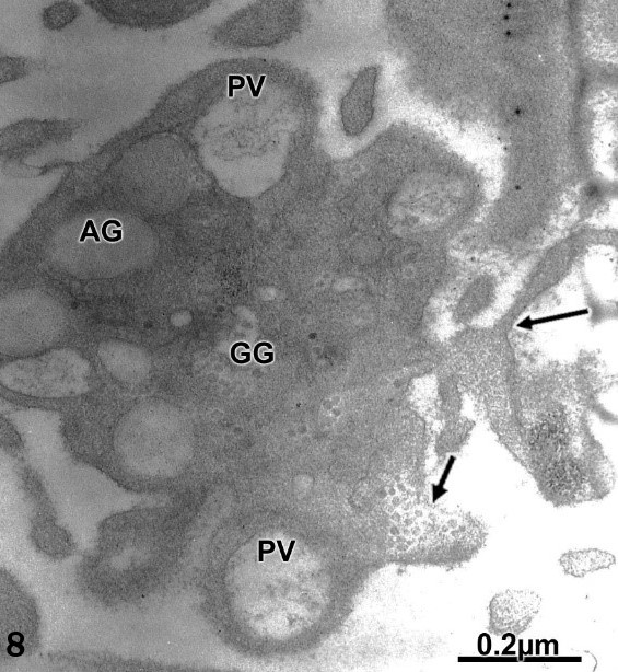
Fig. 8: Phagocytic platelet exhibiting a bifurcated pseudopodia (long arrow) engulfing plasma substance. The platelet outer surface shows low density glycogen granules (short arrow) released toward the blood plasma. Large phagocytic vacuoles (PV) containing granular and fine particles are seen. Note the depleted alpha granules (AG), and low electron and membrane-bound glycogen granules (GG).
Delta granules or 5HT granules were not observed in most platelets examined. This finding can be correlated with the clinically depressed status observed in the patients under study.
Some phagocytic platelets engulfing plasma serum content exhibit a dense granular system, presumably containing ATP, ADP, and calcium within a membrane bound compartment (Fig. 9).

Fig. 9: Phagocytic platelet cytoplasmic segment showing a vacuole containing high electron dense calcium granules (CG). Note the phagocytosis of plasma substance (PS) and cell debris.
Another phagocytic platelets contain and release high electron dense large granules within vacuoles and release them to the blood plasma. Such granules seem to correspond to engulfed lipoprotein macromolecules from the serum plasma. The platelet matrix also shows high density particles corresponding to calcium migrating toward the pseudopods. (Fig. 10).
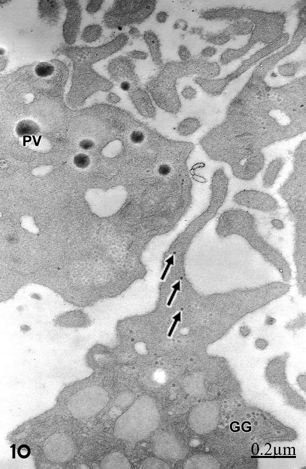
Fig. 10: Phagocytic platelet engulfing high density globular structures which appear enclosed within phagocytic vacuoles (PV) suggesting uptake of plasma lipoprotein characterized by their large size and high electron density. Another activated platelet show high density calcium granules (arrows) dispersed in the cytoplasmic matrix and migrating toward the pseudopods (arrows).
Discussion
The present paper reports the presence of 5HT granules in activated blood platelets of schizophrenic patients further revalidating by means of electron microscopy the hypothesis of blood platelet as a model of serotoninergic central neurons. It is important to emphasize that 5HT granules also were found in active and degenerated platelet. The presence of biogenic amines in schizophrenia has been also examined by different microscopy, biochemical and neuroimagen techniques. Markianos et al. (1992) [13] reported biogenic amine metabolites in plasma of drug-naive schizophrenic patents and associated with patient symptomatology. 5-HT concentration was significantly higher in schizophrenic patients than in depressed patients or in healthy controls, while it was significantly lower in depressed patients than in healthy controls or in schizophrenic patients [14]. van der Heijden et al. (2004) [15] emphasized the relevance of glutamate and serotonin in schizophrenic patients. Cai et al. (2011) [16] also found abnormalities in plasma monoamine metabolism in schizophrenic patients. A critical role for dopaminergic-serotoninergic interactions at the prefrontal cortex (PFC) level in schizophrenia patients have been reported by Bosia et al. (2011) [17]. Rassmusen et al. (2011) [18] observed decreased frontal cortical serotonin2A receptor binding in naïve first-episode schizophrenic patients. The platelet activation process seems to reorganize the platelet cytoskeleton. Actin filaments and microtubules were not well preserved by the glutaraldehyde primary fixative solution used in our electron microscopy study. A process of disassembly of both cytoskeletal structure could take place during the centrifugation for isolation of the platelet fraction. Another possibility to be considered should be the platelet activation process.
The activated surface activity of the limiting plasma, expressed by the presence of shallow and deep invaginations, endocytic vesicles, and phagocytic activity of electron dense plasma substance is probably related with the presence of serotonin, dopamine, norepinephrine levels and their metabolites in plasma of schizophrenic patients [1-6].
The dilated canalicular system containing moderate electron dense substance and showing an outer surface revealed the releasing activity of platelet granular content to the outer blood plasma as illustrated in Fig. 3.
We have herein reported increased amount of clustered and membrane bound glycogen granules termed glycogenosomes. In blood platelet glycogen platelets are considered a source of energy for most platelet function [19], mainly for the release reactions of granular systems.
The glycocalix layer observed in the platelet outer surface is thought to be composed of membrane glycoproteins, glycolipids, mucopolysaccharides, and adsorbed plasma proteins. This glycocalix layer should be further immunocytochemically studied to establish a relationship with the unifying hypothesis of schizophrenia as postulated by Kinney et al. (2010) [20].
In relationship with our study in depressed patients, platelets are usually considered as an easily accessible model of central serotonergic neurons due to similarities in enzymatic and receptor equipment, and to the fact that the uptake of serotonin into platelets closely resembles reuptake of serotonin into serotonergic central neurons [1]. Modai et al. [2] earlier reported the 5-hydroxitriptamine uptake by blood platelets of unipolar and bipolar depressed patients. The study of blood 5-hydroxytryptamine (5-HT) levels and platelet 5-HT pharmacodynamics in patients with a variety of psychiatric and neurologic disorders has generated interesting leads into possible abnormalities of CNS 5-HT neurons in these patients. [10,11]. Pivac et al. [21] also using blood platelets as a peripheral model for central serotonergic neurons suggested that platelet 5-HT concentration and monoamine oxidase (MAO) activity might serve as biological, even trait, markers for particular mental disturbances. All activated platelet examined in the study of depressed patients did not show the presence of 5HT granules, a fact correlated with the low level of plasmatic serotonin in these patients. These findings are in agreement with our preliminary report of Palmar et al. (1997) on the fine structure of blood platelet in depression [7], revealing the contribution of electron microscopy as a potential diagnostic research tool in biological psychiatry.
Some platelets examined in depressed patients showed low electron density of alpha granules indicating their release content through the openings to the platelet outer surface of canalicular system as observed in Figs. 6 and 7. Some platelet showed an increased amount of calcium granules. Intracellular Ca2+ elevation generates a cascade of events that leads to platelet activation and degranulation due to released alpha granules and 5HT granules [22].
We found phagocytic and activated platelets phagocyting high electron dense globules resembling plasma lipoprotein substance. This finding is probably related with the neuroleptic protocol applied to the patients and the changes in plasma lipid and lipoprotein levels during treatment with psychoactive drugs [23].
The platelet activation process seems to reorganize the platelet cytoskeleton. Actin filaments and microtubules were not well preserved by the sodium glutaraldehyde primary fixative buffer solution used in our electron microscopy study. A process of disassembly of both cytoskeletal structure could take place during the centrifugation method for isolation of the platelet fraction. These factor deserve further investigation to understand the relationship of damage of cytoskeleton and the pathogenesis of bipolar syndrome. The interactions with neuroleptic treatment should also be considered.
Conclusions
Platelet fractions in schizophrenic patients showed activated blood platelets characterized by presence of high electron dense 5TH granules. moderately electron dense alpha granules, open and dilated canalicular system containing electron dense substance, dense and clear mitochondria, small glycogenosomes, free dispersed beta-glycogen granules, free ribosomes, pseudopods, outer surface glycocalix layer, shallow and deep invagination of limiting plasma membrane, phagocytic vacuoles, and clear endocytic and endoplasmic vesicles. On the contrary, activated and phagocytic platelets in depressed patients were found exhibiting absence of 5HT granules, disassembly of microtubules and actin filaments, depleted alpha granules, and increased membrane surface activity featured by formation of phagocytic vacuoles and endocytic vesicles. The absence of platelets 5HT granules is in agreement with clinical depressive phase of patients under study and the low concentration of biogenic amines in plasma of depressed patients. The blood platelet fraction examined from schizophrenic patients showed preservation of 5HT granules in agreement with the concentration of biogenic amine metabolites in plasma of schizophrenic patents, which validate the hypothesis of blood platelet as a good model of central serotoninergic neurons.
Acknowledgement
This paper was carried out with the logistic and administrative support of Biological Research Institute, Faculty of Medicine. Zulia University and San Rafael Clinical Home and Castejón Foundation in Maracaibo Venezuela.

Open Access By Aditum Open Access Journals id licensed under Creative Commons Attribution 4.0 International License. Based On a Work at aditum.org