
Clinical Medical Case Reports and Case Series
OPEN ACCESS | Volume 3 - Issue 2 - 2026
ISSN No: 3065-7644 | Journal DOI: 10.61148/3065-7644/CMCRCS


Hassan Aghabararzade Omran1, Narges Mostafaloo2, Farshid Ouliaie3*
1Department of Internal Medicine, Shahid Beheshti Hospital, Babol University of Medical Sciences, Babol, Iran.
2Assistant Professor of Nephrology, Department of Internal Medicine, School of Medicine, Infectious Diseases and Tropical Medicine Research Center, Health Research Institute, Shahid Beheshti Hospital, Babol University of Medical Sciences, Babol, Iran.
3Clinical Research Development Unit of Shahid Beheshti Hospital, Babol University of Medical Sciences, Babol, Iran.
*Corresponding author: Farshid Ouliaie, Clinical Research Development Unit of Shahid Beheshti Hospital, Babol University of Medical Sciences, Babol, Iran.
Received: July 01, 2026 | Accepted: July 16, 2026 | Published: July 20, 2026
Citation: Hassan A Omran, Mostafaloo N, Ouliaie F. (2026) “Vascular wall Injury: A Life-Threatening Complication of Left Central Venous Catheterization for Dialysis: a Case Report and Review of the Literature”, Clinical Medical Case Reports and Case Series, 3(2); DOI: 10.61148/3065-7644/CMCRCS/065.
Copyright: © 2026. Farshid Ouliaie. This is an open access article distributed under the Creative Commons Attribution License, which permits unrestricted use, distribution, and reproduction in any medium, provided the original work is properly cited.
Background: One of the primary issues with hemodialysis is adequate vascular access. Central venous catheters, particularly those on the left side of the body, are linked to more serious side effects like thrombosis, venous stenosis, thoracic duct damage, pneumothorax, and catheter displacement because of their unique anatomy (longer path and steeper angle). These issues raise mortality, medical expenses, and length of stay. Because left central venous catheterization is uncommon and can have serious side effects, this report describes a rare instance of an early complication of this procedure.
Case Description: A 42-year-old man with end-stage renal disease (ESRD) receiving hemodialysis had his right subclavian percutaneous catheter infected, necessitating a catheter exchange and re-catheterization through the left subclavian vein. After catheterization, the patient had one successful dialysis session, but 24 hours after the procedure, he was unable to attend the second dialysis session due to acute symptoms of severe chest pain, palpitations, shortness of breath, and low oxygen saturation. After removal of the secondary permcath, the patient's symptoms stabilized.
Conclusion: Even though central venous catheter placement is frequently done; it is not always safe when done by inexperienced people. In our situation, though, we were certain that the catheter was in the vein, and considering the sequence of events, it seemed improbable that an infection was the cause of his decline. It was unlikely that a venous air embolism would happen this way, and no pneumothorax was discovered. Following a thorough review of the literature, we have concluded that vascular erosion brought on by the central venous catheter most likely caused the patient's decline. Vascular erosion from central venous catheters is an uncommon but dangerous side effect that can be challenging to identify and raises morbidity and death rates.
Dialysis catheter, left central vein, complication, pleural effusion
Background:
Currently, there are three main methods for vascular access in hemodialysis patients: arteriovenous fistula (AVF), arteriovenous graft (AVG), and central venous catheters (CVC)(1). An arteriovenous fistula is preferred because it is more durable and has fewer complications than other methods. However, in many patients, using central venous catheters is unavoidable due to poor vascular conditions or the need for immediate access to dialysis(2). Central venous catheters are usually placed in one of three locations: the internal jugular (preferably the right), subclavian, or femoral vein (3). The choice of catheterization site should be made with consideration of each potential complications, including infection, thrombosis, and venous stenosis(4). Catheterization in the left internal jugular vein or the left subclavian vein presents special anatomic challenges. Studies have shown that catheters placed on the left side are associated with a higher rate of life-threatening complications, including pneumothorax, thoracic duct injury, venous stenosis, and thrombosis, compared with the right side(2). This increased risk is largely due to the specific anatomy of the region, including the steeper angle of the left veins and the longer path to reach the right atrium of the heart(4). In addition, left-sided catheters may also be associated with a higher risk of catheter migration and malposition. These complications not only affect patients' quality of life but can also lead to increased length of stay, medical costs, and even mortality(2).
Given the low prevalence of left central venous catheterization and the occurrence of various life-threatening complications (both early and after years), this case report aims to present a rare case of an early life-threatening complication in the context of left central venous catheterization and reviews similar cases in this field.
Case presentation:
A 42-year-old man with a history of end-stage renal disease (ESRD) who was regularly hemodialyzed via a right subclavian percutaneous catheter presented with fever and chills during dialysis. Luminal blood culture was positive, confirming catheter-related infection. Therefore, the infected percutaneous catheter was removed and the patient was considered for placement of a new catheter in the left subclavian vein.
The procedure for placement of the new catheter was performed under ultrasound guidance and local anesthesia. Blood flow was favorable in both lumens of the catheter. The position of the catheter tip was first checked by aspirated blood gas analysis (confirmation of venous position) and then by chest CT scan. The CT scan reported the position of the catheter tip at the end of the left subclavian vein.
The patient underwent a 4-hour hemodialysis session without complications. However, approximately 24 hours after the procedure, the second dialysis session was not completed due to poor flow, and he was admitted to the hospital with acute symptoms of severe chest pain, shortness of breath, palpitations, and decreased oxygen saturation. Chest radiography revealed cardiomegaly, bilateral pleural effusions with more severity on the right side. Chest CT scan confirmed bilateral pleural effusions (left < right) and pericardial effusion. (fig.1) Differential diagnoses of mechanical catheter complication, aortic dissection, and polyserositis were proposed. Echocardiography did not show evidence of vascular flaps that would suggest rupture or dissection. With the diagnosis of a possible mechanical complication due to the catheter, the decision to remove it was immediately made. The patient was transferred to the operating room and the catheter was removed by the same primary surgeon. Immediately after catheter removal, the patient's pain, respiratory and hemodynamic symptoms improved significantly. A new temporary femoral catheter was placed to continue dialysis. The patient made a complete recovery over the next few days with continued antibiotic therapy and was discharged in good general condition. At three-month follow-ups, the patient had a stable condition and no other complications.
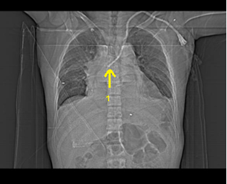
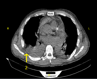
Fig.1. Spiral CT scan of the lungs without contrast shows moderate right (2) and mild left pleural effusion with passive collapse of adjacent segments. Marker 1 shows the catheter tip.
Discussion:
This rare and serious complication of central venous catheter (CVC) placement highlights the importance of close patient monitoring after catheterization in the first 24 hours. Despite being a common procedure, CVC insertion has serious side effects. Pneumothorax, trauma, and vascular perforation are the most frequently reported complications(5). These common causes were methodically ruled out in this case. Further research into more obscure mechanisms is necessary because there was no single trigger for the patient's acute deterioration, regardless of the patient's distress and pleural effusion. A thorough literature analysis shows that catheter tip erosion is likely an underlying pathology(6).
Central venous catheter vascular erosion is a rare but serious complication. Its incidence appears to be between 0.4% and 1% and can be difficult to diagnose, leading to increased morbidity and mortality(7). The proposed pathophysiology involves mechanical trauma from the catheter tip to the endothelium that causes mediastinitis. Over time, this pressure continues to cause wall necrosis and ultimately perforation. This risk is exacerbated by patient movement. Studies have shown that catheters, particularly those inserted through the internal jugular vein, can be significantly displaced by neck flexion (up to 3 cm), changing the tip angle and increasing the risk of wall injury(8). The clinical presentation in this case was consistent with published reports: the onset of symptoms was after a latent period. A high level of suspicion is necessary when a patient presents with acute pain like aortic dissection and cardiorespiratory distress with an indwelling central venous catheter(9). Catheter imaging is necessary, and once diagnosed, immediate removal of the vein is the cornerstone of management, followed by treatment such as thoracentesis according to clinical severity(10). The literature suggests that symptoms occur within the one to 60 hours after catheter insertion. This delay in symptom onset can lead clinicians to iatrogenic causes.
In previous studies, these symptoms occurred longer than in our case (48 hours or later), but in the study by Balasubramanian et al., the patient presented with symptoms similar to our case after 24 hours. This raises the importance of considering vascular erosion as an early case(11).
Symptoms also vary among patients; for example, in the study by Balasubramanian et al, these symptoms included breathlessness, bilateral pleural effusions, pericardial effusion, left-sided basal atelectasis, and emphysema(11), but in the study by PASSARO et al, the patient presented with chest pain only(12). These different symptoms, which are common to other life-threatening diseases, can mislead the physician, but we recommend that physicians consider catheter-induced vascular erosion in patients with catheter placement, especially on the left side, either as an early complication or after weeks of catheter placement(13).
An independent and well-established risk factor for erosion is left-sidedness. The catheter tip is more likely to attach to the left subclavian vein's lateral wall due to the more acute angle created by the winding path. Placing the catheter on the right side is highly advised to lower this risk(14). It is crucial to use a catheter long enough (at least 20 cm for adults) when inserting a vein on the left side to place its tip in the right atrium or subclavian vein, where there is less chance of damaging the vessel wall(15).
The following preventive actions can be taken: Aspiration assessment; Free aspiration of blood from all channels should be checked. If aspiration cannot be regarded as a risk for erosion or malposition, it is important to note that free flow does not necessarily rule out an imminent perforation; postoperative chest radiography is required to confirm tip position, which should ideally be parallel to the SVC wall within the vessel's shadow; the postural flow should not be positional; and fluctuations in aspiration with patient movement indicate an unstable tip that may be vulnerable to erosion and migration(16).
Consequently, catheter erosion is a rare but life-threatening complication of CVC placement. A high level of suspicion, rapid imaging, and immediate intervention are critical for a positive outcome. This indicates that anatomical variations and left-sided placement require greater caution, careful technique, and operative care to avoid catastrophic outcomes(17).
Table 1 compares other case reports in which catheterization caused vascular erosion:
Table.1: A review of previous articles
|
Authors |
Year of study |
age |
Gender |
Country |
Clinical signs & Symptom |
Cause |
|
S.Balasubramanian et al |
2014 |
57 |
man |
UK |
Breathlessness, bilateral pleural effusions, pericardial effusion, left-sided basal atelectasis, emphysema |
Central venous catheter vascular erosion |
|
K. QUILLEN et al(18) |
1995 |
46 |
man |
USA |
chest pain, dyspnea, pleural effusion, hemothorax |
Central venous catheter vascular erosion |
|
M.E. PASSARO et al |
1994 |
45 65 82 |
Man Man Woman |
USA |
Case1: chest pain Case2: anterior chest pain radiating down arm Case3: chest pain |
Central venous catheter vascular erosion |
|
L. BARDOSI et al(19) |
1988 |
69 |
woman |
UK |
pleural effusion, hemothorax, |
Central venous catheter vascular erosion |
Conclusion:
This report highlights the potentially life-threatening nature of the seemingly simple placement of central venous catheters. Complex vascular anatomy can lead to misplacement, even when standard protocols are followed. Clinical vigilance, rapid recognition, and immediate intervention are critical to prevent a catastrophe. This case highlights the need for very careful review of venographic images and consideration of additional confirmatory methods in equivocal cases.
Abbreviations:
ESRD end-stage renal disease
AVF arteriovenous fistula
AVG arteriovenous graft
CVC central venous catheters
SVC superior vena cava
Acknowledgements: The authors sincerely express their gratitude and appreciation for the patient's cooperation and patient consent to the publication of this report. We also express our deepest gratitude to all members of the medical and nursing team who played a role in the diagnosis and care of this patient. The authors thank and appreciate the cooperation of Clinical Research Development Unit of Shahid Beheshti Hospital of Babol.
Author contributions: HA prepared the original draft preparation. FO contributed to the investigation and revision of the manuscript. NM contributed to editing of the manuscript.
Funding: None.
Data availability: The data of this study are available from the corresponding author on
reasonable request.
Declarations:
Ethics Statement: All procedures in this study were conducted in accordance with the ethical principles outlined in the Declaration of Helsinki. Written informed consent was obtained from the patient for the publication of his medical information and images. In this form, the objectives of the study, diagnostic-therapeutic procedures, potential benefits, and risks of disclosing information were fully and simply explained to the patient, and his right to withdraw from the study at any stage without affecting his treatment was emphasized. In order to maintain the confidentiality of patient information, all personally identifiable data (such as name, exact age, location, and case number) have been removed, and none of the attached images contain patient identification information. This case report study has the code of ethics IR.MUBABOL.HRI.REC.1404.100 from the Iranian Research Ethics Organization.
Consent for publication: Written informed consent was obtained from the patient involved in this report for the publication of his personal and clinical information.
Competing of Interest: The authors have no conflicts of interest to report.


Open Access By Aditum Open Access Journals id licensed under Creative Commons Attribution 4.0 International License. Based On a Work at aditum.org


