Clinical Medical Case Reports and Case Series
OPEN ACCESS | Volume 1 - Issue 2 - 2026
ISSN No: 3065-7644 | Journal DOI: 10.61148/3065-7644/CMCRCS


A. Ibnouzaki1,2, F. Bennaoui1,2, A. Lalaoui1,2, K. Abilalaa1,2, G. Kassal1,2, N. El Idrissi Slitine1,2*, F.M.R. Maoulainine1,2
1Neonatal Intensive Care Unit, Mohammed VI University Hospital, Marrakech, Morocco.
2Laboratory of Childhood, Health and Development, Cadi Ayyad University.
*Corresponding author: N. El Idrissi Slitine, 1Neonatal Intensive Care Unit, Mohammed VI University Hospital, Marrakech, Morocco.
Received: April 05, 2026 | Accepted: April 13, 2026 | Published: April 16, 2026
Citation: A. Ibnouzaki, F. Bennaoui1, A. Lalaoui, K. Abilalaa, G. Kassal, N. El Idrissi Slitine, F.M.R. Maoulainine. (2026) “Incidental Discovery of an Amniotic Band in a Newborn: A Case Report”, Clinical Medical Case Reports and Case Series, 3(1); DOI: 10.61148/3065-7644/CMCRCS/062.
Copyright: © 2026. N. El Idrissi Slitine. This is an open access article distributed under the Creative Commons Attribution License, which permits unrestricted use, distribution, and reproduction in any medium, provided the original work is properly cited.
Amniotic band syndrome (ABS) is a rare condition, with an incidence of about 1 in 1,200 to 1 in 15,000 live births. It results from fibrous strands of the amniotic membrane that can trap fetal parts, causing congenital anomalies of varying severity. We report the case of a full-term male newborn admitted at 3 days of life for neonatal jaundice in the context of early-onset bacterial infection. Physical examination revealed a circumferential amniotic band located at the distal third of the right leg, initially without functional impairment. The clinical outcome was complicated by signs of distal vascular compression, prompting surgical intervention consisting of simple release of the band, with a favorable evolution.
Amniotic band; congenital anomaly; neonatal surgery; newborn; vascular compression
Amniotic bands, also known as amniotic band syndrome (ABS), constitute a rare condition with an estimated incidence ranging from 1 in 1,200 to 1 in 15,000 live births (1). They are characterized by fibrous strands originating from the amniotic membrane that may entrap fetal parts, leading to congenital anomalies of highly variable severity (2). Diagnosis is usually made during the antenatal period through routine morphological ultrasound examination. However, localized or less severe forms may escape prenatal detection and be identified only during neonatal clinical assessment (4). The severity of involvement depends on the anatomical location of the band, the degree of constriction, and the presence or absence of neurovascular compromise (5). This case aims to emphasize the importance of meticulous neonatal examination at birth and the need for ongoing clinical surveillance to prevent functional complications.
Case report
We report the case of a 3-day-old male newborn, born at term by vaginal delivery, with a birth weight of 1,900 g. Antenatal follow-up was insufficient, and no prenatal morphological ultrasound identified any congenital anomalies. On admission to the neonatal unit, the infant presented with jaundice. Physical examination revealed a circumferential amniotic band located at the distal third of the right leg, responsible for a cutaneous indentation without initial signs of distal ischemia or functional deficit. The remainder of the clinical examination was unremarkable. The newborn was hospitalized for management of neonatal jaundice associated with early-onset bacterial infection. Close clinical monitoring of the amniotic band was initiated. On day 14 of hospitalization, the clinical course was marked by the development of swelling of the right foot associated with delayed capillary refill, suggesting distal vascular compromise (Fig1). Surgical management was therefore indicated and consisted of simple release of the amniotic band. Postoperative evolution was favorable, with resolution of compression signs and preservation of limb function (Fig2).
Discussion
This case illustrates a localized form of amniotic band that was initially minimally symptomatic but later progressed to vascular compromise, thereby requiring surgical intervention. The pathophysiology of amniotic bands is classically explained by two main theories. The exogenous theory, which is the most widely accepted, attributes band formation to early rupture of the amnion, resulting in free-floating fibrous strands capable of constricting fetal parts. The endogenous theory proposes an intrinsic defect in embryonic development or localized vascular disruption, which may account for complex forms without identifiable amniotic rupture (2). To date, no single theory fully explains the entire clinical spectrum, suggesting that the underlying mechanisms are likely multifactorial (3). Although ABS may be suspected on prenatal ultrasound through visualization of its consequences such as limb defects or segmental anomalies the bands themselves are often difficult to detect using standard ultrasonography (4). Advanced imaging modalities, including three-dimensional ultrasound and fetal magnetic resonance imaging (MRI), have demonstrated improved diagnostic accuracy in selected cases (6). Prenatal diagnosis allows anticipation of postnatal management and, in selected cases, consideration of fetal intervention. Nevertheless, as illustrated in the present case, minor forms may remain undiagnosed, highlighting the crucial role of systematic neonatal clinical examination. Management depends primarily on the degree of constriction, functional impairment, and the presence of vascular or neurological compromise. Simple forms without complications may be managed conservatively with close monitoring, whereas the appearance of vascular or neurological compromise necessitates early surgical intervention (7). Several recent studies report favorable functional outcomes following simple release of the band, particularly when performed before irreversible damage occurs. In the present case, multidisciplinary collaboration between neonatologists and pediatric surgeons enabled timely intervention and resulted in a favorable functional outcome.
Conclusion
The incidental discovery of an amniotic band in a newborn underscores the importance of thorough and repeated clinical examination at birth. Even in the absence of initial functional impairment, close follow-up is essential to detect early signs of complications. Timely and appropriate surgical management generally leads to favorable functional outcomes.
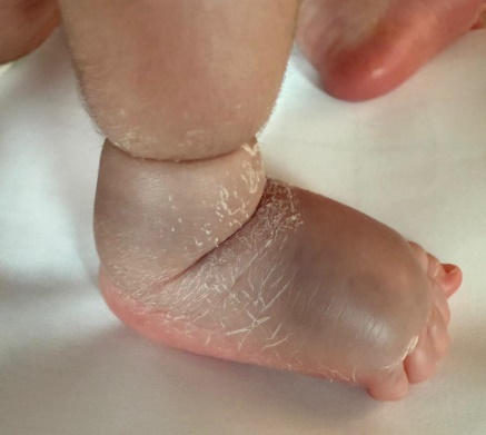
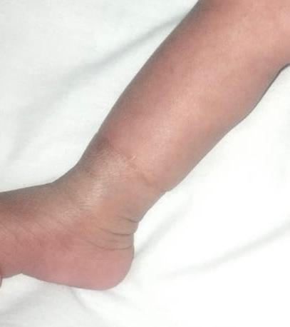
Figure 1: Complicated amniotic band syndrome Figure 2: After surgical treatment.
Consent
Informed consent for publication of their clinical details and/or clinical images was obtained from the patient’s parents.
Conflict of interest statement
None


Open Access By Aditum Open Access Journals id licensed under Creative Commons Attribution 4.0 International License. Based On a Work at aditum.org