
Clinical Case Reports and Clinical Study
OPEN ACCESS | Volume 14 - Issue 2 - 2026
ISSN No: 2766-8614 | Journal DOI: 10.61148/2766-8614/JCCRCS


Dr. GHOUZLANI IMANE*, Dr S. BENSIMIMOU, Pr M. LAHJAOUJ, Pr M. LOUDGHIRI, Pr W. BIJOU, Pr Y. OUKESSOU, Pr S. ROUADI, Pr R.L. ABADA, Pr M. ROUBAL, Pr M. MAHTAR
SERVICE D’ORL ET CHIRURGIE CERVICO-FACIALE, HOPITAL 20 AOUT CASABLANCA.
*Corresponding author: Dr. GHOUZLANI IMANE, SERVICE D’ORL ET CHIRURGIE CERVICO-FACIALE, HOPITAL 20 AOUT CASABLANCA.
Received: August 20, 2026 | Accepted: September 29, 2026 | Published: October 14, 2026
Citation: GHOUZLANI IMANE, S. BENSIMIMOU, M. LAHJAOUJ, M. LOUDGHIRI, W. BIJOU, Y. OUKESSOU, S. ROUADI, R.L. ABADA, M. ROUBAL, M. MAHTAR. (2026) “Oncocytic Carcinoma of the Parotid Gland With Extensive Parapharyngeal Extension and Suspected Cervical Nodal Involvement: A Case Report and Review of the Literature” Clinical Case Reports and Clinical Study, 13(5); DOI: 10.61148/2766-8614/JCCRCS/249.
Copyright: © 2026 GHOUZLANI IMANE. This is an open access article distributed under the Creative Commons Attribution License, which permits unrestricted use, distribution, and reproduction in any medium, provided the original work is properly cited.
Oncocytic carcinoma (OC) of the salivary glands is an exceptionally rare, high-grade malignancy that accounts for approximately 0.18% of all epithelial salivary gland tumors and around 5% of oncocytic salivary neoplasms. Its clinical, radiological, and even cytological overlap with benign oncocytic lesions frequently leads to delayed or unexpected diagnosis. We report the case of a 46-year-old man with type 2 diabetes managed with oral antidiabetic agents, who presented with a 10-month history of a slowly enlarging, painless left parotid swelling without inflammatory signs or facial nerve dysfunction. Clinical examination revealed a firm, mobile, 2 cm left parotid mass, no palpable cervical lymphadenopathy, and a homolateral oropharyngeal bulge. Contrast-enhanced cervical computed tomography (CT) and parotid magnetic resonance imaging (MRI) showed a heterogeneous, infiltrative left parotid process extending into the pre- and retrostyloid parapharyngeal space, with associated parapharyngeal and level II lymphadenopathy and mass effect on the oro- and nasopharynx; the morphological and dynamic features were considered suspicious for malignancy. The patient underwent parotidectomy, during which the tumor was removed in two fragments. Definitive histopathological examination confirmed oncocytic carcinoma. A follow-up MRI documented ipsilateral cervical lymphadenopathy, and the patient was scheduled for radical neck dissection. This case highlights the diagnostic pitfalls of OC, the importance of correlating imaging with definitive histology, and the central role of complete surgical resection with adequate nodal management and consideration of adjuvant radiotherapy.
Salivary gland carcinomas are uncommon neoplasms that make up only a small fraction of head and neck malignancies, and the parotid gland is by far their most frequent site of origin [1,2]. Population-based and institutional series confirm both the rarity and the marked histological heterogeneity of these tumors, with more than 20 recognized malignant epithelial subtypes and considerable variability in biological behavior [1,2].
Within this already scarce group, oncocytic carcinoma (OC) is one of the rarest entities. It represents approximately 5% of oncocytic salivary gland neoplasms, roughly 0.5% of epithelial salivary gland malignancies, and about 0.18% of all epithelial salivary gland tumors [4,5].
OC is defined as a proliferation of cytomorphologically malignant oncocytes—large epithelial cells with abundant granular eosinophilic cytoplasm reflecting an accumulation of mitochondria—arranged in an adenocarcinomatous, infiltrative architecture [3,5]. The terms oncocytic carcinoma, oncocytic adenocarcinoma, malignant oncocytoma, and malignant oxyphilic adenoma have historically been used interchangeably [3]. Because a purely oncocytic malignancy is difficult to define and molecularly poorly characterized, the entity has remained taxonomically contentious, and its status was revisited in the fifth edition of the World Health Organization (WHO) Classification of Head and Neck Tumours [1]. Regardless of nomenclature, the malignant nature of the lesion is established by histological criteria—cellular pleomorphism, mitotic activity, necrosis—and by infiltrative behavior including perineural, vascular, or lymphatic invasion, destruction of adjacent structures, and regional or distant metastasis [5,8].
The principal clinical challenge posed by OC is that its presentation and imaging features closely mimic those of benign oncocytic lesions such as oncocytoma and Warthin tumor, and even fine-needle aspiration cytology may be misleading [5,12]. As a result, the diagnosis is often made only on definitive histopathology after surgery. We report a case of parotid OC with extensive parapharyngeal involvement and radiologically suspicious cervical lymphadenopathy in a middle-aged diabetic man, and we review the literature on the presentation, diagnosis, and management of this uncommon malignancy.
Case Presentation
Clinical history. A 46-year-old man, known to have type 2 diabetes mellitus controlled with oral antidiabetic agents, presented to our department with a slowly progressive swelling of the left parotid region. The history of the disease dated back approximately 10 months to the appearance of a mass in the left parotid area that had gradually increased in size. There were no overlying inflammatory signs, no local pain, and no facial nerve dysfunction. The patient reported no fever, no weight loss, and no prior history of head and neck irradiation or salivary gland disease. There was no reported tobacco or alcohol dependence.
Physical examination. Inspection of the face and neck showed an asymmetry of the left parotid region, with a visible swelling filling the preauricular and retromandibular areas and effacing the retromandibular groove, without overlying skin discoloration or inflammatory changes (Figure 1). On palpation, the left parotid mass was firm, measuring approximately 2 cm at its palpable superficial component, and mobile relative to both the superficial and the deep planes. Facial nerve function was intact in all branches. There was no palpable cervical lymphadenopathy on either side of the neck. Oropharyngeal examination disclosed a homolateral (left) bulging of the lateral oropharyngeal wall/soft palate region, consistent with deep parapharyngeal extension of the parotid process (Figure 2). The remainder of the otorhinolaryngological and general examination was unremarkable.
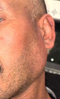
Figure 1: Preoperative clinical photograph (left lateral view) showing the swelling of the left parotid region, with fullness of the preauricular and retromandibular areas and effacement of the retromandibular groove, without inflammatory skin changes.
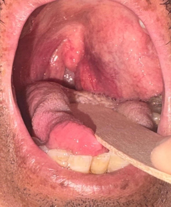
Figure 2: Preoperative intraoral photograph showing homolateral (left) bulging of the oropharyngeal wall and soft palate, reflecting deep parapharyngeal extension of the parotid tumor.
Imaging. Contrast-enhanced cervical CT (spiral, millimetric acquisition with multiplanar reconstruction) demonstrated a lesional process of the left parotid gland, spontaneously hypodense (estimated density around 15 HU), moderately and heterogeneously enhancing after contrast, invading the retro- and prestyloid space and measuring 47 × 32 mm in the axial plane with a craniocaudal extension of approximately 60 mm. A bulky left parapharyngeal adenopathy measuring 31 × 26 mm exerted a mass effect and displaced the left aspect of the naso- and oropharynx. No contralateral cervical adenopathy was identified; a few sub-centimetric nodes with a benign fatty hilum were noted. Bone-window analysis showed no aggressive osseous lesion. The overall CT impression was a suspicious left parotid process of 60 × 47 × 32 mm invading the retro- and prestyloid space, associated with parapharyngeal adenopathy and mass effect on the pharynx (Figure 3).
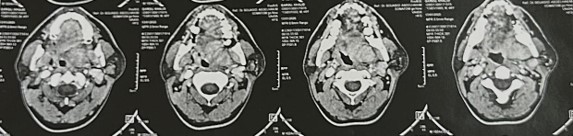
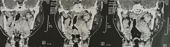
Figure 3: Contrast-enhanced cervical CT (axial and coronal reconstructions) showing the heterogeneously enhancing left parotid lesion extending into the pre- and retrostyloid parapharyngeal space, with adjacent parapharyngeal adenopathy and mass effect on the pharynx.
The difference in lesion dimensions between CT and MRI was considered in the context of different imaging modalities, acquisition planes, and delineation of the infiltrative component; the MRI measurements included the more extensive infiltrative soft-tissue component.
MRI. Parotid MRI (axial, coronal, and sagittal T1 and T2 sequences; axial T2 STIR; axial diffusion-weighted imaging; and post-gadolinium T1 sequences) confirmed an irregular, ill-defined left parotid tissue process, in mild heterogeneous T2 hyperintensity, mild T1 hypointensity, and mild diffusion hyperintensity with an intermediate apparent diffusion coefficient (ADC), enhancing heterogeneously after gadolinium and measuring 75 × 65 mm with a craniocaudal extension of 66 mm. Medially, the process infiltrated the pre- and retrostyloid space with invasion of parapharyngeal fat and exerted a marked mass effect on the soft palate and on the lateral wall of the nasopharynx and oropharynx, resulting in significant narrowing of the aerodigestive lumen. Laterally, it infiltrated the left jugal subcutaneous fat with a discrete bulging of the cheek. A left level IIA laterocervical lymph node measuring 11 mm in short axis was described. The pharynx and larynx were otherwise normal, the contralateral parotid and both submandibular glands were unremarkable, and the jugulocarotid vascular axes were free. The MRI conclusion was a locally advanced left parotid tissue process whose morphological and dynamic features were suspicious for malignancy, to be correlated with histology (Figure 4).
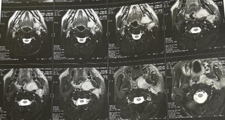
Figure 4: Parotid MRI. Representative axial T2-weighted images showing an irregular, ill-defined, heterogeneously enhancing left parotid mass infiltrating the parapharyngeal space with mass effect on the soft palate and pharyngeal wall.
Surgical management. After multidisciplinary discussion, the patient underwent parotidectomy with facial nerve identification and preservation, through a modified Blair incision with elevation of a cervicofacial flap. Because of the size of the lesion and its deep parapharyngeal extension, the tumor was delivered in two fragments (piecemeal) rather than as a single specimen. The superficial component was resected first: after dissection of its capsule from the surrounding parenchyma, it was mobilized and removed from the parotid bed, exposing the underlying operative field (Figure 5). The second, deeper component, arising from the deep lobe and extending medially into the pre- and retrostyloid parapharyngeal space, appeared as a lobulated, well-vascularized, purplish mass; it was progressively delivered from the parapharyngeal fat, with control of the adjacent vascular structures with a vessel loop and preservation of the facial nerve branches and of the jugulocarotid axis (Figure 6). Macroscopic examination of the resected specimen showed a lobulated, dark reddish-brown, fleshy mass with an irregular surface (Figure 7). Facial nerve function was assessed postoperatively.
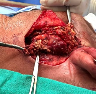
Figure 5: Intraoperative photograph after removal of the first (superficial) tumor fragment, showing the left parotid operative field with the residual deep tumor component still lying in the pre- and retrostyloid space.
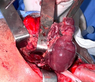
Figure 6: Intraoperative photograph showing delivery of the second, deep-lobe tumor fragment from the parapharyngeal space. The lesion appears as a lobulated, well-vascularized, purplish mass; the adjacent vascular structures are controlled with a vessel loop.
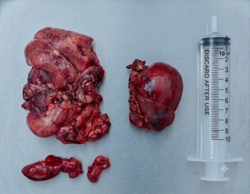
Figure 7: Gross surgical specimen after parotidectomy. The tumor was removed in two fragments; a syringe is included for scale. Note the lobulated, hemorrhagic, fleshy cut surface of the oncocytic carcinoma.
Histopathology. Definitive histopathological examination of the resected specimen concluded that the lesion was an oncocytic carcinoma of the parotid gland. The tumor was composed of large polygonal cells with abundant granular eosinophilic cytoplasm and an infiltrative growth pattern, consistent with a malignant oncocytic neoplasm. Immunohistochemistry and, where available, antimitochondrial staining are the recommended ancillary tools to confirm the oncocytic nature of the lesion and to exclude oncocytic mimics [5,11].
Postoperative course and follow-up. The immediate postoperative course was uneventful, with primary wound healing and no salivary fistula. Clinically, the parotid swelling had disappeared, the facial contour was restored, and facial symmetry was preserved at rest (Figure 8). Intraoral examination showed marked regression of the homolateral oropharyngeal bulging, with a symmetric soft palate and lateral pharyngeal wall and restoration of a normal oropharyngeal lumen (Figure 9), confirming effective removal of the deep parapharyngeal component. A follow-up MRI performed after surgery documented ipsilateral (left) cervical lymphadenopathy. In light of the radiologically suspicious for nodal involvement neck and the high-grade nature of the tumor, the patient was scheduled for a radical neck dissection, with adjuvant radiotherapy to be discussed at the multidisciplinary tumor board according to the final pathological findings.
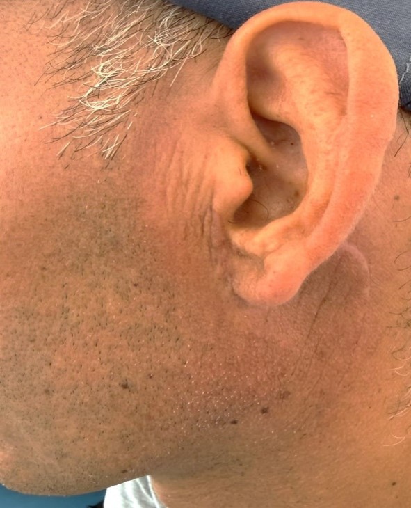
Figure 8: Postoperative clinical photograph (left lateral view) showing disappearance of the parotid swelling, restoration of a normal facial and retromandibular contour, and preserved facial symmetry at rest.
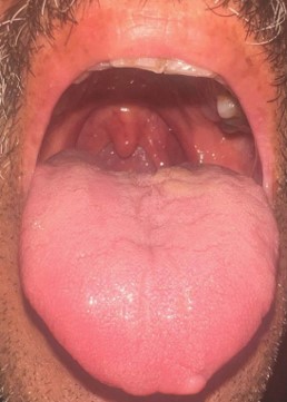
Figure 9: Postoperative intraoral photograph showing regression of the left oropharyngeal bulging, with a symmetric soft palate and lateral pharyngeal wall and a normally patent oropharyngeal lumen.
Discussion
Epidemiology and terminology
OC of the salivary glands is among the rarest malignant salivary neoplasms. Early literature reviews counted only a few dozen well-documented cases: Guclu et al. found approximately 41 reported cases at the time of their review [6], and later series continued to describe the tumor only in small numbers [9,10]. In a 16-year institutional review of parotid oncocytic neoplasms, oncocytoma was the most frequent morphology while frankly malignant OC represented less than 10% of the cohort, underscoring how uncommon the malignant variant is relative to its benign counterparts [7]. Large salivary series similarly place OC well below 1% of all salivary gland tumors [8]. The terms oncocytic carcinoma, oncocytic adenocarcinoma, malignant oncocytoma, and malignant oxyphilic adenoma have been applied to the same entity over time [3]. Because molecular studies have not identified a specific alteration defining a purely oncocytic carcinoma, and because many oncocytic malignancies actually represent oncocytic subtypes of other carcinomas, the fifth edition of the WHO Classification approached the entity cautiously, and its independent status remains debated [1].
Clinical presentation
Most reported patients with parotid OC are older adults, typically in the sixth to eighth decades, with a slight male predominance in several series [7,10]. Our patient was comparatively young at 46 years. The most common clinical picture is a painless, slowly enlarging parotid mass, exactly as observed here over a 10-month course [4,5]. Facial nerve palsy, when present, is an alarming sign that suggests an aggressive or advanced tumor; its absence, as in our patient, does not exclude malignancy [10]. A distinctive feature of the present case is the deep parapharyngeal extension producing a homolateral oropharyngeal bulge—an uncommon but important physical finding that should prompt cross-sectional imaging and raise suspicion of a deep-lobe or parapharyngeal-space tumor rather than a simple superficial parotid lesion.
Imaging features and pitfalls
Imaging cannot reliably distinguish OC from benign oncocytic lesions or from other salivary malignancies, and this is a recurring theme in the literature [14,15]. On MRI, malignant parotid tumors tend to show ill-defined margins, heterogeneous signal, and restricted or intermediate diffusion; ADC analysis has been proposed to help separate benign from malignant parotid lesions, although overlap persists and no single threshold is diagnostic [14]. Our case illustrates a classic pitfall: the CT described a relatively well-limited process, whereas the MRI—with superior soft-tissue contrast—demonstrated an irregular, ill-defined, infiltrative mass with parapharyngeal and subcutaneous fat invasion and an intermediate ADC, features that shifted the radiological impression toward malignancy. This discordance reinforces current recommendations that MRI is the reference modality for characterizing salivary tumors and for mapping deep extension, while contrast-enhanced CT of the neck and chest complements staging of nodal and distant disease [15]. Imaging nonetheless has well-recognized limitations in detecting perineural spread and microscopic infiltration, which may be underestimated preoperatively [15].
Diagnosis and histopathology
The definitive diagnosis of OC rests on histopathology. Fine-needle aspiration cytology is useful in the parotid work-up but is limited for oncocytic lesions, because malignant and benign oncocytes share abundant granular cytoplasm and cytological atypia may be subtle; misclassification is well documented [12]. Frozen section can identify an infiltrative malignant epithelial lesion but rarely provides a precise subtype [5]. On permanent sections, malignancy is established by cellular pleomorphism, increased mitoses, necrosis, and, most importantly, infiltrative growth with perineural, vascular, or lymphatic invasion and destruction of adjacent tissues [5,8]. Immunohistochemistry is an indispensable adjunct: antimitochondrial antibody staining confirms the oncocytic (mitochondria-rich) nature of the cells, while a panel including cytokeratins and proliferation markers such as Ki-67 helps grade the lesion and exclude mimics [5,9,11]. The differential diagnosis is broad and includes oncocytoma, nodular oncocytic hyperplasia, Warthin tumor, and oncocytic variants of mucoepidermoid carcinoma and salivary duct carcinoma, all of which must be excluded before a diagnosis of primary OC is accepted [1]. Metastatic oncocytic or clear-cell carcinomas (for example of renal origin) should also be considered in the appropriate clinical context [3].
Biological behavior and prognosis
OC is regarded as a high-grade malignancy with a propensity for local recurrence and for both regional and distant metastasis. Reported metastatic sites include cervical lymph nodes, lungs, kidneys, bone, and—exceptionally—the central nervous system, including a rare intradural spinal metastasis [8,13]. Nodal metastases may appear late in the disease course: Gallego et al. described cervical nodal disease confirmed four years after the initial parotidectomy, illustrating the need for prolonged follow-up [4]. In one of the larger single-institution parotid OC series, Huang et al. reported a five-year local control of approximately 67%, overall survival of about 69%, and freedom from distant metastasis around 61%, with clinical N category, local recurrence, and distant metastasis emerging as significant prognostic factors [10]. In the present case, the combination of a large size, parapharyngeal and subcutaneous fat infiltration, and radiologically suspicious cervical lymphadenopathy places the patient in a high-risk category and justifies aggressive locoregional treatment.
Management
Surgery is the cornerstone of treatment for salivary gland malignancy, and for parotid cancer the reference procedure is a parotidectomy aiming for clear margins with removal of the intraparotid lymph nodes; the facial nerve is preserved whenever it is not directly infiltrated [16,18]. In our patient the tumor was removed in two fragments because of its size and deep extension (Figures 5 and 6). Piecemeal resection is oncologically less desirable than en-bloc excision, as it may compromise margin assessment and carries a theoretical risk of tumor spillage; when it occurs, close follow-up and multidisciplinary discussion of adjuvant treatment are recommended [16]. This point is worth emphasizing transparently in any report of such a case.
Management of the neck depends on nodal status and on the risk of occult metastasis. In clinically node-positive (cN+) salivary gland cancer, ipsilateral neck dissection is recommended; in cN0 necks, elective dissection is advised except for tumors at low risk of occult spread, with the decision driven mainly by histological grade and T category [17]. Because OC is a high-grade tumor, and because our patient developed radiologically evident ipsilateral cervical adenopathy, ipsilateral neck dissection was planned in view of the radiologically suspicious cervical lymphadenopathy and the high-grade nature of the tumor. Current recommendations support addressing levels II–IV in high-grade histologies, with extension to levels I and V according to clinical involvement [17].
Adjuvant radiotherapy is recommended in the presence of adverse pathological features—high grade, positive or close margins, nodal metastasis, extranodal extension, perineural invasion, or advanced T stage—and should be discussed at a multidisciplinary tumor board in light of the final pathology [18]. Given the high-grade nature of OC, the tumor size, the parapharyngeal extension, the piecemeal resection, and the radiologically suspicious nodal disease, this patient is likely to meet several indications for postoperative radiotherapy. Systemic therapy has a limited, non-standardized role in salivary gland carcinoma, and inclusion in clinical trials is encouraged where available [18]. Because of the risk of late recurrence and distant metastasis inherent to OC, long-term structured surveillance is essential [4,10]. Finally, the patient’s diabetes, managed with oral antidiabetic agents, is relevant chiefly to perioperative optimization and wound healing rather than to tumor biology, and does not modify the oncological strategy.
Conclusions
Oncocytic carcinoma of the parotid gland is an exceptionally rare, high-grade salivary malignancy whose clinical and radiological features overlap substantially with those of benign oncocytic lesions, so that the diagnosis is frequently established only on definitive histopathology supported by immunohistochemistry. This case, in a comparatively young diabetic man, is notable for extensive parapharyngeal extension manifesting as a homolateral oropharyngeal bulge and for radiologically suspicious cervical lymphadenopathy. It underscores several practical lessons: the value of MRI in detecting infiltrative behavior that CT may miss; the need for complete, ideally en-bloc, surgical resection with careful margin control; the importance of appropriate neck management when cervical nodal involvement is suspected; and the role of adjuvant radiotherapy for high-risk features. Because oncocytic carcinoma can recur and metastasize late, prolonged multidisciplinary follow-up is mandatory.
Additional Information
Disclosures. Human subjects: Consent was obtained by all participants in this study. Conflicts of interest: In compliance with the ICMJE uniform disclosure form, all authors declare no competing financial interests. Financial relationships: All authors have declared that they have no financial relationships at present or within the previous three years with any organizations that might have an interest in the submitted work. Other relationships: All authors have declared that there are no other relationships or activities that could appear to have influenced the submitted work. Informed consent was obtained from the patient for publication of this case report and the accompanying images.



Open Access By Aditum Open Access Journals id licensed under Creative Commons Attribution 4.0 International License. Based On a Work at aditum.org


