
Clinical Case Reports and Clinical Study
OPEN ACCESS | Volume 14 - Issue 2 - 2026
ISSN No: 2766-8614 | Journal DOI: 10.61148/2766-8614/JCCRCS


I. Ghouzlani*, R. Chelly, S. Bensimimou, M. Lahjaouj, M. Loudghiri, W. Bijou, Y. Oukessou, S. Rouadi, R. L. Abada, M. Roubal, M. Mahtar
Department of Otorhinolaryngology and Head and Neck Surgery, Hôpital 20 Août 1953, Ibn Rochd University Hospital, Hassan II University, Casablanca, Morocco.
*Corresponding author: I. Ghouzlani, Department of Otorhinolaryngology and Head and Neck Surgery, Hôpital 20 Août 1953, Ibn Rochd University Hospital, Hassan II University, Casablanca, Morocco.
Received: August 20, 2026 | Accepted: September 29, 2026 | Published: October 14, 2026
Citation: I. Ghouzlani, R. Chelly, S. Bensimimou, M. Lahjaouj, M. Loudghiri, W. Bijou, Y. Oukessou, S. Rouadi, R. L. Abada, M. Roubal, M. Mahtar. (2026) “Extranodal NK/T-Cell Lymphoma, Nasal Type, Unmasked after Septoplasty in a Patient with a Fifteen-Year History of Maxillo-Mandibular Osteitis: a Case Report” Clinical Case Reports and Clinical Study, 13(5); DOI: 10.61148/2766-8614/JCCRCS/249.
Copyright: © 2026 GHOUZLANI IMANE. This is an open access article distributed under the Creative Commons Attribution License, which permits unrestricted use, distribution, and reproduction in any medium, provided the original work is properly cited.
Background. Extranodal NK/T-cell lymphoma, nasal type (ENKTL-NT) is a rare, aggressive, Epstein–Barr virus (EBV)-associated malignancy with a predilection for midfacial structures. Its early symptoms are indistinguishable from chronic inflammatory sinonasal disease, and diagnosis is characteristically delayed. We report a case in which the tumour was uncovered only after routine septal surgery, following more than a decade of treatment for presumed chronic osteitis.
Case presentation. A 45-year-old North African man had undergone two operations (2011 and 2016) for maxillary and mandibular osteitis. Facial computed tomography in May 2025 showed osteosclerosis and cortical irregularity of the alveolar and palatine processes of the left maxilla, a discontinuity of the palatine process creating an oro-nasal communication, rhizalysis of teeth 21 and 23, and infiltration of the left naso-genian soft tissues. Septoplasty with inferior turbinoplasty was performed in August 2025 for chronic nasal obstruction. Postoperatively he developed a progressively enlarging swelling of the left upper buccal vestibule with obliteration of the ipsilateral nasal vestibule. Biopsies showed a largely crushed large-cell lymphoid proliferation infiltrating the squamous mucosa and minor salivary gland tissue; immunohistochemistry demonstrated diffuse CD45, CD56 and granzyme B expression, focal CD3 positivity, and negative CD20 and AE1/AE3 cytokeratin, establishing the diagnosis of ENKTL-NT. 18F-FDG PET/CT showed a hypermetabolic naso-labial mass centred on the left nasal vestibule (SUVmax 11.26; MTV40 28 cm³) involving the cartilaginous septum, limen nasi, inferior turbinate, nasolacrimal duct and palate, with no nodal or visceral disease (stage IE; PINK score 0). Three cycles of chemotherapy followed by involved-field radiotherapy achieved complete clinical and metabolic remission. Conclusions. A chronic destructive midfacial lesion that behaves atypically — particularly one managed for years as osteitis or chronic rhinosinusitis — should prompt deep, repeated biopsies with full immunohistochemistry and EBER in situ hybridisation. Turbinate and septal specimens should be submitted routinely for histopathology. Localised disease treated with asparaginase-based chemotherapy combined with radiotherapy now carries a high complete-response rate.
Background
Extranodal NK/T-cell lymphoma, nasal type (ENKTL-NT) is a malignant proliferation of natural killer cells or, less often, cytotoxic T lymphocytes, invariably associated with Epstein–Barr virus (EBV) and recognised as a distinct entity in the fifth edition of the World Health Organization Classification of Haematolymphoid Tumours [1]. Its geographical distribution is strikingly uneven: it accounts for fewer than 1% of non-Hodgkin lymphomas in Europe and North America, compared with 3–8% in East Asia and Latin America, where it is the commonest subtype of peripheral T-cell lymphoma [2,3]. In North Africa it remains exceptional, the published experience being limited to isolated reports and small series [4–7].
The clinical onset is deceptive. Nasal obstruction, purulent rhinorrhoea, foul-smelling crusting and epistaxis are readily attributed to chronic rhinosinusitis, nasal polyposis or infection, and are treated as such for months or years [8,9]. The angiocentric and angiodestructive growth pattern subsequently produces ischaemic necrosis and progressive destruction of midline structures — septum, turbinates, hard palate, labial and genian soft tissues — the picture historically described as “lethal midline granuloma” [8,10]. At that stage the differential diagnosis includes granulomatosis with polyangiitis, invasive fungal disease, tuberculosis, cocaine-induced necrosis and odontogenic osteomyelitis [8].
We report a patient in whom ENKTL-NT was diagnosed in the immediate aftermath of a septoplasty, against a background of maxillo-mandibular osteitis operated on twice and evolving for more than a decade, and we discuss the diagnostic pitfalls, the contribution of metabolic imaging and current treatment strategies.
Case Presentation
History
A 45-year-old man, with no reported tobacco or alcohol use and no known immunosuppression, had been followed for many years for a chronic destructive process of the midface. He had been operated on twice — in 2011 and again in 2016 — in a maxillofacial surgery department for maxillary and mandibular osteitis, with transient improvement followed by local recurrence on each occasion.
Contrast-enhanced facial computed tomography performed in May 2025 showed osteosclerosis with cortical irregularity of the alveolar and palatine processes of the left maxilla, together with a discontinuity of the palatine process with corticated margins and diastasis, creating a communication between the oral mucosa and the nasal cavity. There was a periradicular granuloma of tooth 21, root exposure with rhizalysis of teeth 21 and 23, and infiltration of the overlying left naso-genian soft tissues without a demonstrable collection. The mandible was intact. Circumferential mucosal thickening of the right maxillary sinus, polypoid thickening of the left maxillary sinus and opacification of several ethmoid cells were noted. The globes, optic nerves, extraocular muscles, deep facial spaces, major salivary glands and temporomandibular joints were unremarkable, and no cervical lymph node of significant size was identified. The report concluded that the bone changes should be interpreted against previous imaging (Figure 2).
Because of disabling chronic nasal obstruction, septoplasty with inferior turbinoplasty was carried out in our department in August 2025. The resected specimens were not submitted for histopathological examination.
Postoperative course and examination
In the immediate postoperative period the patient developed a painless, non-fluctuant swelling of the left upper buccal vestibule which enlarged progressively, together with obliteration of the left nasal vestibule, persistent unilateral nasal obstruction, infiltrative swelling of the upper lip and ipsilateral epiphora. There was no fever, night sweats or significant weight loss.
On examination there was diffuse infiltration of the upper lip and of the left nasolabial fold, with effacement of the normal contours and distortion of the left nostril. Intraoral examination revealed a violaceous, infiltrative and exophytic swelling of the upper vestibule overlying teeth 21 to 23, with root exposure and a persistent oro-nasal communication. Anterior rhinoscopy showed the left nasal vestibule to be filled by a friable, crusted lesion that bled on contact. No cervical lymphadenopathy was palpable, and examination of the other nodal areas and of the abdomen was normal (Figure 1).
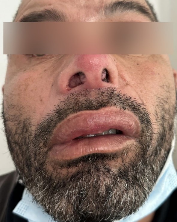
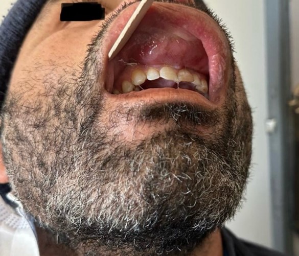
Figure 1. Clinical appearance at diagnosis. (A) Basal view showing infiltration of the upper lip and of the left naso-labial region, with an ulcerated crusted lesion filling the left nasal vestibule. (B) Intraoral view showing a violaceous exophytic swelling of the left upper buccal vestibule overlying teeth 21–23, with root exposure. The ocular region has been obscured.
Histopathology
Four biopsy fragments were taken from the vestibular swelling. Histological examination showed squamous mucosa infiltrated by a largely crushed tumoral proliferation of lymphoid appearance; in the preserved areas the cells were large, with eosinophilic cytoplasm and hyperchromatic nuclei, and mitotic figures were numerous. The stroma was scanty. Minor salivary gland parenchyma was also infiltrated by the tumour. The initial report concluded that this was a large-cell lymphoid proliferation requiring immunohistochemical characterisation.
Immunohistochemistry showed diffuse expression of CD45, CD56 and granzyme B by the tumour cells, focal CD3 positivity, and negativity for CD20 and for AE1/AE3 cytokeratin. The morphological and immunophenotypic features were consistent with extranodal NK/T-cell lymphoma, nasal type. In situ hybridisation for EBV-encoded small RNAs (EBER) could not be performed, as the technique was not available at the time of diagnosis.
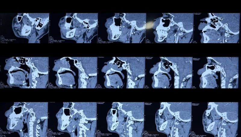
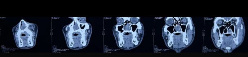
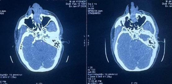
Figure 2. Contrast-enhanced facial computed tomography. (A) Sagittal reformats showing osteosclerosis and cortical irregularity of the alveolar and palatine processes of the left maxilla, with discontinuity of the palatine process. (B) Coronal reformats showing the oro-nasal communication and infiltration of the left naso-genian soft tissues. (C) Axial slices.
Staging
18F-FDG PET/CT was performed in February 2026 (213 MBq, capillary blood glucose 1.04 g/L, weight 71 kg). It demonstrated a hypermetabolic soft-tissue mass centred on the left nasal vestibule, with a maximum standardised uptake value (SUVmax) of 11.26 and a metabolic tumour volume (MTV40) of 28 cm³. Medially, the mass extended to the cartilaginous nasal septum, the left limen nasi and the inferior turbinate, with invasion of the left nasolacrimal duct. Anteriorly and inferiorly it invaded the soft tissues of the upper lip and the palate, abutting the maxillary bone without clearly demonstrable bone lysis. There was no suspicious hypermetabolism in the cervical, axillary, mediastinal or abdominopelvic nodal areas, and no pleuropulmonary, hepatic or bone marrow involvement. Bilateral adrenal hypermetabolism (SUVmax 2.81) was considered non-specific. The study concluded that there was an isolated hypermetabolic naso-labial mass infiltrating the palate (Figure 3).
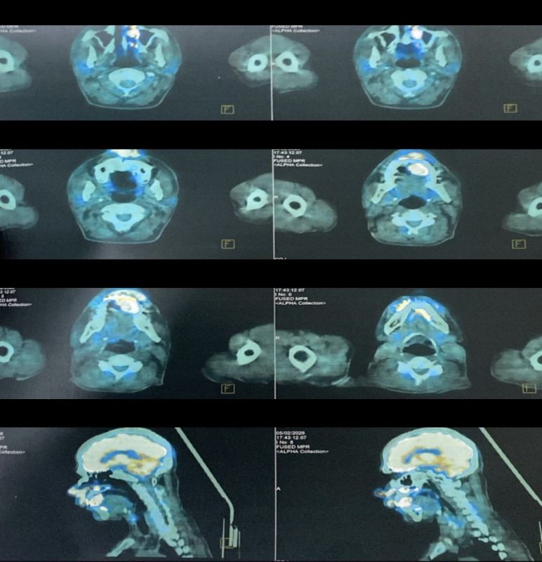 Figure 3. Baseline 18F-FDG PET/CT, fused images. Axial slices at the level of the nasal vestibule and of the oropharynx show intense uptake within the left naso-labial mass (SUVmax 11.26; MTV40 28 cm³); the sagittal reformats (bottom row) show anterior and inferior extension towards the upper lip and the hard palate, with no hypermetabolic nodal or visceral disease elsewhere.
Laboratory work-up included a full blood count, serum lactate dehydrogenase and β2-microglobulin and HIV serology. The disease was classified as Ann Arbor stage IE. The Prognostic Index of Natural Killer lymphoma (PINK) score was 0 — age under 60 years, localised stage, no distant nodal involvement and nasal location — placing the patient in the low-risk group [12].
Treatment and outcome
The case was discussed at a multidisciplinary oncology meeting. The patient received three cycles of chemotherapy followed by involved-field radiotherapy to the left nasal cavity and ipsilateral maxilla.
The labial and vestibular swelling and the nasal vestibular lesion regressed completely (Figure 4). End-of-treatment 18F-FDG PET/CT showed no residual pathological uptake, confirming complete metabolic remission. The patient remains in complete remission and is under clinical, endoscopic and metabolic imaging surveillance.
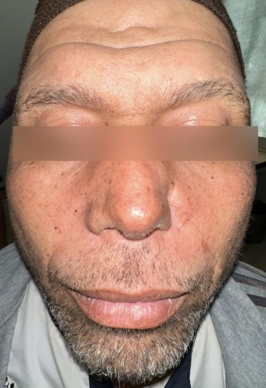
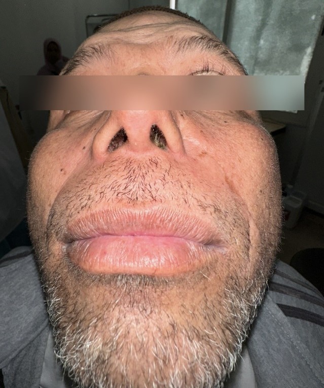
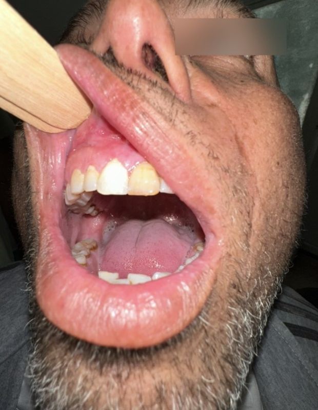
Figure 4. Clinical appearance after chemotherapy and radiotherapy. (A) Frontal and (B) basal views showing complete regression of the labial and naso-labial infiltration and of the left nasal vestibular lesion. (C) Intraoral view showing regression of the vestibular swelling, with residual root exposure of teeth 21–23. The ocular region has been obscured.
Discussion
A lymphoma concealed by a “benign” midline disease
The most instructive feature of this case is the length and the nature of the pre-diagnostic pathway. For close to fifteen years the destructive midfacial process was attributed to chronic maxillo-mandibular osteitis, prompting two operations, and subsequently to symptomatic septal deviation. Such trajectories are frequently described in ENKTL-NT: non-specific symptoms precede ulceration and local destruction by months to years, and the disease is commonly labelled chronic rhinosinusitis, fungal sinusitis, odontogenic osteomyelitis or granulomatosis with polyangiitis [8–10]. Andreou et al. emphasise that oral warning signs — palatal involvement, gingival ulceration, tooth mobility and exfoliation, and oral malodour — are often overlooked [8]. In our patient the oro-nasal communication, the rhizalysis of teeth 21 and 23 and the naso-genian soft-tissue infiltration already visible on the May 2025 CT are, in retrospect, entirely compatible with tumour infiltration, which illustrates how difficult it is to separate a chronic infective process from a lymphomatous one on imaging alone.
This confusion is not anecdotal. Maghrebian and Mediterranean series report diagnostic delays of several months, usually after repeated courses of antibiotics or anti-inflammatory drugs and, in some cases, after a first non-diagnostic biopsy [4–7]. In a series of 15 patients, Tababi et al. stressed the need to repeat sampling in any destructive sinonasal lesion of undetermined cause [6].
Presentation after septal surgery
The postoperative onset deserves comment. The vestibular swelling appeared in the immediate aftermath of septoplasty with turbinoplasty, which initially suggested an infective or inflammatory complication of the procedure. Two non-exclusive hypotheses may be advanced: that the disease was already present but subclinical at the time of surgery and simply continued its natural course; or that tumour growth was accelerated by surgical trauma and the accompanying local inflammatory response. Either way, this observation argues for the routine histopathological examination of every turbinectomy and septal resection specimen — particularly when the mucosa appears macroscopically unusual — and for prompt re-evaluation of any postoperative swelling with an atypical course. In our patient the surgical specimens were discarded, and an opportunity for earlier diagnosis was probably lost.
Histological pitfalls and the role of EBER
The histological diagnosis of ENKTL-NT is notoriously difficult. Extensive ischaemic necrosis related to angiodestruction, frequent crush artefact in small specimens and an abundant reactive inflammatory infiltrate account for the high proportion of non-diagnostic biopsies [8,10]. In our case the proliferation was described as “largely crushed”, and the diagnosis rested on the few preserved areas. Deep, multiple, adequately sized biopsies taken from non-necrotic tissue are therefore essential.
The immunophenotype observed here — CD45+, CD56+, granzyme B+, focal CD3+, CD20−, AE1/AE3− — is characteristic. CD3 positivity on paraffin sections usually reflects cytoplasmic CD3ε, the surface chain being absent from NK cells; negative CD20 and negative cytokeratins exclude B-cell lymphoma and undifferentiated carcinoma respectively, the principal differential diagnoses at this site [1,3]. Demonstration of EBV by EBER in situ hybridisation nevertheless remains the reference criterion required by the 2022 WHO classification, and its absence is the main limitation of our report [1,3]. Access to this technique should be regarded as a priority in any centre managing sinonasal tumours.
Staging and the contribution of metabolic imaging
18F-FDG PET/CT is now the reference investigation for initial staging and response assessment in ENKTL-NT, whose lesions are consistently FDG-avid [3,11]. In our patient it delineated local extension — cartilaginous septum, limen nasi, inferior turbinate, nasolacrimal duct and palate — and, more importantly, established that the disease was strictly localised, with no nodal or visceral hypermetabolism. This directly determined management, since radiotherapy is central to the treatment of localised disease [11,17]. The prognostic value of baseline metabolic parameters, in particular SUVmax and metabolic tumour volume, has been reported by several groups, with higher SUVmax associated with shorter survival, although these data remain heterogeneous and no consensus threshold exists [3].
Prognostic assessment
The outlook in ENKTL-NT was long regarded as dismal, with median survival under one year in older series treated with CHOP-like regimens, largely because tumour cells express P-glycoprotein and are therefore resistant to anthracyclines [3,11]. Several prognostic models have been proposed: the Korean model of Lee et al., combining B symptoms, stage, lactate dehydrogenase and regional nodal involvement [13]; and the PINK score, developed in the era of non-anthracycline-based therapy and based on age over 60 years, stage III/IV disease, distant nodal involvement and extranasal location [12]. Extranasal disease in particular carries a significantly worse prognosis than the classical nasal form [14]. Our patient combined favourable features — age under 60, stage IE, nasal location and no nodal involvement — giving a PINK score of 0 and low-risk classification, consistent with the complete remission achieved.
Treatment
Treatment of localised disease now rests on non-anthracycline chemotherapy containing L-asparaginase or pegaspargase combined with high-dose involved-field radiotherapy, generally 50–56 Gy [11,17,18]. Three combination strategies have been evaluated: radiotherapy followed by chemotherapy, concurrent chemoradiotherapy, and the so-called sandwich approach in which radiotherapy is bracketed by chemotherapy cycles [11]. The GELOX regimen (gemcitabine, L-asparaginase, oxaliplatin) sandwiched with 56 Gy achieved a complete response rate of 74% and an overall response rate of 96%, with 5-year overall survival of 85% in the updated analysis [11,15]. Substituting pegaspargase for L-asparaginase (P-GEMOX) simplifies administration and improves immunological tolerance [11]. Concurrent chemoradiotherapy combining 50 Gy with pegaspargase achieved a 90% complete response rate at the end of the concurrent phase and 100% at the end of treatment in a two-centre phase II study [16]. Kwong et al. showed that sequential and concurrent strategies yielded comparable survival [17].
In our patient, three cycles of chemotherapy followed by radiotherapy conform to this combined-modality principle and produced complete clinical and metabolic remission. Surveillance should be intensive during the first two years, when most relapses occur, and should combine clinical examination, nasal endoscopy, metabolic imaging and — where available — quantification of circulating EBV DNA, whose kinetics provide a sensitive marker of tumour burden and of relapse [3,18].
Conclusions
ENKTL-NT remains a difficult diagnosis, and delay is the rule rather than the exception. Our case illustrates a particularly misleading presentation, in which a destructive midfacial lesion was managed for more than a decade as chronic maxillo-mandibular osteitis, the diagnosis being reached only when a vestibular swelling appeared after septoplasty. Any chronic, destructive or ulcero-proliferative midline facial lesion that fails to respond to conventional treatment should lead to deep and repeated biopsies with complete immunohistochemistry and testing for EBV, and turbinate and septal specimens should be submitted routinely for histopathology. Staging with PET/CT and the combination of asparaginase-based chemotherapy with involved-field radiotherapy now yield high complete-response rates in localised disease, as illustrated by the favourable outcome in our patient.
Declarations
Ethics approval and consent to participate. [Complete according to journal requirements.]
Consent for publication. Written informed consent was obtained from the patient for publication of this case report and of the accompanying clinical and radiological images. All identifying data have been removed. A copy of the consent form is available for review by the Editor-in-Chief of this journal.
Availability of data and materials. All data generated or analysed during this study are included in this published article.
Competing interests. The authors declare that they have no competing interests.
Funding. No specific funding was received for this work.
Authors' contributions. IG managed the patient, collected the data and drafted the manuscript. RC and SB participated in the management of the patient and in data collection. ML, ML, WB, YO, SR, RLA and MR critically revised the manuscript for important intellectual content. MM supervised the work and gave final approval. All authors read and approved the final manuscript.
Acknowledgements. The authors thank the Departments of Pathology, Radiology, Nuclear Medicine and Haematology–Oncology of Ibn Rochd University Hospital for their collaboration.



Open Access By Aditum Open Access Journals id licensed under Creative Commons Attribution 4.0 International License. Based On a Work at aditum.org


