
Clinical Case Reports and Clinical Study
OPEN ACCESS | Volume 13 - Issue 4 - 2026
ISSN No: 2766-8614 | Journal DOI: 10.61148/2766-8614/JCCRCS


Yonas Kinfemichael Gebre
Alatyon General Hospital Hawassa, Ethiopia.
Corresponding author: Yonas Kinfemichael Gebre, Alatyon General Hospital Hawassa, Ethiopia.
Received: September 05, 2026 | Accepted: September 18, 2026 | Published: September 28, 2026
Citation: Yonas K Gebre. (2026) “Sheehan Syndrome in Ethiopia: A Case Series Highlighting Diverse Clinical Presentations and Diagnostic Challenges” Clinical Case Reports and Clinical Study, 13(5); DOI: 10.61148/2766-8614/JCCRCS/252.
Copyright: © 2026 Yonas Kinfemichael Gebre. This is an open access article distributed under the Creative Commons Attribution License, which permits unrestricted use, distribution, and reproduction in any medium, provided the original work is properly cited.
Sheehan syndrome is a rare hypopituitarism, develops as a result of ischemic pituitary necrosis due to severe postpartum hemorrhage. SS is a very significant cause of maternal morbidity and mortality in developing countries and manifestation varies based on the affected hormones.
We report three cases of Sheehan syndrome in women aged 35, 25 and 35 years who presented to our hospital with variable clinical manifestations of hypopituitarism. All three patients were from the southern part of Ethiopia and had a history of severe postpartum hemorrhage following their most recent deliveries.
Case 1: A 35-year-old woman presented 5 months after a twin delivery with features suggestive of chronic hypopituitarism. Her pregnancy and delivery had been complicated by massive postpartum hemorrhage requiring blood transfusion. Following delivery, she was unable to breastfeed and subsequently developed amenorrhea. Clinical examination revealed features suggestive of anterior pituitary hormone deficiency, including loss of axillary and pubic hair. Hormonal evaluation demonstrated deficiencies involving multiple pituitary axes. Pituitary MRI revealed a partially empty sella, supporting the diagnosis of Sheehan syndrome. She was started on appropriate hormonal replacement therapy, with subsequent clinical improvement.
Case 2: A 25-year-old woman presented 11 months after her last delivery with progressive symptoms of adrenal insufficiency. She initially experienced asthenia, anorexia, and weight loss, followed by dizziness, nausea, persistent vomiting, and abdominal pain. Despite repeatedly seeking medical care, the underlying diagnosis was not initially recognized. On presentation to our hospital, she developed bradycardia and persistent hypotension despite fluid resuscitation and vasopressor therapy. Further evaluation and review of her obstetric history revealed a previous episode of severe postpartum hemorrhage requiring blood transfusion. Hormonal evaluation demonstrated multiple pituitary hormone deficiencies consistent with Sheehan syndrome. Pituitary MRI showed an empty sella. Following glucocorticoid and thyroid hormone replacement, her hemodynamic status improved markedly.
Case 3: A 35-year-old woman was diagnosed with Sheehan syndrome 1 year after her last delivery, which had been complicated by massive postpartum hemorrhage. She presented with features of chronic hypopituitarism, including failure of lactation and amenorrhea, and was found to have profound hyponatremia on laboratory evaluation. Additional hormonal investigations demonstrated deficiencies of multiple anterior pituitary hormones. Pituitary MRI revealed an empty sella, supporting the diagnosis. She was treated with intravenous 0.9% normal saline and oral salt supplementation for severe hyponatremia, together with appropriate hormonal replacement therapy. Her clinical condition and electrolyte abnormalities subsequently improved.
Across all three cases, the diagnosis was supported by the combination of a history of severe postpartum hemorrhage, clinical features of hypopituitarism, hormonal abnormalities, and characteristic pituitary MRI findings. The interval between the last delivery and diagnosis ranged from 5 months to 1 year.
Sheehan syndrome remains an important but often underdiagnosed cause of panhypopituitarism in resource-limited settings. A history of postpartum hemorrhage should prompt a high index of suspicion when women present with nonspecific symptoms. Early clinical recognition, appropriate hormonal evaluation, brain MRI, and timely treatment are essential to prevent life-threatening complications, particularly adrenal crisis. Patient education and increased awareness among primary care physicians are crucial for early diagnosis and referral.
Sheehan syndrome
2. Background
Sheehan's syndrome (SS) is characterized by various degrees of hypopituitarism, and develops as a result of ischemic pituitary necrosis due to severe postpartum hemorrhage. [1] SS is a very significant cause of maternal morbidity and mortality in developing countries although it is a rarity in developed countries in which obstetrical care has been improved. According to a large retrospective study, the current incidence in the developed world is 5.1 per 100,000 women [2] [3]. However, it continues to be reported widely in undeveloped or developing nations where massive postpartum hemorrhage is still a common occurrence. [4].
The pituitary gland undergoes physiological enlargement during pregnancy, with a physiological increase in lactotroph cell mass and hypertrophy of its anterior portion, as determined by autopsies and magnetic resonance imaging (MRI) [5] [6]. Increased pituitary volume, small sella size, disseminated intravascular coagulation and autoimmunity are the proposed factors in the pathogenesis of SS. [7] Sheehan’s syndrome can present during the postpartum period or several months or years following delivery. A long delay in arriving at a diagnosis is not uncommon in SS: study in Kashmir India, the interval between the start of symptoms and ultimate diagnosis ranged between one week and 20 years (mean 5.94 ±5.05, median 4 years). [8]
The syndrome manifested with clinical feature of hypopituitarism such as lactation failure, amenorrhea, genital and axillary hair loss, asthenia, sign of premature aging, and dry skin. It can also presented as an emergency condition with circulatory failure, severe Hyponatremia, diabetic insipidus, Hypoglycemia and congestive heart failure. [9]
3. Case Presentation
Case 1: Chronic Sheehan syndrome after twin Delivery
A 35-year-old woman from Southern Ethiopia, Oromia region, presented with a complaint of vomiting of ingested matter, loss of appetite, significant weight loss of 15 kg, and generalized fatigue of 5 months' duration. In association, she also has decreased libido, progressive loss of axillary and pubic hair, failure to lactate, and amenorrhea. In addition, she has hoarseness of voice, headache, and constipation.
She was a para III mother whose most recent pregnancy, 6 months before presentation, resulted in the vaginal delivery of twins. The postpartum period was complicated by massive postpartum hemorrhage, requiring transfusion of 5 units of whole blood. She remained hospitalized for one month before being discharged in stable condition.
Otherwise, no blurring of vision or diplopia. She had no history of head trauma, surgery, or irradiation, and no history of polyuria or polydipsia.
On physical examination, she was chronically sick-looking and emaciated. Her blood pressure was 115/85 mmHg, pulse rate 82 beats per minute, respiratory rate 20 breaths per minute, and temperature 36.7 °C; oxygen saturation was 96% on atmospheric air. Her weight was 46kg.
She had dry, coarse skin and sparse axillary and pubic hair. Examination of other systems was unremarkable.
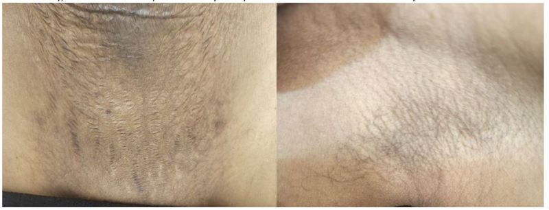
Figure 1: showing sparse axillary and Pubic Hair
Laboratory findings showed the following: white blood cell count 5000 cells/μL, hematocrit 41.3%, creatinine 0.8 mg/dl, normal liver function tests, sodium 140 mEq/l, potassium 4.12 mEq/L, random blood sugar 97 mg/dL, Estradiol 15.52 Mui/ml, FSH 3.955 Mui/ml, LH 2.786 Mui/ml, Prolactin 3.788 Mui/ml, free T3 1.804 µIU/mL, Free T4 7.14 Mui/ml, TSH 1.036, Serum cortisol 9.3 µIU. Shown on table 1 with respective reference ranges.
Table 1 Laboratory findings of the patient’s hormone profile at presentation
|
Plasma Hormones |
Patient’s Value |
Reference Value |
|
: |
1.036 µIU/mL |
0.3-45 µIU/mL |
|
Plasma Cortisol |
9.3 µIU/mL |
25-350 µIU/mL |
|
Free Thyroxine 3 |
1.804 pmol/L |
2-4.1 pmol/L |
|
Free Thyroxine 4 |
7.14 pmol/L |
9-17.5 pmol/L |
|
Estradiol (mui/ml) |
15.52 mui/ML |
Women ovulation peak 18-147Mui/l |
|
Prolactin(mui/ml) |
3.788 ng/ml |
Women of Menstrual cycle 5-35 ng/ml |
|
FSH(mui/ml) |
3.955 mUI/ml |
Women ovulation peak 6.3-24 mUI/l |
|
LH(mui/ml) |
2.78 mUI/ml |
1–12 mIU/mL follicular phase |
Brain magnetic resonance imaging (MRI) revealed decreased pituitary volume with a concave superior contour related to previous peripartal apoplexy (Figure 2).
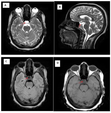
Figure 2: Brain magnetic resonance imaging shows an empty Sella.
A, T2-weighted Axial B, T2 Sagittal C, FLAIR Axial D, T1 axial (The arrow indicates The pituitary gland volume has decreased and has concave superior surface with the visualized parenchyma T2W hyper intense, T1W isointense avid post contrast enhancement.)
As the clinical manifestations related to the patient’s previous obstetric history suggested Sheehan’s syndrome, relevant available hormone studies were performed. Show on the table 1 above.
She was started on Levothyroxine 100mcg per day, Prednisolone 5mg per day and Oral contraceptive Pills (COC) which are going to be titrated based on clinical response on her subsequent follow up.
The patient has shown satisfactory progress, with no new episodes of decompensation at 3 months after hospital discharge, vomiting and fatigue subsided ;her appetite had returned and has 7kg weight gain, a good quality of life and improved libido.
Case 2: Chronic Sheehan Syndrome with Imminent Adrenal crisis
A 25-year-old lady from the southernmost part of Ethiopia presented with a complaint of vomiting of ingested matter for 11 months associated with epigastric burning sensation, loss of appetite, and easy fatigability and progressive weight loss.
She had repeatedly visited a nearby clinic and was managed for dyspepsia and gastroenteritis, took several courses of antibiotics but with no lasting improvement. She was told to have gallstone disease and was scheduled for surgery.
On Physical examination, she looks acutely sick on a chronic basis and sleepy with BP-80/60mmHg, PR- 50-54 beats/min, RR-14/min PSO2-94% on Atmospheric air, 36 °C and weight was 47kg. She had a puffy face and mild epigastric and periumbilical tenderness. Other systems didn’t have remarkable findings. . On search for causes of decreased mentation, she was also found to have low blood sugar level to the level below the recording limit of point of care glucometer. She was given 3 vial of 40% dextrose IV push and putted on 10% dextrose infusion. She had records of leukopenia as determined on 1st day and 3rd days of admission which improved on 6th day of antibiotic therapy. Other laboratory tests are shown in the table below
Table 2: Laboratory Test results
|
Date |
CBC |
Renal Function Tests(mg/dl) |
Liver Enzymes(Iu/l) |
Electrolyte(mmol/l) |
CRP |
RBS and Hgb A1c |
|
1st day of admission |
WBC-2.8 Hgb-8.6 Plt-226 |
Cr-0.84 Urea-4.81 |
AST-41.16 ALT-15.79 ALP-108.9 |
K-3.3 Na-140.6 Cl- 106.3 Ca+2-1.03 |
|
41.15mg/dl 5.8%
|
|
2nd Day of admission |
|
|
|
K+-2.89 Na+-143.5 Cl- 109.3 Ca-0.98 |
|
|
|
3rd Day of admission |
WBC-2.8 Hgb-10.2 Plt-284 |
Cr- 1.15 Urea- 7.62 |
|
K+-3.39 Na+-146.7 Cl- 111.8 Ca-1.03 |
45.8 |
|
|
5th Day of admission |
|
|
|
|
|
|
|
6th Day of admission
|
WBC-17.8 Hbg-10.3 Plt-234 |
Cr- 0.94 Urea- 8.39 |
AST-180.2 ALT-59.2 ALP-149.2 |
K+-3.86 Na+-148.3 Cl- 111.5 Ca-0.92 |
|
|
Echocardiography performed on the second day of admission revealed Normal Echocardiograph study with ejection fraction of 64%; however, marked bradycardia with a heart rate of 32 beats/min was noted during the examination. Abdominopelvic ultrasonography performed on the day of admission demonstrated long-segment wall thickening of the descending colon and rectum, more prominent in the rectum. Chest and abdominopelvic CT scans done on second day to clarify causes of bowel wall thickening demonstrated findings consistent with colitis.(showed on the Figure 3)
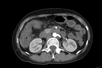
Figure 3: Abdominal Ct scan Image showing diffuse bowel thickening
Despite intravenous fluid resuscitation and blood transfusion, the patient remained persistently hypotensive. Vasopressor support with norepinephrine was initiated at 0.1 μg/kg/min and gradually escalated to 0.3 μg/kg/min, at which point her blood pressure stabilized within the normotensive range. For the above given the clinical suspicion of sepsis with a gastrointestinal source was intervened, empiric antimicrobial therapy with Meropenem 1 g intravenously every 8 hours was commenced.
Repeat ultrasonography after 4 days of oral vancomycin with an impression of C. difficile associated diarrhea (short of C.diff test assay) and intravenous meropenem showed resolution of bowel wall thickening and there which after which ileocolonoscopic exam was reserved for financial reason. Upper gastrointestinal endoscopy revealed a few erosions in the gastric antrum, which didn’t fully explain the patient's whole clinical presentation
However, despite appropriate antimicrobial treatment and hemodynamic support, the patient remained vasopressor-dependent. Concurrent hypokalemia was corrected with intravenous potassium chloride supplementation. The patient was given a stress dose of intravenous hydrocortisone (50 mg/6 h).
Later on a thorough re-evaluation, including a more detailed obstetric history revealed that symptoms occurred following delivery of a 3000 g male neonate, which was complicated by excessive bleeding and required blood transfusion. After delivery, she didn’t breastfeed and never had menses. She also complains of loss of sexual libido and amenorrhea.
The detailed clinical reevaluation and hormonal assay (showed on the table 3) uncovered a previously overlooked diagnosis that explained the patient's refractory shock. Additionally, Brain MRI was done and revealed the partially empty sella. (Showed on figure 4)
Based on the revised clinical impression which Indicates Chronic Sheehan syndrome with imminent adrenal crisis, levothyroxine and estrogen containing oral pill was added as she was already started on hydrocortisone for refractory shock.
Table 3: Laboratory Hormones Profile of case 2 patient
|
Hormones |
Patient's Value |
Reference Value |
|
Thyroid-Stimulating Hormone (TSH) |
1.412 µIU/mL |
0.30–4.50 µIU/mL |
|
Plasma Cortisol |
112 nmol/L* |
138–690 nmol/L (morning) |
|
Free Thyroxine 4 (FT4) |
6.12 pmol/L |
9.0–17.5 pmol/L |
|
Prolactin |
0.465 ng/mL |
Women (menstrual cycle): 5–35 ng/mL |
|
Follicle-Stimulating Hormone (FSH) |
0.721 mIU/mL |
Women (ovulation peak): 6.3–24.0 mIU/mL |
|
Luteinizing Hormone (LH) |
0.157 mIU/mL |
Women (follicular phase): 1–12 mIU/mL |
Following hormone replacement therapy, the patient demonstrated remarkable clinical improvement, with progressive stabilization of blood pressure, resolution of Bradycardia, and successful reduction of vasopressor requirements, supporting the endocrine origin of her hemodynamic instability.
Finally, she was strictly instructed on the nature of her illness and was given started on prednisolone 5 mg in the morning and levothyroxine 75 μg/day to be titrated based on clinical response.
On subsequent clinical assessment which was after 2-month, abdominal pain, nausea, vomiting, and diarrhea had subsided; her appetite had returned and she has weight gain 5kg to become 52kg.
Her Vital signs were BP 100/60mmHg PR 91 beats per minutes and Unremarkable Physical examinations. On investigations, WBC-5300cell/ μl, Hgb-12.9g/dl Plt-239 x 103cell/ μl, RBS- 72mg/dl, Liver enzymes were with in normal range and Creatinine 0.73mg/dl
Electrolytes were Normal (Na+-143mmol/l K+-3.53mmol/l), Ca++-1.09mmol/l Cl—106.8mmol/l)
Table 4 Laboratory Hormones Profile of case 2 patient on her 2nd visit
|
Hormones |
Patient's Value 2nd time |
Reference Value |
|
Thyroid-Stimulating Hormone (TSH) |
0.78 µIU/mL |
0.30–4.50 µIU/mL |
|
Plasma Cortisol |
35.7 nmol/L |
25–350 nmol/L (morning) |
|
Free Thyroxine 4 (FT4) |
18.9 pmol/L |
9.0–17.5 pmol/L |
|
Prolactin |
1.643 ng/mL |
Women (menstrual cycle): 5–35 ng/mL |
|
Follicle-Stimulating Hormone (FSH) |
0.030 mIU/mL |
Women (ovulation peak): 6.3–24.0 mIU/mL |
|
Luteinizing Hormone (LH) |
0.030 mIU/mL |
Women (follicular phase): 1–12 mIU/mL |
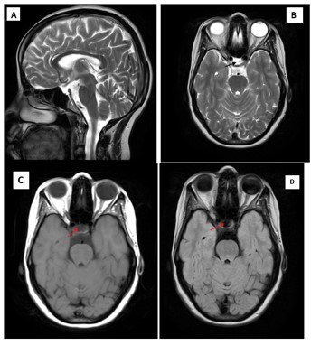
Figure 4 Brain magnetic resonance imaging shows an empty Sella.
A, T2-weighted Sagittal B, T2-weighted Axial C, T1-weighted Axial D, FLAIR Axial (The arrows showed Partially empty sella)
Case 3: Chronic Sheehan Syndrome with Profound Hyponatremia
She was a 35 years old Para VII mother who came from Southeast Ethiopia. She presented with abdominal cramp, multiple episodes of vomiting of ingested matter, throat pain and intermittent mucoid diarrhea of 5 days duration. She has been having intermittent headache, light headedness, fatigability, blurring of vision, loss of libido, progressive weight loss and amenorrhea over the past 1 year.
She had a delivery complicated by bleeding at which time she was transfused with 2 units of blood at local hospital and has not breast fed her infant.
On examination, her vital signs were as follows: blood pressure 100/70 mmHg, pulse rate 60 beats/min, respiratory rate 20 breaths/min, and temperature 36.7°C.
Head and neck examination revealed periorbital puffiness. Examination of the integumentary system demonstrated sparse axillary and pubic hair. The remainder of the physical examination was unremarkable.
Initial laboratory investigations revealed a white blood cell count of 4,500 cells/μL, hematocrit of 31.7%, and a platelet count of 224 × 10⁹/L. The serum creatinine was 1.05 mg/dL, and liver function tests were within normal limits. Random blood glucose was 89 mg/dL.
Electrolyte analysis demonstrated profound hyponatremia with a serum sodium level of 111.1 mEq/L and mild hypokalemia with a potassium level of 3.46 mEq/L.
Hormonal evaluation revealed estradiol 15.52 mIU/mL, FSH 4.47 mIU/mL, LH 3.35 mIU/mL, and prolactin 0.86 mIU/mL. Thyroid function tests showed free T3 of 1.827 pmol/L, free T4 of 6.7 pmol/L, and TSH of 2.4 mIU/L. Serum cortisol was 7.5 µg/dL. Brain magnetic resonance imaging (MRI) revealed an empty sella turcica
Table 5 Laboratory hormones result of Case 3 Patient
|
Plasma Hormone |
Patient's Value |
Reference Value* |
|
Thyroid-Stimulating Hormone (TSH) |
2.4 µIU/mL |
0.3-4.5µIU/mL |
|
Serum Cortisol |
7.5 µg/dL |
25-350 µIU/Ml Morning |
|
Free Triiodothyronine (FT3) |
1.827 pmol/L |
2 – 4.1 pmol/L |
|
Free Thyroxine (FT4) |
6.7 pmol/L |
9-17.5 pmol/L |
|
Estradiol |
15.52 mIU/mL |
Women ovulation peak 18-147Mui/l |
|
Prolactin |
0.868 ng/mL |
Women of Menstrual cycle 5-35 ng/ml |
|
Follicle-Stimulating Hormone (FSH) |
4.47 mIU/mL |
Women ovulation peak 6.3-24 mUI/l |
|
Luteinizing Hormone (LH) |
3.35 mIU/mL |
1–12 mIU/mL follicular phase |
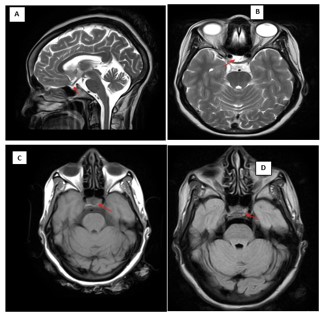
Figure 4 Brain MRI A: T2 weighted Sagittal B T2 weighted Axial C, T1 weighted Axial D: FLAIR Axial (The arrows in the above figure indicates an empty Sella)
On the second day of admission, the patient's management plan was revised following reassessment and review of the clinical findings and investigation results. Hormone replacement therapy was initiated with levothyroxine 100 μg orally once daily and hydrocortisone 100 mg intravenously twice daily. Additional treatment included pantoprazole 40 mg orally twice daily, oral sodium supplementation (½ teaspoon of table salt daily) for persistent hyponatremia, and a combined oral contraceptive (one tablet daily) as hormone replacement therapy. On her 3rd day of admission, serum electrolyte was repeated serum potassium level (K+) was 3.78 mEq/L and serum Sodium (Na+) was 138.0 mEq/L. She was discharged with an advice on her illness and levothyroxine 100 μg orally once daily and hydrocortisone was change into Prednisolone 5mg po daily. She presented on follow up no new complaint, symptoms subsided and started to gain weight.
4. Discussion
Sheehan's syndrome is generally diagnosed several years postpartum and results from partial or complete deficiency of one or more anterior pituitary hormones. Its clinical manifestations vary according to the specific hormone deficiencies. Prolactin deficiency can cause lactation failure, whereas gonadotropin deficiency commonly causes amenorrhea or loss of genital hair. Corticotrophin deficiency can result in generalized fatigue, weakness, hypoglycemia, or dizziness. Growth hormone deficiency may cause fatigue and decreased quality of life. Symptoms of central hypothyroidism resemble those of primary hypothyroidism; however, patients with central hypothyroidism have low triiodothyronine and thyroxine levels with normal or inappropriately low thyroid-stimulating hormone levels. [10] Anemia and other hematological abnormalities are common in patients with Sheehan's syndrome. The response of these abnormalities to replacement of thyroxine and glucocorticoids is not clear. [11]. Because hormone-secreting cells are located near the vasculature, growth hormone and prolactin secretion are most commonly affected, followed by follicle-stimulating hormone and luteinizing hormone; severe pituitary necrosis also affects secretion of thyroid-stimulating hormone and adrenocorticotropic hormone. The time between postpartum hemorrhage and symptom onset can be several years. [12] [13]
Hemorrhagic shock during pregnancy is a key entry point in diagnosis. The diagnosis of Sheehan’s syndrome is determined by the patient’s history and physical examination, confirmed by laboratory tests and MRI imagine. Failure to lactate is often a common initial complaint in patients with Sheehan syndrome. [14] A woman with Sheehan’s syndrome with undiagnosed hypopituitarism might be apparently asymptomatic until her body is exposed to stressful situations like surgery or infection many years after her delivery, and then she presents with adrenal crisis. [15]
The patients were aged 35, 25, and 35 years, respectively, and all were from the southern part of Ethiopia. The intervals between their last deliveries and the diagnosis of Sheehan’s syndrome were 5 months, 11 months, and 1 year, respectively, with a mean interval of 9.3 months. All patients had a history of massive postpartum hemorrhage and exhibited physical signs suggestive of Sheehan’s syndrome. Failure of lactation was one of the most common presenting features. In addition, gonadal hormone deficiency manifested primarily as amenorrhea, decreased libido, and loss of axillary and pubic hair. All patients had undergone diagnostic evaluations at other healthcare facilities before presenting to our hospital. However, the diagnosis was delayed, which may be attributed to limited awareness of panhypopituitarism and its diverse and nonspecific clinical manifestations. Inadequate history-taking and physical examination may have further contributed to the difficulty in recognizing this rare disorder. The second patient had a particularly progressive presentation suggestive of secondary adrenal insufficiency. She initially developed asthenia, anorexia, and weight loss, followed by dizziness, nausea, persistent vomiting, and abdominal pain as her condition progressively deteriorated. Despite repeatedly seeking medical care, the underlying diagnosis remained unrecognized until she presented to our hospital.
Pituitary crisis is a potentially life-threatening complication of chronic pituitary hormone deficiency. It may develop after a period of vague or nonspecific symptoms and can be precipitated by factors such as electrolyte disturbances, infection, trauma, or other forms of physiological stress. Acute pituitary hormone deficiency commonly leads to volume depletion and reduced cardiac output, resulting in hemodynamic instability. With prompt hormonal replacement and restoration of intravascular volume, cardiovascular function can usually improve rapidly. [16] Consistent with this presentation, the second patient developed bradycardia and persistent hypotension despite adequate fluid resuscitation and vasopressor therapy. Although the initial diagnosis was not recognized, subsequent clinical re-evaluation, the diagnosis was reconsidered after her history of severe postpartum hemorrhage requiring blood transfusion was revealed. Following the initiation of appropriate hormonal replacement therapy, she demonstrated a marked clinical response, with improvement in her hemodynamic status. This case highlights the importance of considering pituitary crisis in patients with unexplained or refractory hypotension, particularly when there is a history of significant postpartum hemorrhage suggestive of postpartum pituitary dysfunction.
Interestingly, Sheehan syndrome can cause a wide range of hematological manifestations through unknown complex hormonal effects; the most frequent one is anemia. Pancytopenia is rarely present in Sheehan’s syndrome and is demonstrated in only a few case reports worldwide. [17] The second patient CBC profile showed bi-cytopenia, characterized by leukopenia and moderate normochromic normocytic anemia, which improved following hormone replacement therapy. Similarly, the third patient had mild normochromic normocytic anemia, whereas no hematological abnormalities were detected in the first case.
Second the patient also had hypoglycemic episodes at initial presentation, which were initially treated with bolus dextrose push but recurred. Then she was treated with dextrose infusion and subsequently normalized with the commencement of hormone replacement therapy.
Other laboratory investigations may reveal electrolyte disturbances, with hyponatremia being one of the most common abnormalities observed in patients with Sheehan syndrome. Hyponatremia may be an initial presenting feature of acute Sheehan syndrome, occurring in approximately 60% of reported cases. It is also a frequent electrolyte abnormality in patients with chronic disease, with reported prevalence ranging from 33% to 69%. [12]
The cause of hyponatremia in Sheehan's syndrome is controversial. Explanations include impaired free-water retention secondary to elevated antidiuretic hormone secretion as well as diminished free water clearance in the kidney secondary to the lack of cortisol thought to modulate distal tubular function in the presence of low levels of antidiuretic hormone. Other workers have found blunted aldosterone responsiveness to salt restriction in this condition, which was variable with glucocorticoid and/or thyroid hormone supplementation. The renin-aldosterone axis has been assumed to be independent of pituitary control, but there may be a regulating pituitary factor. [18] Whereas hypokalemia in patients with Sheehan syndrome is usually associated with gastrointestinal losses, such as those resulting from diarrhea and vomiting, rather than adrenal insufficiency, since adrenal aldosterone production is not dependent on pituitary hormones. In our case series, the second patient developed moderate hypokalemia, which was managed with potassium chloride (KCl) supplementation.
The third patient, in contrast, presented with profound hyponatremia, which was treated with intravenous 0.9% normal saline and oral salt supplementation. These contrasting electrolyte abnormalities highlight the variable metabolic manifestations of Sheehan’s syndrome and the importance of recognizing electrolyte disturbances as potential clues to underlying hypopituitarism.
Pituitary MRI is an important diagnostic tool in Sheehan syndrome, as it commonly demonstrates partial or complete empty sella. Imaging studies suggest that the pituitary gland may initially become enlarged during the first month after delivery, followed by gradual shrinkage over the subsequent months. This progressive loss of pituitary tissue can result in the development of an empty sella, typically becoming evident within 6 months to 1 year after delivery. In patients with established Sheehan syndrome, an empty sella is a characteristic and frequently observed MRI finding. [19] [20] In our case series, all three patients demonstrated either an empty or partially empty sella on pituitary MRI, which, together with their clinical presentation, history of postpartum hemorrhage, and hormonal abnormalities, strongly supported the diagnosis of Sheehan syndrome.
The treatment includes the replacement of deficient pituitary hormones. An important point in patients with SS is the progressive nature of the disease. In patients presenting with acute adrenal failure, GC treatment should be started immediately after getting serum samples for cortisol measurement. In the long-term replacement, the patient should be monitored for optimal GC dose to prevent the unwanted effects of excess GC dose such as osteoporosis, glucose intolerance and weight gain while keeping the quality of life of the patient well. Hydrocortisone may be preferred although it cannot completely mimic the normal physiology of HPA axis. [21] Its recommend using HC, usually 15–20 mg total daily dose in single or divided doses. Patients using divided doses should take the highest dose in the morning at awakening and the second in the afternoon. [22]
Levothyroxine replacement should not be started before GC replacement or ruling out adrenal insufficiency. Dose adjustment should be done according to free T4 levels rather than TSH. In elderly and patients with coronary artery disease, LT4 should be given at low doses and increased slowly. GH may increase the requirement of LT4 dose or may unmask a mild secondary hypothyroidism. [21]
For our patients, hormone replacement therapy was tailored according to their age, fertility preferences, and financial capacity, using the available and affordable treatment options.
5. Conclusion
Sheehan syndrome remains the leading cause of panhypopituitarism in developing countries with inadequate obstetric care. However, it is frequently underdiagnosed. With this case report, we try to highlight the importance of maintaining a high index of suspicion among primary care physicians working in resource limited settings when women with a history of postpartum hemorrhage present with nonspecific symptoms before deciding to treat the patients symptomatically. Comprehensive clinical evaluation is essential for early diagnosis, timely referral for further investigations such as hormonal panels and brain MRI, and prompt initiation of treatment to prevent life threatening adrenal crisis. None of our three patients were aware of Sheehan syndrome, even though they had significant obstetric histories, and one experienced a severe adrenal crisis. We want to emphasize that women with a history of postpartum bleeding should be informed about the risk of Sheehan syndrome. Primary care physicians should educate patients about possible symptoms and the importance of seeking medical attention early. Sheehan syndrome should be considered a top differential diagnosis in these cases, and proper treatment should be initiated early to protect the patients from deadly complications.
6. Abbreviations
CBC: Complete Blood count KCL: Potassium chloride; LDL: Low-density lipoprotein; MCV: Mean corpuscular volume; MRI: Magnetic resonance imaging; NS: Normal saline. GC: Glucocorticoid SS: Sheehan’s syndrome COC: Combined Oral Contraceptive CT Scan: Computed C. decficille.
7. Acknowledgements
We would like to acknowledge Alatyon General Hospital, Department of Internal Medicine, for their support and contribution to this case series. We would also like to extend our special thanks to our patients for their cooperation and for granting permission to use their clinical information and MRI images for the purpose of this case series.
8. Authors contributions; Mamaru Sintayehu, Corresponding author, editing
Yonas kinfemichael, Corresponding author, drafting, editing,
Kerebih Molla; editing
Yonas Kassahu; Editing
Hailegebriel Demelash; data extraction
9. Availability of data and materials
The data sets generated during the study are available from the corresponding author upon request.
10. Funding
There was no funding in the writing and publishing of this case series
10. Declarations
Ethics approval and consent to participate
The patients provided written informed consent for the publication of their medical information and relevant clinical images in this case series.
Consent for publication
Written informed consent was obtained from the patients for publication of this case report and any accompanying images. A copy of the written consents are available for review by the Editor-in-Chief of this journal.
Competing interests
The authors declare that they have no competing interests.



Open Access By Aditum Open Access Journals id licensed under Creative Commons Attribution 4.0 International License. Based On a Work at aditum.org


