
Clinical Case Reports and Clinical Study
OPEN ACCESS | Volume 13 - Issue 4 - 2026
ISSN No: 2766-8614 | Journal DOI: 10.61148/2766-8614/JCCRCS


Hamawa S.S1*., Filgona J2., Sudi I.Y3 and Ibrahim T3
1Department of Zoology, Adamawa State University, Mubi.
2Department of Microbiology, Adamawa State University, Mubi.
3Department of Biochemistry, Adamawa State University, Mubi.
*Corresponding author: Hamawa Stephen Sanda, Department of Zoology, Adamawa State University, Mubi.
Received: August 22, 2026 | Accepted: September 01, 2026 | Published: September 14, 2026
Citation: Hamawa S.S., Filgona J., Sudi I.Y and Ibrahim T. (2026) “Epidemiological Study of Schistosomiasis and Transmission Potential of the Snail Intermediate Host in Hong Local Government, Adamawa State, Nigeria” Clinical Case Reports and Clinical Study, 13(4); DOI: 10.61148/2766-8614/JCCRCS/251.
Copyright: © 2026 Hamawa Stephen Sanda. This is an open access article distributed under the Creative Commons Attribution License, which permits unrestricted use, distribution, and reproduction in any medium, provided the original work is properly cited.
Schistosomiasis remains one of the most important neglected tropical diseases affecting rural communities in Nigeria, particularly in areas with poor sanitation and frequent human contact with freshwater bodies. This study investigated the epidemiology of schistosomiasis and the transmission potential of freshwater snail intermediate hosts in the Hong Local Government AreaThe study aimed to determine the prevalence of schistosomiasis among residents, identify the species and distribution of snail intermediate hosts, and assess environmental factors associated with disease transmission. A cross-sectional survey design was adopted involving parasitological examination of urine and stool samples collected from selected communities and schools within the study area. Snail sampling was carried out from streams, ponds, and irrigation sites using scoop nets and hand-picking methods, while cercarial shedding techniques were used to determine snail infectivity. Findings from related studies in Adamawa State and other endemic regions indicate that species such as Bulinus globosus, Bulinus truncatus, and Biomphalaria pfeifferi are important intermediate hosts responsible for sustaining transmission of Schistosoma parasites in freshwater habitats. Human infection is commonly associated with activities such as farming, fishing, swimming, and domestic use of contaminated water sources. Environmental conditions, including water temperature, vegetation, rainfall pattern, and irrigation practices, also contribute significantly to snail abundance and disease transmission. The study is expected to provide baseline epidemiological data on schistosomiasis in Hong LGA and contribute to effective control strategies through improved public health education, snail control measures, environmental sanitation, and mass drug administration programs. The findings will also assist health authorities and researchers in understanding the transmission dynamics of schistosomiasis in rural communities of Adamawa State.
Schistosomiasis; Transmission Potential
Schistosomiasis is a disease spread by snails that is caused by Schistosoma blood flukes. Millions of people in sub-Saharan Africa are affected by the most neglected tropical disease. Schistosomiasis has been found in Latin America, Asia, and the Middle East (WHO, 2020). It is one of the most hazardous tropical infections, only surpassed by intestinal helminthes and malaria in terms of serious, difficult-to-treat illness (WHO, 2024).
Schistosomiasis affects over 200 million people worldwide and remains a major public health concern in tropical regions. Schistosomiasis flukes employ snails, invertebrates in the Phylum Mollusca, as intermediate hosts (WHO 2024). Except for slugs, this group of species has a distinctive morphology called "shell," which is a key trait of the group. In 76 countries where Schistosomiasis is endemic, 700 million people are at risk, and 200,000 die each year (WHO 2024). Human schistosomiasis has two primary types in sub-Saharan Africa. They include intestinal and genitourinary schistosomiasis. Because they may be found in freshwater environments and other ecological niches, including wetlands, rice paddies, dams, and ponds, snails occupy a variety of habitats. The intermediate host for Schistosoma species, including Biomphalaria S. mansoni, Oncomelania, S. japonicum, Bulinus, S. haematobium, S. intercalatum, S. guineensis, Neotricula aperta, and S. mekongi, is Biomphalaria. The parasite lifecycle involves humans and freshwater snails (e.g., Bulinus, Biomphalaria) as intermediate hosts. Infected humans release eggs into water, which hatch into miracidia that infect snails. These snails subsequently release cercariae, the infective stage for humans. Transmission potential refers to the capacity of infected snails to produce and release cercariae, thereby sustaining disease transmission. Snail hosts are central to schistosomiasis epidemiology because they: Support asexual multiplication of the parasite, amplify infection intensity through cercarial release, and determine spatial and temporal transmission patterns. Human infection risk is directly linked to cercarial density in water bodies, which originates from infected snails. Determinants of Transmission Potential of Snails include: nutrients (e.g., algae) for growth and reproduction, well-nourished snails produce more cercariae, and resource-rich environments increase transmission potential. Studies show that cercarial production increases with food availability, indicating that not all infected snails contribute equally to transmission. Snail density leads to resource competition. Competition reduces individual snail growth and parasite output. Transmission may decline despite high snail numbers; thus, transmission potential is not always proportional to infected snail density, contradicting traditional epidemiological assumptions. Larger snails produce more cercariae. Transmission is influenced by snail biomass rather than just abundance. Population structure (size distribution) is a key factor. Environmental conditions such as Water quality, Vegetation, Temperature, and human and animal activity affect snail distribution and infection rates. Habitat variability leads to spatial heterogeneity in transmission risk. Predators and competitor snail species can reduce host snail populations. Biological control methods may lower transmission. However, partial reduction of snails can sometimes increase per-snail cercarial output. Mathematical models suggest that ecological interactions must be considered in control strategies.
Description of the Study Area
This research was carried out in Kulinyi and Pella district in Hong Local Government Area of Adamawa state, Nigeria. Hong LGA is located between latitudes 10° 0’ 00’’ N and 10° 35’ 00’’ N and longitudes 12° 35’ 00’’ E and 13° 20’ 00’’ E. It has a total land area of 2,419.11 km2 (Bashir and Raji, 1999). Hong is one of the 21 Local Government Areas of Adamawa State, created in 1987 during the defunct Gongola State. Other districts in Hong include: Dugwaba, Gaya, Hildi, Hong, and Uba. Hong occupies a very fertile land that receives much rainfall, mostly beginning in April and stopping in October or early November. There are rivers, ponds, streams, and dams for agricultural activities such as fishing and other domestic purposes, which are mostly carried out throughout the seasons. The primary occupations of the inhabitants of Hong Kong are mainly farming, fishing, and trading (Joshua, 2017).
Ethical approval
An introduction letter was requested from the office of the Head of the Department of Zoology. Before the commencement of the investigation, ethical clearance and approval were obtained from the Health Department, Hong local government area. Informed consent was sought and obtained from the village head, school authorities, the parents /guardians, and the participants, before the commencement of the study.
Sample size determination.
The minimum sample size was calculated according to the method described by Anikwe et al (2020). The minimum sample size (N) was calculated using the formula:
N=Z2pq/e2 Where Z is the standard deviation at 1.96 (which corresponds to 95% confidence interval). Prevalence from previous study = (0.45), the probability of the event occurring, and q = (0.05), the probability of the event not occurring. E is the desired level of precision, also known as sampling error: 5(0.05) %.
N= (1.96)2×0.45×0.5 =346
(0.05)2
The minimum sample size for this study is 346, but 400 samples were collected
Sample collection
Stratified random sampling techniques were used to select locations within the study area based on proximity to the water body. Systematic random sampling was also used in selecting the participants and snail intermediate hosts. A well-structured questionnaire was used to collect information on sex, age, source of drinking and domestic water, water contact activities, and parent occupation during sample collection.
All the study participants were instructed on how to aseptically collect a stool and urine sample into the sample bottle provided. Approximately 5–10 ml of urine and 3–5 g of faeces were collected. 5ml of 10% formaldehyde was added to the stool sample as a preservative. Each urine sample was preserved by adding 5 drops of 1% domestic bleach (sodium chloride) (Bala et al., 2012).
All collections were done between 10:00 am and 2:00 pm (10:00 am hours and 14:00hours) to coincide with the period when excretion of Schistosoma eggs is highest. The samples collected were well packaged to avoid breakage and transported back to the Parasitology Laboratory, Department of Zoology, Adamawa State University, Mubi, for examination.
Table 1: Locations where samples were collected and their coordinates in the study Area.
|
S/N |
Location |
Ward |
Latitude |
Longitude |
|
1 |
Ngalbi |
Thilbang |
N10º10'43" |
E12º 54'58" |
|
2 |
Zhedinyi |
Daksiri |
N10º8'22" |
E12º58'43" |
|
3 |
Mullah |
Husherizum |
N10º8'53" |
E13º4'20" |
|
4 |
Bakwa |
Daksiri |
N10º8'6" |
E13º1'1" |
|
5 |
Dagwaba |
Daksiri |
N10º7'49" |
E12º55'58" |
|
6 |
Lebeng |
Daksiri |
N10º8'25" |
E12º58'24" |
|
7 |
Kwakwa`ah s |
Husherizum |
N10º10'45" |
E13º2'55" |
|
8 |
Kala`a |
Bangshika |
N10º14'44" |
E13º1'19" |
|
9 |
k/kuka |
Bangshika |
N10º14'17" |
E13º0'7" |
|
10 |
Delhi |
Husherizum |
N10º10'27" |
E13º5'33" |
|
11 |
Dilwachira |
Husherizum |
N10º12'32" |
E13º6'22" |
|
12 |
Bangshika |
Bangshika |
N10º12'5" |
E12º59'29" |
|
13 |
Dzumah |
Thilbang |
N10º10'26" |
E12º56'28" |
|
14 |
Gashaka |
Thilbang |
N10º10'35" |
E12º58'35" |
Urine and Stool Sample Analysis
Each urine sample was appropriately labelled and visually screened. The visual screening was done by careful observation of the urine sample for a specific colour. The urine colour chart ranges from i. light-yellow normal colour of urine indicating urine free of any trace of Microhaematuria or proteinuria, (ii) Light –brown urine with presence of low proteinuria and microhaematuria (iii) dark brown urine with medium presence of proteinuria and microhaematuria (iv) bloody brown urine with visible haematuria. (Tulu et al., 2014).
Microscopic examination.
The urine samples were processed using concentration or sedimentation techniques as described by Filgona and Hamawa (2023). The sediment containing eggs at the bottom of the tube, while the supernatant was poured out carefully without disturbing the sediment.
The sediment was gently mixed by swirling the tube, and a disposable Pasteur pipette was transferred to a clean, grease-free microscope slide. A coverslip was placed over the sediment on the slide and examined under a light microscope, using low-power (10x) and high-power (40x) objectives as described by Nwoko et al. (2022).
Stool Sample Analysis.
Stool samples from the participant were examined visually for any visible signs of the presence of blood or mucus. (Macalanda et al, 2024).
Sedimentation techniques were used as described by Hamawa et al (2024). The stool sample was mixed with a formaldehyde solution, and the mixture was strained through gauze or a sieve, and the filtrate was collected into a test tube. The filtrate was centrifuged at 1500 rmp for 5 minutes and allowed to stand undisturbed for 20–30 minutes. The supernatant was decanted, and the sediment was examined microscopically. The prepared slide was examined under a light microscope using both low-power (10x) and high-power (40x) objectives to identify the characteristic oval-shaped eggs of Schistosoma mansoni, which possess a prominent lateral spine (Hamawa et al., 2024).
Snail Sampling and Identification.
Snail samples were collected from rivers, seasonal streams, ponds, and other micro-habitats where human activities are carried out within the Kulinyi and Pella District. The snail samples were collected using a long-handled scoop net and a pair of forceps. The collection was conducted at each site, and the number of snail species encountered was recorded (Buba et al,2023). Each of the districts was stratified into two strata, and from each stratum, four sampling points were selected for snail collection. The collected snail samples were kept in a specimen container labelled based on the site of collection and transported to the laboratory for further examination. The snails were identified based on their morphological characteristics and keys for the identification of African freshwater snails of medical and veterinary importance, as described by Brown (1994).
Cercariae shedding and identification
Cercarial release from snails was conducted as described by Sharif et al. (2010). Identified snails were placed individually in wide-mouth glass specimen bottles containing 30 mL of de-chlorinated water and exposed to sunlight (no direct sunlight) between 9:00 am and 1:00 pm daily for possible cercarial shedding (Sharif et al., 2010). A volume of 15 ml of water from each specimen bottle was poured into a Petri dish and examined for cercariae under a stereoscopic microscope, as described by Uthpala et al. (2010). Snails that did not release cercariae on the first day of exposure were re-exposed every subsequent day until the seventh day, after which they were discarded. The absence of cercarial shedding on the 7th day of exposure to sunlight indicated the absence of infection. Water in the specimen bottles housing the snails was changed daily to avoid buildup of toxicity arising from the snails’ metabolic wastes. Cercariae released by the snails were mounted on a grease-free glass slide for examination (Uthpala et al., 2010). Identification of cercariae to the species level was performed using standard morphological characteristics and measurements (Brown, 1994). Body shape: Usually elongated or oval. The body may be transparent or slightly opaque. And divided into the Anterior region (head region) and the posterior region connected to the tail. Tail Used for swimming and movement in water. May be a simple tail or forked tail (furcocercous), as seen in Schistosoma cercariae. Tail length varies among species. An oral sucker located at the anterior end, it functions in attachment and penetration into the host. This involved narcotizing the cercariae with a 0.35% NaCl solution (Brown, 1994).
Data Analysis
Data was entered into Microsoft Excel (version 2013). Descriptive statistics were used to compute the data. The prevalence was calculated and expressed as % =number infestednumber Examined
 ×100 (Sahoo et al., 2023).
T-test, Chi-square test, and ANOVA were used to compare the differences in the prevalence of intestinal and urinary schistosomiasis based on sex, age group, and parents’ occupation among the study population.
Odds ratio (OR), at 95% confidence interval (CI), and relative risk were used to determine the association between the prevalence of intestinal and urinary schistosomiasis and the risk factors using Epi Info, and Shannon diversity index was used to determine diversity and species richness of snail intermediate host in the study locations.
Result
A total of four (400) participants were recruited for the study. Of these participants, 200 were from Kulinyi and Pella districts of Hong LGA, respectively. Samples from the identified communities in four wards of Kulinyi and Pella districts were collected (from July 2025 to January, 2026). These wards include: Husherizum, Banshika, Daksiri, and Thilbang wards. A total of 400 samples, each for urine and stool, were obtained for screening. The overall prevalence of intestinal and urogenital schistosomiasis was 8.75% (70) out of 800 samples examined. (Table 1). This is statistically significant at P-value 0.000 (p<0.05).
Prevalence of Intestinal (S. mansoni) and Urogenital (S. haematobium) Schistosomiasis based on locations revealed that Husherizum ward had the highest prevalence with 27.0%, while Thilbang had the lowest prevalence with 9.0%. (Table 2)
Prevalence of Schistosoma haematobium based on sex and location in the study area revealed that Male had the highest rate of infection with 22.5%, while females had 9.0%. Husherizum ward had the highest prevalence in males with 32.0% and females with 14.0%, while Thilbang ward had the least prevalence of 12.0% in males and 6.0% in females, respectively (Table 3).
The overall prevalence of S. mansoni was 3.5%. Males had the highest rate of infection with 3.0%, while females had 0.5%. Husherizum ward had the highest prevalence in males with 6.0% and females with 2.0%, while Thilbang ward had the no prevalence with 0.0% in both males and females, respectively. (Table 4)
Table 1: overall prevalence of intestinal and urogenital schistosomiasis among inhabitants of Kulinyi and Pella districts, Hong LGA, Adamawa State, Nigeria.
|
Samples |
Number examined |
Number infected (%) |
Number uninfected (%) |
Odd ratio |
95%CI |
Risk ratio |
|
|
Urine |
400 |
63(15.8) |
337(84.25) |
10.50 |
4.7428-23.2261 |
9.00 |
|
|
Stool |
400 |
07(1.8) |
393(98.25) |
0.10 |
0.0431-0.2108 |
0.11 |
|
|
Total |
800 |
70 (8.75) |
730 (91.25) |
|
|
|
|
Table 2: prevalence of intestinal and urogenital schistosomiasis based on locations.
|
Location |
S.mansoni |
S.haematobium |
Total |
||
|
Number examined |
Number infected (%) |
Number examined |
Number infected (%) |
Prevalence (%) |
|
|
Husherizum |
100 |
4 |
100 |
23 |
27(13.5) |
|
Bangshika |
100 |
1 |
100 |
14 |
15(7.5) |
|
Daksiri |
100 |
2 |
100 |
17 |
19(9.5) |
|
Thilbang |
100 |
0 |
100 |
09 |
09(4.5) |
|
Total |
400 |
07 |
400 |
63 |
70(8.8) |
Table 3: prevalence of Schistosoma haematobium based on sex and locations in the study area.
|
Wards |
Males |
|
Females |
|
||||
|
Num. examined |
Num. infected |
Prevalence (%) |
Num. examined |
Num. infected |
Prevalence (%) |
Total |
||
|
Husherizum |
50 |
16 |
32 |
50 |
7 |
14 |
23 |
|
|
Bangshika |
50 |
11 |
22 |
50 |
3 |
6 |
14 |
|
|
Daksiri |
50 |
12 |
24 |
50 |
5 |
10 |
17 |
|
|
Thilbang |
50 |
6 |
12 |
50 |
3 |
6 |
9 |
|
|
Total |
200 |
45 |
22.5 |
200 |
18 |
9 |
63(15.8) |
|
Table 4: prevalence of Schistosoma mansoni based on sex and location in the study area.
|
Wards |
Males |
Females |
|
||||
|
Number examined |
Number infected |
Prevalence (%) |
Number examined |
Number infected |
Prevalence (%) |
Total |
|
|
Husherizum |
50 |
03 |
6 |
50 |
01 |
2 |
4 |
|
Bangshika |
50 |
01 |
2 |
50 |
00 |
0 |
1 |
|
Daksiri |
50 |
02 |
4 |
50 |
00 |
0 |
2 |
|
Thilbang |
50 |
00 |
0 |
50 |
00 |
0 |
0 |
|
Total |
200 |
06 |
3 |
200 |
01 |
0.5 |
7(1.8) |
Schistosoma haematobium prevalence in the study area based on age group showed that age group 12-17 years had the highest prevalence (18.6%), while age group 24 and above years of age had the least prevalence (1.64%). Age group 12-17 years had the highest odds (3.0) of being infected with urinary schistosomiasis, and age group above 24 years had the lowest odds (0.15). (Table 5)
Prevalence of S. mansoni infection based on age group showed that age group 12-17 years had the highest rate of infection (4.4%), while age group 5-11 years had the lowest prevalence (1.6%). The age groups of 18-23 years and 24 years and above had 0.0% prevalence, respectively (Table 6)
In terms of snail (intermediate host) distribution and abundance for S. mansoni and S. haematobium, a total of one thousand two hundred and eighty-eight (1,288) freshwater snails were collected from some streams, ponds/dams, and wetlands/rice paddies in the study area during the period of collection. The distribution and abundance of schistosome intermediate hosts for S. mansoni and S. haematobium in the study area as shown in Table 7. Bulinus species constitutes the highest number of the species collected, 1220(94.7%). Daksiri ward had the highest number of Bulinus species (470) with a relative abundance (RA) of 38.5, while Thilbang ward had the least number of Bulinus species (150) with a relative abundance of 12.3, respectively. Biomphalaria species recovered during sample collection were 68(5.3%) out of the 1,288 snails collected. They were found to be more abundant in Husherizum ward with the relative abundance (RA) of 47.0. Thilbang ward had the least (0.0%) with no Biomhalaria species found during the period of collection (Table 4.7). Out of the one thousand two hundred and eighty-eight (1288) freshwater snail intermediate hosts for S. mansoni and S. haematobium collected from various habitats, Bulinus species were found to be more abundant in ponds/dams (630) with a relative abundance of 51.6%, as well as in wetlands/rice paddies (408) with a relative abundance of 33.4%, respectively. The least number of Bulinus species were found in streams (182) with a relative abundance of 14.9.
Biomphalaria species, on the other hand, were found to be more abundant in streams (44) with a relative abundance of 64.7, as well as in wetlands/rice paddies (18) with a relative abundance of 26.5. Biomphalaria species were least abundant in pond/dams (6) with a relative abundance of 8.8, respectively (Table 8). The two species of freshwater snails were evenly distributed across the various habitats in the study area.
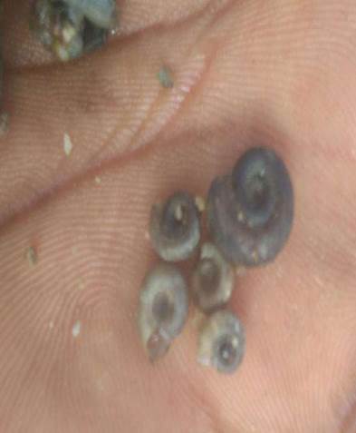
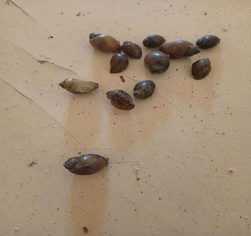
Plate 1: Biomphalria Snail species 2: Bulinus snail Species
Table 6: Prevalence of Schistosoma haematobium based on Age group in the study area.
|
Age group (yrs) |
Num. examined |
Num. infected |
Prevalence (%) |
Odd ratio |
95%CI |
Risk ratio |
|
5-11 |
129 |
9 |
6.9 |
0.75 |
0.3435-1.6375 |
0.76 |
|
12-17 |
113 |
20 |
18.6 |
3.0 |
1.5104-5.9112 |
2.69 |
|
18-23 |
97 |
7 |
7.9 |
0.55 |
0.2102-1.433 |
0.56 |
|
24-above |
61 |
1 |
1.64 |
0.15 |
0.0208-1.1471 |
0.17 |
|
Total |
400 |
37 |
9.25 |
|
|
|
Table 7: Prevalence of Schistosoma mansoni based on Age group in the study area.
|
Age group (yrs) |
Num. examined |
Num. infected |
Prevalence (%) |
Odd ratio |
95%CI |
Risk ratio |
|
5-11 |
129 |
2 |
1.6 |
0.12 |
0.0284-0.5068 |
0.13 |
|
12-17 |
113 |
5 |
4.4 |
0.39 |
0.1508-1.0441 |
0.42 |
|
18-23 |
97 |
0 |
0 |
|
|
|
|
24-above |
61 |
0 |
0 |
|
|
|
|
Total |
400 |
7 |
1.75 |
|
|
|
Table 8: Distribution and abundance of schistosome intermediate host for S. mansoni and S. haematobium.
|
Location |
Snail species |
|
||
|
Bulinus |
|
Biomphalaria |
|
|
|
Number collected |
Relative abundance |
Number collected |
Relative abundance |
|
|
Husherizum |
340 |
28 |
32 |
47 |
|
Bangshika |
260 |
21.3 |
8 |
11.8 |
|
Daksiri |
470 |
38.5 |
28 |
41.2 |
|
Thilbang |
150 |
12.3 |
0 |
0 |
|
Total |
1,220 |
|
68 |
|
|
Shannon H |
1.35 |
|
1.20 |
|
|
Richness |
0.84 |
|
0.86 |
|
|
Evenness |
0.77 |
|
0.83 |
|
Table 9: Distribution of schistosome intermediate host for S. mansoni and S. haematobium based on habitat type in the study area.
|
Habitat type |
Snail species |
|||
|
Bulinus spp |
Relative abundance |
Biomphalaria spp |
Relative abundance |
|
|
Streams |
182 |
14.9 |
44 |
64.7 |
|
Ponds/dams |
630 |
51.6 |
6 |
8.8 |
|
Wetlands/rice paddies |
408 |
33.4 |
18 |
26.5 |
|
Total |
1220 |
|
68 |
|
A higher rate of cercariae shed was observed in Bulinus spp 118(9.7%) out of the 1220 Bulinus snail samples examined, compared to Biomphalaria spp., where only 2(2.9%) shed cercariae out of the 68 Biomphalaria snail samples examined (Table 4.9).
Table 10: Infectivity status of snail intermediate host encountered in Kulinyi and Pella districts of Hong LGA, Adamawa State, Nigeria.
|
Snail species |
Number examined |
Number infected |
Percentage infected % |
Risk ratio |
95%CI |
|
Bulinus spp. |
1220 |
118 |
9.7 |
3.29 |
0.8305-13.021 |
|
Biomphalaria spp |
68 |
2 |
2.9 |
0.30 |
0.0768-1.204 |
|
Total |
1288 |
120 |
9.3 |
|
|
|
|
|
|
|
|
|
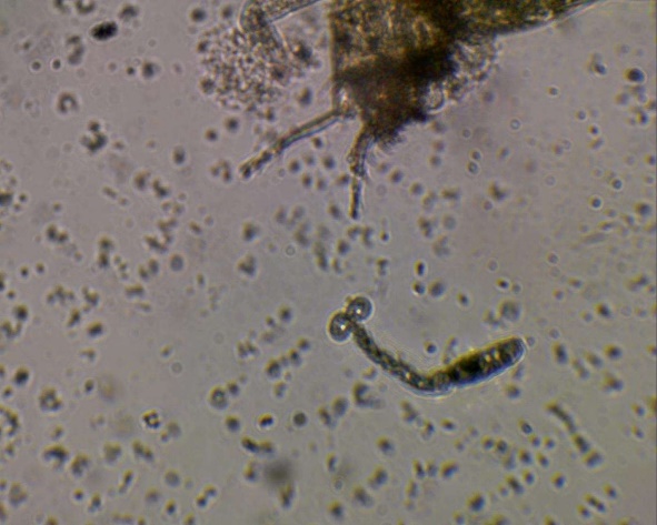
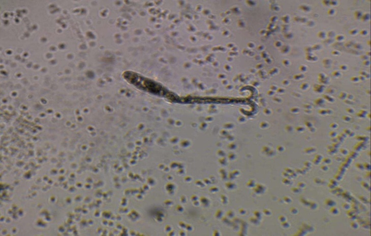
Plate 3: forked-tailed cercaria shed by Bulinus snail species
Discussion
The present study on the epidemiology of Schistosomiasis and the transmission potential of snail intermediate hosts in Hong Local Government Area, Adamawa State, Nigeria, revealed that schistosomiasis remains an important public health problem in rural communities where residents depend heavily on natural water bodies for domestic, agricultural, and recreational activities. The occurrence of infection among the study population indicates continuous transmission within the area and suggests the presence of favorable ecological conditions that support both the parasite and its snail intermediate hosts.
The prevalence recorded in this study may be associated with frequent human-water contact activities such as farming, fishing, irrigation, swimming, washing, and fetching water from streams and ponds. Similar findings have been reported in several endemic communities in northern Nigeria where poor sanitation, inadequate potable water supply, and occupational exposure contribute significantly to disease transmission (Daniel et al, 2024; Mereta et al,2019; Na'acha et al, 2021). Children and young adults are usually more affected because of their increased contact with contaminated water during swimming and other outdoor activities (Birma et al, 2017; Anagbogu et al, 2026).
The detection of freshwater snails such as Bulinus globosus, Bulinus truncatus, and Biomphalaria pfeifferi in the study area confirms the availability of competent intermediate hosts necessary for the completion of the life cycle of Schistosoma species. The abundance of these snails in slow-moving or stagnant water bodies with aquatic vegetation supports earlier reports that environmental conditions such as temperature, rainfall, pH, and vegetation influence snail survival and reproduction. Areas with irrigation activities and permanent water bodies often provide ideal habitats for snail breeding, thereby increasing transmission potential (Taofiq et al, 2017; Buba et al, 2023; Birma et al, 2017; Daniel etal, 2024).
The cercarial shedding observed among some of the collected snails demonstrates active transmission of schistosome parasites within the aquatic environment. Snails shedding cercariae indicate that infected individuals in the community are contaminating water sources through urination or defecation, allowing miracidia to infect susceptible snails and continue the transmission cycle. The presence of infected snails, therefore, represents a direct epidemiological risk to people using these water bodies.
Variation in infection prevalence among communities may be linked to differences in environmental sanitation, water usage patterns, socio-economic status, health awareness, and access to medical care. Communities located closer to rivers, ponds, and irrigation channels are more likely to experience higher infection rates due to increased exposure to cercaria-infested water. Seasonal changes may also influence transmission, as snail population density and cercarial release tend to increase during favorable climatic conditions.
The findings of this study agree with previous studies conducted in different parts of Nigeria and other African countries, where schistosomiasis transmission was strongly associated with the distribution of snail intermediate hosts and poor environmental hygiene (Mathew et al, 2025; Isa et al,2019; Hamawa et al, 2024). The persistence of the disease in Hong Local Government Area highlights the need for integrated control measures that combine mass drug administration with praziquantel, public health education, provision of safe drinking water, improved sanitation, and snail control programs. Furthermore, environmental management strategies such as clearing aquatic vegetation, proper waste disposal, and reducing human contamination of water bodies may help interrupt transmission. Health education campaigns should focus on increasing community awareness regarding the mode of transmission, preventive practices, and the importance of early diagnosis and treatment.
In conclusion, the study demonstrates that the Hong Local Government Area possesses ecological and socio-environmental conditions favorable for the continued transmission of Schistosomiasis. The presence of infected snail intermediate hosts and the observed human infection rates indicate ongoing transmission within the area. Effective and sustainable control measures are therefore essential to reduce disease burden and prevent further spread of schistosomiasis in the affected communities.



Open Access By Aditum Open Access Journals id licensed under Creative Commons Attribution 4.0 International License. Based On a Work at aditum.org


