
Clinical Case Reports and Clinical Study
OPEN ACCESS | Volume 13 - Issue 4 - 2026
ISSN No: 2766-8614 | Journal DOI: 10.61148/2766-8614/JCCRCS


Kaikat Zahira1,2, Bennaoui Fatiha 1,2*, Abi Al Alaa Khalid1,2, Lalaoui Abdessamad1,2, Kassal Ghizlane1, El Idrissi Slitine Nadia1,2, Fadl Mrabih Rabou Maoulainine1,2
1Neonatal Intensive Care Unit, Mother-Child Hospital, CHU Mohammed VI, Marrakech, Morocco.
2Childhood, Health and Development Research LABORATORY, Cadi-Ayyad University, Marrakech, Morocco.
*Corresponding author: Bennaoui Fatiha, Neonatal Intensive Care Unit, Mother-Child Hospital, CHU Mohammed VI, Marrakech, Morocco.
Received: July 10, 2026 | Accepted: July 18, 2026 | Published: July 22, 2026
Citation: Zahira K, Fatiha B, Alaa Khalid AA, Abdessamad L, Ghizlane K, Slitine Nadia EI, Rabou Maoulainine FM. (2026) “Right-Sided Congenital Diaphragmatic Hernia Associated with Severe Intrauterine Growth Restriction: Diagnostic Challenges and Therapeutic Limitations of Neonatal Resuscitation — A Case Report” Clinical Case Reports and Clinical Study, 13(4); DOI: 10.61148/2766-8614/JCCRCS/244.
Copyright: © 2026 Bennaoui Fatiha. This is an open access article distributed under the Creative Commons Attribution License, which permits unrestricted use, distribution, and reproduction in any medium, provided the original work is properly cited.
Right-sided congenital diaphragmatic hernia (CDH) is a rare malformation, accounting for 10% to 15% of CDH cases, and is often associated with a poorer prognosis due to intrathoracic liver herniation. The concomitant presence of severe intrauterine growth restriction (IUGR) further complicates neonatal intensive care management. We report the case of a male newborn delivered at 37 weeks and 5 days of gestation by cesarean section for acute fetal distress, with severe IUGR (birth weight, 1,600 g). Immediate respiratory distress (Silverman score, 3/10) with absent breath sounds on the right side led to admission to the neonatal intensive care unit, where right-sided CDH was confirmed on chest radiography. Despite a medical stabilization protocol including protective ventilation and targeted hemodynamic support, the infant developed refractory pulmonary arterial hypertension and severe hypoxemia, and died 48 hours after birth, before surgical repair could be considered. This case illustrates the diagnostic and therapeutic challenges of right-sided CDH complicated by severe IUGR and underscores the limitations of current resuscitation protocols when pulmonary hypoplasia and pulmonary hypertension are severe.
Right-sided congenital diaphragmatic hernia; Intrauterine growth restriction; Persistent pulmonary hypertension; Neonatal resuscitation; Pulmonary hypoplasia; Golden hour; Neonatal mortality
Congenital diaphragmatic hernia (CDH) is a rare anatomical malformation characterized by failure of the diaphragm to close during embryogenesis, allowing abdominal viscera to migrate into the thoracic cavity and impairing fetal lung development. The left-sided (Bochdalek) form is by far the most common, while the right-sided form remains rare, accounting for only 10% to 15% of cases [1,2].
From a pathophysiological standpoint, right-sided CDH is characterized by frequent ascension of the liver into the thoracic cavity, which increases the risk of bilateral pulmonary hypoplasia and persistent pulmonary hypertension of the newborn (PPHN)[1,2]. Although prenatal diagnosis using ultrasound and fetal MRI has improved considerably, the right-sided form still poses significant diagnostic challenges in the delivery room, as the herniated liver can obscure the diaphragmatic defect on standard radiographs and mimic a common thoracic opacity, sometimes delaying critical management [3].
prognosis of these newborns now depends on a standardized treatment strategy in the neonatal intensive care unit that prioritizes rigorous cardiorespiratory stabilization before surgical treatment is considered[4].
We report the case of a newborn with right-sided congenital diaphragmatic hernia complicated by severe intrauterine growth restriction. The objective of this report is to illustrate the diagnostic and therapeutic challenges of this rare condition, to analyze its multidisciplinary management during the neonatal period, and to compare our experience with recent data from the literature.
Case Report
We report the case of a male newborn, the product of a well-monitored, singleton first pregnancy, delivered at an estimated gestational age of 37 weeks and 5 days based on an early first-trimester ultrasound. The mother, aged 24 years, had no history of diabetes or parental consanguinity; she had iron-deficiency anemia and was receiving iron supplementation. The perinatal history was notable for premature rupture of membranes (PROM) of 3 days’ duration, with associated suspicion of maternal infection.
Delivery was by cesarean section for acute fetal distress. The amniotic fluid was clear. The Apgar score was 4/10 at 1 minute and 5/10 at 5 minutes. The infant had severe intrauterine growth restriction (IUGR), with a birth weight of 1,600 g. Respiratory distress was present from birth, with nasal flaring, intercostal retractions, and moderate xiphoid retraction, corresponding to a Silverman score of 3/10. The infant was immediately transferred to the neonatal intensive care unit for further management.
On admission, the newborn was cyanotic and tachypneic, with a pulse oxygen saturation of 88%, but was hemodynamically stable. The chest appeared symmetrical, with vesicular breath sounds absent over the right hemithorax on auscultation; the abdomen was soft. Initial clinical examination found no other congenital anomalies.
Given the unilateral absence of right-sided breath sounds and the respiratory distress, an emergency frontal chest radiograph was obtained. It showed gastrointestinal opacities occupying the right hemithorax, associated with leftward mediastinal shift and ipsilateral pulmonary hypoplasia, confirming the diagnosis of right-sided congenital diaphragmatic hernia (Figure 1).
Transthoracic echocardiography performed on day 1 of life showed situs solitus, pulmonary arterial hypertension, and a right-sided liver within the thoracic cavity (Figure 2). Transfontanellar ultrasound was normal, and initial laboratory results were unremarkable.
Therapeutic management consisted of multidisciplinary medical stabilization in the neonatal intensive care unit. A double-lumen drainage tube was placed to decompress the intrathoracic digestive tract. Respiratory support was provided by noninvasive oxygen therapy, and probabilistic antibiotic therapy with a third-generation cephalosporin and intravenous gentamicin was started for suspected early-onset neonatal bacterial infection.
Despite aggressive resuscitation, optimization of protective ventilation settings, and maximal hemodynamic support, the infant’s cardiorespiratory status continued to deteriorate because of refractory hypoxemia and severe, treatment-resistant pulmonary arterial hypertension. The patient died 48 hours after birth, before the stability criteria required for surgical treatment could be met.
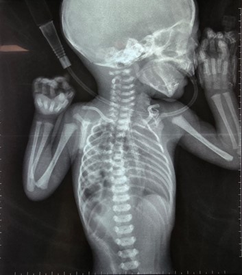
Figure 1: Frontal chest radiograph showing gastrointestinal opacities occupying the right hemithorax, leftward mediastinal shift, and ipsilateral pulmonary hypoplasia, consistent with right-sided congenital diaphragmatic hernia.
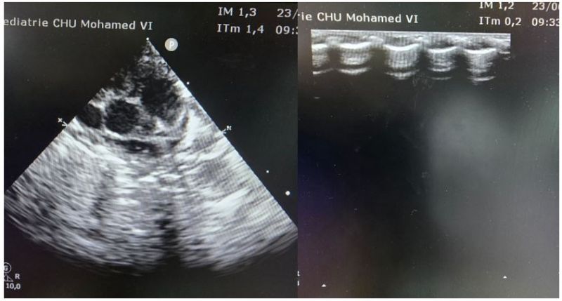
Figure 2: Transthoracic echocardiographic images obtained on day 1 of life, showing situs solitus with a right-sided intrathoracic liver. [Authors: please confirm/complete the specific echocardiographic views and any additional findings shown in each panel.]
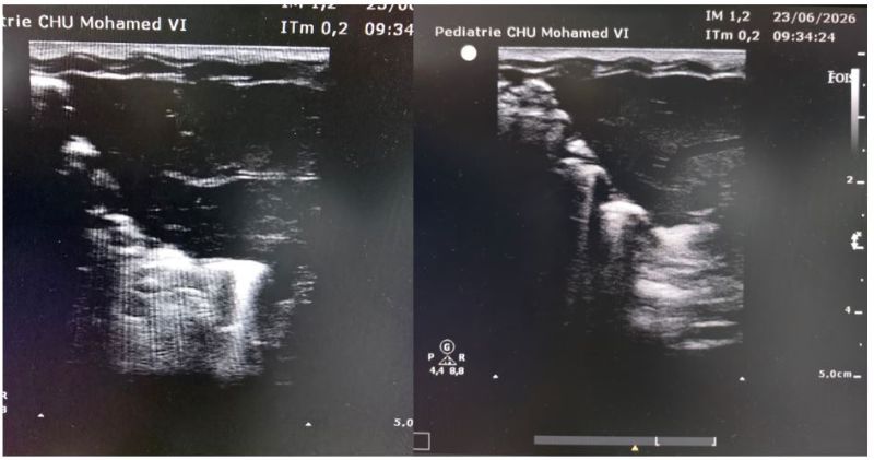
Figure 3: [Legend to be provided by the authors — imaging modality, view, and relevant findings for each panel were not specified in the source material.]
Discussion
Right-sided congenital diaphragmatic hernia is a distinct and particularly complex anatomo-clinical entity, accounting for only 10% to 15% of all CDH cases[1,2,5]. From a diagnostic standpoint, assessing its severity during the fetal and neonatal periods presents specific challenges. In our case, the coexistence of severe intrauterine growth restriction (birth weight of 1,600 g at 37 weeks and 5 days) illustrates a major issue recently highlighted in the literature[6]: overall fetal hypotrophy can lead to underestimation of true lung size relative to eutrophic fetuses on prenatal ultrasound biometry, potentially resulting in a falsely reassuring or, conversely, an overly pessimistic prenatal assessment of pulmonary hypoplasia. [Authors: please verify and, if needed, replace this citation with the specific study documenting IUGR-related bias in prenatal lung-size measurements such as the observed-to-expected lung-to-head ratio.]
In terms of immediate management, the clinical approach to this newborn adhered to the evolving concept of the “Golden Hour” in neonatal resuscitation, whereby standardized, rapid stabilization in the delivery room and neonatal unit is prioritized[7]. Current international and European consensus protocols emphasize that CDH should no longer be viewed as a surgical emergency, but rather as a medical emergency, with priority given to cardiorespiratory stabilization over immediate surgical repair[4,8]. These guidelines recommend strictly avoiding bag-mask ventilation, to prevent distension of the herniated intrathoracic gastrointestinal loops and further pulmonary compression, and favor early endotracheal intubation and protective, gentle ventilation strategies together with early use of targeted vasoactive agents to counter the vicious cycle of hypoxemia and pulmonary arterial hypertension[8,9].
Management of this newborn also had to account for an additional comorbidity: a history of premature rupture of membranes 3 days before delivery. Pulmonary inflammation associated with early-onset neonatal bacterial infection may exacerbate pulmonary vascular dysfunction and worsen pulmonary arterial hypertension[8], potentially compounding the underlying hypoplasia in this patient.
The combined impact of severe IUGR and right-sided CDH proved particularly detrimental in this case. Right-sided CDH has historically carried a poorer prognosis than the left-sided form because of the frequent intrusion of the liver into the fetal thoracic cavity[1,2,5]. In our patient, this severity was considerably compounded by the association with severe IUGR, which drastically reduced physiological reserve and cardiorespiratory adaptive capacity, illustrated by the extent of pulmonary hypoplasia and pulmonary arterial hypertension observed. The death that occurred prior to surgery is consistent with the current consensus that the immediate prognosis of CDH depends primarily on the degree of underlying pulmonary hypoplasia and the responsiveness of the pulmonary vascular bed to hypoxemia, rather than on the timing of surgical repair itself[4,8].
Conclusion
Right-sided congenital diaphragmatic hernia associated with severe intrauterine growth restriction is an extremely severe clinical condition, associated with a very high rate of early neonatal mortality. The death of this newborn before surgical treatment could be undertaken is a reminder that, despite major advances in modern neonatal resuscitation protocols and rigorous application of the “Golden Hour” concept, bilateral pulmonary hypoplasia and refractory pulmonary arterial hypertension remain the primary factors limiting survival. This case underscores the need for further research into prenatal prognostic markers that combine fetal lung volume assessment with individualized growth curves, and highlights the importance of transparent reporting of fatal outcomes to improve understanding of current therapeutic limitations and to refine the counseling provided to parents at the time of prenatal diagnosis.
Conflict of Interest Statement
The authors declare that they have no conflict of interest.



Open Access By Aditum Open Access Journals id licensed under Creative Commons Attribution 4.0 International License. Based On a Work at aditum.org


