Case Reports International Journal
OPEN ACCESS | Volume 3 - Issue 2 - 2025
ISSN No: 3065-6710 | Journal DOI: 10.61148/ 3065-6710/CRIJ
Dr ibtissam larhrabli*; dr naanani othmane ; pr lahjaouj meryem ; pr m. Loudghiri ; pr w. Bijou ; pr y. Oukessou ; pr s. Rouadi ; pr rl abada ; pr m. Roubal ; pr m. Mahtar
ENT Head and Neck Surgery Department, Ibn Rochd University Hospital, Faculty of Medicine and Pharmacy, Hassan II University, Casablanca, Morocco.
*Corresponding author: Dr Ibtissam Larhrabli, Ent Head and Neck Surgery Department, Ibn Rochd University Hospital, Faculty of Medicine and Pharmacy, Hassan II University, Casablanca, Morocco.
Received: June 18, 2025
Accepted: June 27, 2025
Published: July 01, 2025
Citation: Dr Ibtissam Larhrabli, dr naanani othmane, pr lahjaouj meryem, pr m. Loudghiri, pr w. Bijou, pr y. Oukessou, pr s. Rouadi, pr rl abada, pr m. Roubal, pr m. Mahtar. (2025) “Dysphonia revealing laryngeal amyloidosis.” Case Reports International Journal, 3(1); DOI: 10.61148/CRIJ/020.
Copyright: © 2025 Dr Ibtissam Larhrabli. This is an open access article distributed under the Creative Commons Attribution License, which permits unrestricted use, distribution, and reproduction in any medium, provided the original work is properly cited.
Localized amyloidosis is characterized by the accumulation of fibrillar proteins in a specific location of the body without affecting the entire organism [1]. Laryngeal amyloidosis represents an uncommon variant of localized amyloidosis. It is likely that this entity is little known and frequently underdiagnosed. It should be considered when unexplained persistent dysphonia is present [2]. Positive identification, which always relies on histology, is frequently complex and delayed. Despite the rarity of this condition, our observation could guide the informed practitioner in the early detection of this disease and help prevent complications related to its systemic spread. [3-4]
localized amyloidosis
Introduction:
Localized amyloidosis is characterized by the accumulation of fibrillar proteins in a specific location of the body without affecting the entire organism [1]. Laryngeal amyloidosis represents an uncommon variant of localized amyloidosis. It is likely that this entity is little known and frequently underdiagnosed. It should be considered when unexplained persistent dysphonia is present [2]. Positive identification, which always relies on histology, is frequently complex and delayed. Despite the rarity of this condition, our observation could guide the informed practitioner in the early detection of this disease and help prevent complications related to its systemic spread. [3-4]
Observation:
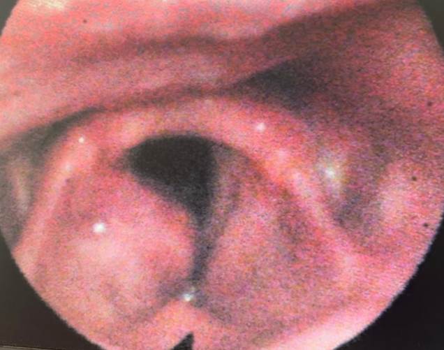
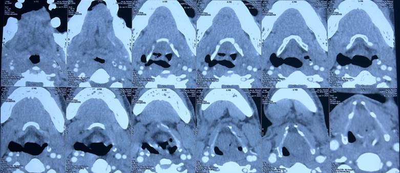
Patient aged 44, chronic smoker for 20 years at a rate of 20 PA without any other particular pathological history, presented with chronic dysphonia resistant to medical treatment for 5 years, causing several general practitioner and ENT consultations, worsening during exercise, a nasofibroscopy was performed on him objectifying a slight swelling of the right ventricular band hiding the epilateral vocal cord, no mass (figure 1), then the patient underwent a CT scan of the larynx objectifying: an asymmetry of the two vocal cords including the hypertrophied right measuring 14mm thick against 5mm on the left without visible mass or nodule, right laryngocele (figure 2).
An LES was performed with deep biopsies at the level of the right CV and BV resulting in: Non-AA laryngeal amyloidosis (figure 3). No other location was objective after a complete assessment requested by the internists, the patient having refused surgery (by laser), was referred to internal medicine for further management with good progress: improvement of dysphonia, but same appearance on nasofibroscopy.
Discussion:
Laryngeal amyloidosis is a disease related to the extracellular deposition in different organs of an amyloid substance, consisting of different protein precursors [1]. The existence of localized amyloidosis generally corresponds to the in-situ production of light chains that deposit near their place of synthesis.
The most common forms are localized amyloidosis of the eyelids, tracheobronchial and vertebral amyloid tumors [2-3]. The first observation of laryngeal amyloidosis was reported in 1875 by Burrow and Neuman [2] represents 0.17 to 1.5% of benign laryngeal tumors. Dysphonia is found in 75%, other signs may be associated: dyspnea, stridor, dry cough, sleep apnea syndrome or dysphagia. Direct laryngoscopy may suggest a neoplastic etiology in the face of a nodular appearance found in 44% of cases. Diagnosis is based on anatomopathological examination with Congo red staining. Once the diagnosis has been established, the nature of the deposits must be specified by an immunohistochemical examination which will allow classification according to the protein nature of the precursor [4]. The diagnosis of laryngeal amyloidosis must seek a systemic extension (renal, cardiac or cutaneous) before retaining the diagnosis of a localized form. The treatment of localized forms essentially involves local surgical procedures, performed by microinstruments or by laser, Wooks and Mcllwain and Talbot use the CO2 laser with a good result [5].
And corticosteroid therapy [2]. Kennedy reported five cases of massive forms of laryngeal amyloidosis with supraglottic localization for which he performed a partial laryngectomy [6-7].

Figure 1: Direct nasofibroscopy showing swelling of the right ventricular band hiding the homolateral vocal cord.

Figure 2: Axial cervical CT scan showing asymmetry of the two vocal cords, including the hypertrophied right one measuring 14 mm thick compared to 5 mm on the left with no visible mass or nodule.

non-AA laryngeal amyloidosis
Conclusion: rare benign pathology, but systemic amyloidosis must be eliminated before retaining the diagnosis of the localized form,
Several therapeutic modalities: the best must be chosen according to the situation in order to preserve the function of the larynx.