
Archives of Clinical Surgery
OPEN ACCESS | Volume 1 - Issue 2 - 2026
ISSN No: - | Journal DOI: 10.61148/ACS


Boudhar H*, Lahjaouj M, Loudghiri M bijou W, Oukessou Y, Abada RL, Rouadi S, Roubal M and Mahtar M
ENT Head and Neck Surgery Department, 20 August Hospital, Ibn Rochd University Hospital, Faculty of Medicine and Pharmacy, Hassan II University of Casablanca, Casablanca, Morocco.
*Corresponding author: Boudhar H, ENT Head and Neck Surgery Department, 20 August Hospital, Ibn Rochd University Hospital, Faculty of Medicine and Pharmacy, Hassan II University of Casablanca, Casablanca, Morocco.
Received: September 01, 2026 | Accepted: September 10, 2026 | Published: September 17, 2026
Citation: Boudhar H, Lahjaouj M, Loudghiri M bijou W, Oukessou Y, Abada RL, Rouadi S, Roubal M and Mahtar M. (2026) “Herpes Zoster Oticus Presenting With Peripheral Facial Palsy and Multiple Cranial Nerve Involvement: A Case Report”, Archives of Clinical Surgery, 1(2); DOI: 10.61148/ACS/006.
Copyright: © 2026. Boudhar H. This is an open access article distributed under the Creative Commons Attribution License, which permits unrestricted use, distribution, and reproduction in any medium, provided the original work is properly cited.
Collet-Sicard syndrome is characterized by unilateral paralysis of the lower cranial nerves IX, X, XI, and XII, resulting from lesions involving the skull base, particularly the jugular foramen and hypoglossal canal [1]. It was first described in 1915 in a World War I soldier whose radiographic examination revealed metallic bullet fragments at the site of the lesions. The most common etiologies include basilar skull fractures and carotid artery dissections. Malignant conditions are also frequently implicated, including parotid tumors, glomus jugulare tumors, hypoglossal nerve schwannomas, skull base tumors, and metastatic lesions. Other reported causes include jugular vein phlebitis and iatrogenic injuries related to internal jugular vein catheterization, cerebral angiography, cardiac surgery, and cerebral vessel clamping [2].
paralysis
Collet-Sicard syndrome is characterized by unilateral paralysis of the lower cranial nerves IX, X, XI, and XII, resulting from lesions involving the skull base, particularly the jugular foramen and hypoglossal canal [1]. It was first described in 1915 in a World War I soldier whose radiographic examination revealed metallic bullet fragments at the site of the lesions. The most common etiologies include basilar skull fractures and carotid artery dissections. Malignant conditions are also frequently implicated, including parotid tumors, glomus jugulare tumors, hypoglossal nerve schwannomas, skull base tumors, and metastatic lesions. Other reported causes include jugular vein phlebitis and iatrogenic injuries related to internal jugular vein catheterization, cerebral angiography, cardiac surgery, and cerebral vessel clamping [2]. Infectious and inflammatory etiologies are less common but have been described, including polyarteritis nodosa, Trousseau syndrome, otitis media, Lyme disease, and varicella-zoster infection [2]. To date, only one case of skull base osteomyelitis complicated by Collet-Sicard syndrome has been reported prior to the present case.
Case report:
A 67-year-old woman with a history of poorly controlled type 2 diabetes mellitus (hemoglobin A1c, 11.2%) was admitted with a 3-day history of worsening dysphagia and a 7-days history of right-sided diffuse redness and erosive, inflamed areas across the concha and inner folds, a hemorrhagic crusted nodular lesion at the lobule, and scattered fresh blood spots consistent with an active inflammatory and vesicular skin process affecting the auricle.
3 days later she developed numbness involving the right side of her face, difficulty closing her right eye, and drooling from the right corner of her mouth. These symptoms were initially considered consistent with Bell’s palsy and were treated with corticosteroids. She subsequently developed a persistent raspy voice. Her dysphagia progressively worsened, with an inability to swallow either pureed food or water.
On admission, physical examination revealed a right-sided facial droop, with inability to raise the right eyebrow or close the right eyelid. Rightward deviation of the tongue was also noted. Examination of the right ear demonstrated marked erythema and edema involving both the external auditory canal and tympanic membrane. A modified barium swallow study revealed impaired peristalsis, pooling within the vallecula and piriform sinuses, and silent aspiration.
Laboratory investigations were notable for mild leukocytosis (10.39 k/mm³; reference range, 3.10–10.20 k/mm³) and significantly elevated inflammatory markers, including an erythrocyte sedimentation rate of 88 mm/h (reference range, 0–10 mm/h) and a C-reactive protein level of 27.0 mg/L (reference, ≤3 mg/L). Culture of the external auditory canal grew pan-sensitive Pseudomonas aeruginosa. Biopsy of the right external auditory canal demonstrated granulation tissue with mixed acute and chronic inflammatory infiltrates, without evidence of cellular atypia or malignancy.
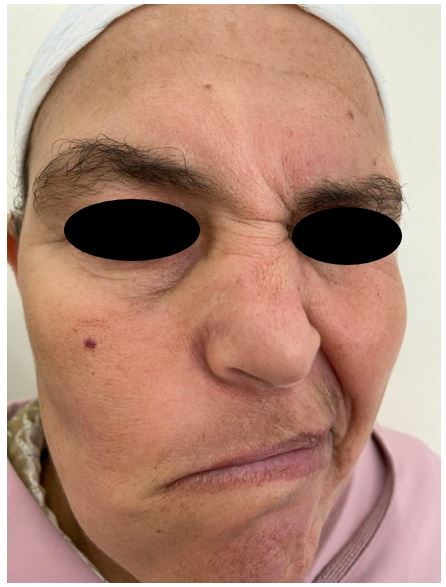
Figure 1. Clinical presentation of right-sided facial nerve palsy in a patient with Collet-Sicard syndrome secondary to skull base osteomyelitis.
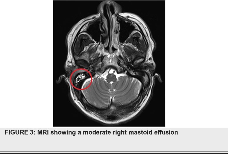
Initial computed tomography (CT) of the head showed no acute intracranial abnormality or lesion involving the internal auditory canal. However, it demonstrated near-complete opacification of the right external auditory canal, effacement of the right tympanic membrane, and enhancement involving the right carotid space and adjacent right parapharyngeal muscles. Magnetic resonance imaging (MRI) of the brain demonstrated a moderate right mastoid effusion (Figure 3). Magnetic resonance angiography of the cervical arterial vasculature showed no significant abnormalities. CT of the cervical soft tissues revealed inflammatory enhancement extending from the right external auditory canal into the right carotid space.
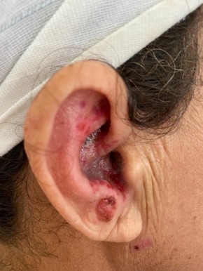
Figure 2. marked edema, erythema, and inflammatory aspect of the pinna.
Given the characteristic imaging findings and markedly elevated inflammatory markers, a diagnosis of skull base osteomyelitis was established.. The patient’s neurological manifestations were therefore attributed to right-sided skull base osteomyelitis secondary to zona, resulting in Collet-Sicard syndrome.
The patient was initially treated with intravenous IV acyclovir and switched to oral 14 days later to complete the course. Because of her severe dysphagia and aspiration risk, a percutaneous gastrostomy tube was placed
At follow-up 2 months after, the patient demonstrated complete resolution of her neurological symptoms and auricle lesions. her swallowing function had also significantly improved, allowing her to resume oral intake. The gastrostomy tube was therefore subsequently removed.
Discussion:
This case represents an unusual disease course, particularly given that only a few similar cases have been reported to date. The patient's symptoms developed following an outbreak of herpes zoster oticus affecting the pinna and external auditory canal, with vesicular lesions preceding the onset of otologic symptoms. The resulting viral-induced tissue damage and local immunosuppression likely created a favorable environment for secondary bacterial superinfection, leading to the development of otitis externa, with Pseudomonas aeruginosa identified as the causative pathogen. The infection subsequently progressed locally to involve the right lateral skull base, resulting in skull base osteomyelitis. The patient’s poorly controlled diabetes mellitus likely contributed to the persistence and progression of the infection.
The infectious process also extended to the right internal auditory canal and cerebellopontine angle, which may explain the patient’s right-sided facial weakness through involvement of cranial nerve VII. Although internal carotid artery stenosis has been reported in other cases of infectious Collet-Sicard syndrome [3], this was unlikely to be responsible for the neurological manifestations in our patient. The patient developed palsies involving cranial nerves IX, X, XI, and XII, consistent with Collet-Sicard syndrome resulting from skull base osteomyelitis extending to the jugular foramen and hypoglossal canal.
An additional distinctive feature of this case is that the skull base infection developed secondary to zona, which typically originates in the auricle [4]. In contrast, previously reported cases of infectious Collet-Sicard syndrome have more commonly been attributed to otitis media. Sibai et al. described a patient who developed a cranial base infection with mass effect following incompletely treated otitis media [4]. Similarly, Blazina et al. reported a case of Collet-Sicard syndrome secondary to chronic otitis media caused by Pseudomonas aeruginosa [5].
Other infectious etiologies were also considered in our patient. Testing for herpes simplex virus (HSV), human immunodeficiency virus (HIV), Borrelia burgdorferi, and varicella-zoster virus was performed, given that these infections have previously been associated with cranial nerve palsies. Kahane et al. described a patient with jugular foramen syndrome associated with a palatolaryngeal herpetic eruption, aseptic meningitis, and elevated serum antibodies against varicella-zoster virus [7]. Kondo et al. reported a case of herpes zoster presenting as meningoencephalitis with involvement of cranial nerves IX to XI [8]. Miyazaki et al. described a patient with hoarseness and dysphagia resulting from pharyngeal and vocal cord palsies associated with Ramsay Hunt syndrome [9]. In another case, a young patient with Lyme disease developed lower cranial nerve palsies [10].
Conclusion:
As illustrated by this case, Ramsey hunt syndrome may progress to skull base osteomyelitis with subsequent involvement of multiple cranial nerves. Recognizing this infectious etiology is crucial, as the neurological manifestations may mimic cerebrovascular disease or other more common neurological conditions, potentially resulting in delayed diagnosis and initiation of appropriate treatment. Clinicians should therefore consider Collet-Sicard syndrome and other cranial neuropathies as potential complications of herpes zoster oticus and skull base osteomyelitis, particularly in elderly patients with poorly controlled diabetes mellitus.


Open Access By Aditum Open Access Journals id licensed under Creative Commons Attribution 4.0 International License. Based On a Work at aditum.org


