Ophthalmology and Vision Care
OPEN ACCESS | Volume 6 - Issue 1 - 2026
ISSN No: 2836-2853 | Journal DOI: 10.61148/2836-2853/OVC


Vinay Gupta
Assistant Professor, Department of Ophthalmology, IGMC, Shimla.
*Corresponding Author: Vinay Gupta, Assistant Professor, Department of Ophthalmology, IGMC, Shimla.
Received: September 09, 2021
Accepted: September 16, 2021
Published: September 24, 2021
Citation: Vinay Gupta. (2021) “Live eye donor - two rare cases in two different situations”, Ophthalmology and Vision Care, 1(4); DOI: http;//doi.org/08.2021/1.1018.
Copyright: © 2021 O. Vinay Gupta. This is an open access article distributed under the Creative Commons Attribution License, which permits unrestricted use, distribution, and reproduction in any medium, provided the original work is properly Cited.
The promotion of eye donation has always been a challenging task for any individual or institute, and hence there is more demand and less supply of corneal tissue. Legally corneal donations are usually cadaveric allograft. But living eye donation though rare is sometimes done. We report two rare cases in which the cornea of the first case was retrieved from living donor, who had traumatic expulsion of right eye in road traffic accident. So, the cornea of this living donor after enucleation of globe was utilized to restore the vision of another corneal blind person. The second case was a patient with failed keratoplasty left eye who was managed by taking graft from his other eye having glaucomatous optic atrophy.
Introduction:
Corneal blindness is one of the major causes of visual deficiency after cataract, glaucoma and age-related macular degeneration (AMD), contributing 5.1% of global blindness [1] There are approximately 6.8 million people who have vision less than 6/60 in at least one eye due to corneal diseases; of these, about a million have bilateral involvement [2]. According to global survey of corneal transplantation and eye banking, there is considerable shortage of corneal graft tissue, with only 1 cornea available for 70 needed. [3] This backlog can only be cleared by making good eye banking services available along with improved collection of eyes and proper facilities for tissue procurement and improved storage. [4]
Living eye donations are extremely rare and not considered viable sources of corneas, as donation would result in blind eye. [5] In auto-keratoplasty, a patient has healthy cornea in one eye and corneal blindness in the other eye and hence patient is a live donor in which the grafts are either exchanged or donor eye is grafted with patch graft. In the first case the cornea was retrieved from living donor, who had traumatic expulsion of right eye in road traffic accident. So, cornea of this living donor after enucleation of globe was utilized to restore the vision of another corneal blind person. The second case was of a patient with failed keratoplasty in left eye who was managed by taking graft from his other eye having glaucomatous optic atrophy.
Case 1
A 40 years old male presented with traumatic expulsion of right eyeball with optic nerve avulsion following motor bike accident. Patient was conscious, well oriented to time, place person with no other vital injury of other parts. On ocular examination right globe was protruding out of orbit with minimal tissue attachment.(Figure 1)Wooden foreign body was present between right lateral orbital wall and right globe.(Figure 2) The cornea of injured eye was clear but pupil was dilated and non-reacting without any perception of light. Lid laceration of 4 x 2cm was present on right upper eyelid. Ecchymosis was present in both upper and lower eyelids.
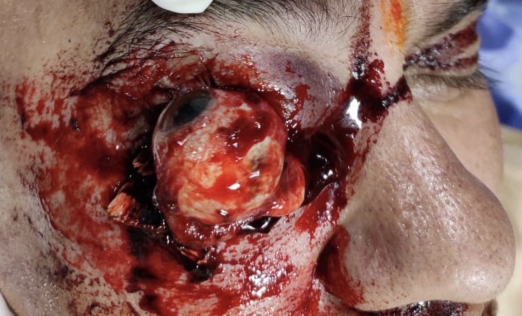
Figure 1: Case 1-Traumatic avulsion of right eye globe.
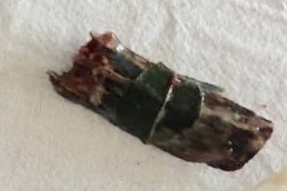
Figure 2: Case 1-Wooden foreign body.
Visual acuity in left eye was 6/6 with normal pupil reactions. Ocular movements and fundus were normal. The CT scan showed protrusion of right globe with optic nerve avulsion. Patient was started on intravenous antibiotics and enucleation of right eye was done along with removal of 5 × 2 cm size foreign body. The right eyelid laceration was repaired. Cornea of patient was preserved after written consent of the patient and used for keratoplasty on another patient having pseudophakic bullous keratopathy right eye. (Figure 3).

Figure 3: Case 1-Live donor at 3 months
Case 2:
A 75 years old male presented with painless progressive loss of vision in right eye for 12 years. The patient had history of trauma in left eye following which he lost vision in this eye. He underwent penetrating keratoplasty in left eye, but 6 months later patient developed pain, redness, watering and diminution of vision left eye. The patient was diagnosed of as graft rejection and put on oral prednisolone and topical prednisolone, G homatropine, G Timolol. The patient was lost to follow up and came after 4 years. On ocular examination there was no perception of light in right eye with afferent pupillary defect. The intraocular pressure was high 32mm of Hg and anterior segment was normal. Fundus examination showed complete glaucomatous optic atrophy.Visual acuity in left eye was HMCF. Cornea was almost opaque due to failed penetrating keratoplasty.The intraocular pressure was normal and B scan showed no gross abnormality of posterior segment.
After informed written consent the patient was planned for autograft from opposite absolute eye having clear cornea. But following graft excision from right eye the expulsive haemorrhage developed despite preoperative I/V Mannitol and Acetazolamide and eye has to be eviscerated. The full thickness corneal graft taken from this eye was put on the other eye. Intraoperative procedure was uneventful in this eye. Post-operatively patient had normal graft host junction, some Descemets folds on the graft, well-formed anterior chamber in left eye and visual acuity of 6/60. (Figure 4) The patient could move independently, which was not possible before surgery. He was started on standard post-operative treatment in left eye for keratoplasty and antibiotics in right eye. At 6 weeks follow up visual acuity in the left eye was 6/24. The cornea was clear, and the graft host junction was well healed up and intraocular pressure was normal.
Figures and legends

Figure 4: Case 2- Postoperative autokeratoplasty at 1 week.
Discussion:
Living eye donation although rare is usually done as an autograft taken from contralateral blind eye with healthy cornea to another corneal blind eye of same patient. The grafts are either exchanged or donor eye is grafted with patch graft. Since this patient has already failed graft in other eye, so putting rejected graft of one eye on the same body would have triggered definite inflammation in this eye. Although the plan on donor eye was to put a patch graft, but patient developed expulsive haemorrhage following trephining. So, the graft was taken, and eye was eviscerated, such absolute eyes for long duration have possibility of developing this complication despite suitable precautions. This could be cosmetically disfiguring to the face, but sight was more important for the patient.
The traumatic damage to eyeball usually spoils the cornea by the time patient reaches to the hospital but the second case not only had healthy cornea but also reached well in time and hence cornea was suitable for transplantation. Although situations were altogether different, this case series presents rare type of eye donations from a living donor.
Globe avulsion is a rare condition usually resulting from severe trauma to the orbit and face. Mid facial injuries with orbital injuries which are often associated with fractures of the orbit floor, medial wall or the roof resulting in herniation of eyeball contents into the surrounding structures. Several theories of eyeball luxation have been proposed. The Morris theory elaborately described the medial entry point and three different mechanisms for eyeball luxation. :(1) propulsion of the globe forward after entering an elongated object to the medial orbit; (2) a wedge-shaped object enters the orbit medially and displaces the globe anteriorly; and (3) optic nerve direct transection by a penetrating object. Song and Carter described abrupt deceleration as a cause of globe luxation. [6, 7] In the present case series, case 1, the patient i.e. live donor had severe ocular injury that the eye was unsalvageable. Therefore, the corneal tissue of this enucleated eye was preserved and penetrating keratoplasty was performed in the other patient having pseudophakic bullous keratopathy in the right eye so that he could self-perform his routine activities.
Autokeratoplasty can be performed as an alternative to conventional allograft transplantation for the management of corneal blindness in patients with a healthy cornea in the fellow blind eye.[8] In case 2, penetrating autokeratoplasty was performed so that visually blind person could be self-dependent. Moreover, the complication of allograft rejection is eliminated and also side effects from long term corticosteroids can be avoided by utilizing autologous tissue. In selected patients, autokeratoplasty can be a useful technique, especially if heavy corneal vascularization or pre-existing glaucoma is present. [9]
Conclusion:
In live eye donor, in autokeratoplasty, a person usually has healthy cornea in one eye and the other eye is blind due to corneal opacity. The grafts are either exchanged or donor eye is grafted with patch graft. Since this patient has already failed graft in other eye, so transplanting this graft to other eye could have made this eye symptomatic due to inflammation. So, the best option was to take the graft and eviscerate this eye. In the other case of present study, the patient not only had the healthy cornea in traumatized globe but also reached hospital well in time, and hence cornea was suitable for transplantation, which is usually spoiled otherwise in these situations. Recent advances in corneal therapies are now focusing on tissue from living-related donors but living eye donation can be a viable option where healthy corneas in blind eyes are otherwise rendered unproductive.


Open Access By Aditum Open Access Journals id licensed under Creative Commons Attribution 4.0 International License. Based On a Work at aditum.org