Veerappan 1, Robin 2, Gokul 3, Krishna Prasad. T 4*
1Associate professor in Anaesthesiology, Shri Sathya Sai Medical College and RI, Ammapettai, 603108, (Sri Balaji Unversity, Deemed To Be University) Kancheepuram, India.
2Resident In surgery, Shri Sathya Sai Medical College and RI, Ammapettai, 603108, (Sri Balaji Unversity, Deemed To Be University) Kancheepuram, India.
3Professor in surgery, Shri Sathya Sai Medical College and RI, Ammapettai, 603108.
4Professor in Anaesthesiology, Shri Sathya Sai Medical College and RI, Ammapettai, 603108, (Sri Balaji Unversity, Deemed To Be University) Kancheepuram, India
*Corresponding Author: Krishna Prasad. T, Professor in Anaesthesiology, Shri Sathya Sai Medical College and RI, Ammapettai, 603108, (Sri Balaji Unversity, Deemed To Be University) Kancheepuram, India.
Received Date: June 15, 2024
Accepted Date: June 22, 2024
Published Date: June 25, 2024
Citation: Veerappan, Robin, Gokul, Krishna Prasad. T. (2024) “Cystoscopic Removal of Unusual Foreign Body in the Bladder.”, International Surgery Case Reports, 6(2); DOI: 10.61148/2836-2845/ISCR/076
Copyright: © 2024. Krishna Prasad. T. This is an open access article distributed under the Creative Commons Attribution License, which permits unrestricted use, distribution, and reproduction in any medium, provided the original work is properly cited.
There were many reported cases of foreign bodies in the bladder, however, this case is interesting because of its presentation and its approach. A young boy reported to OPD with lower urinary tract symptoms for the past week. Radiological evaluation revealed coiled-up foreign body in the urinary bladder which was suspected to be a displaced DJ stent. The patient was taken up for surgery using a urethrocystoscopy and surprisingly a coiled insulated electric wire was found in the bladder. While we did the literature search, it suggested that open surgery is the usual standard approach, here in our case, we removed the wire through the urethral route despite having sharp edges of the wire pointing outwards.
bladder; OPD
Case presentation:
A 17-year-old boy was brought to our outpatient department by his mother with a history of lower abdomen pain of one-month duration. He also complained of passing reddish-coloured urine which was associated with a burning discomfort upon voiding for the last week. The lower abdominal pain was dull and persistent which was not radiating. The urine was reddish colored. He did not give any history of fever, chills, loin pain, groin pain, pyuria, or altered bowel habits. He had no previous medical history. The mother informed us that the boy had been consuming alcohol daily for the last three years and she was unable to get him to stop. The boy also agreed to the same and admitted to consuming around 180ml of whiskey or brandy daily. He had visited a doctor for the same urinary complaint five days back and was clinically diagnosed with a urinary tract infection and started on medication.
The patient however complained that there was no improvement in symptoms despite taking all the prescribed medication for the last five days. Upon examination, he was moderately built and was found to be anemic. His vitals were stable. His abdomen was scaphoid in shape and soft on palpation; however, he had suprapubic tenderness. There was no guarding or rigidity. Bowel sounds were heard normally. The penis was normal, but mild erythema was noted at the urethral meatus. No discharge was seen from the meatus.
Samples were sent for blood and urine routine examination. Blood investigations were normal, however, the urine routine showed 10-15 red blood cells/hpf and 10-12 pus cells/hpf.
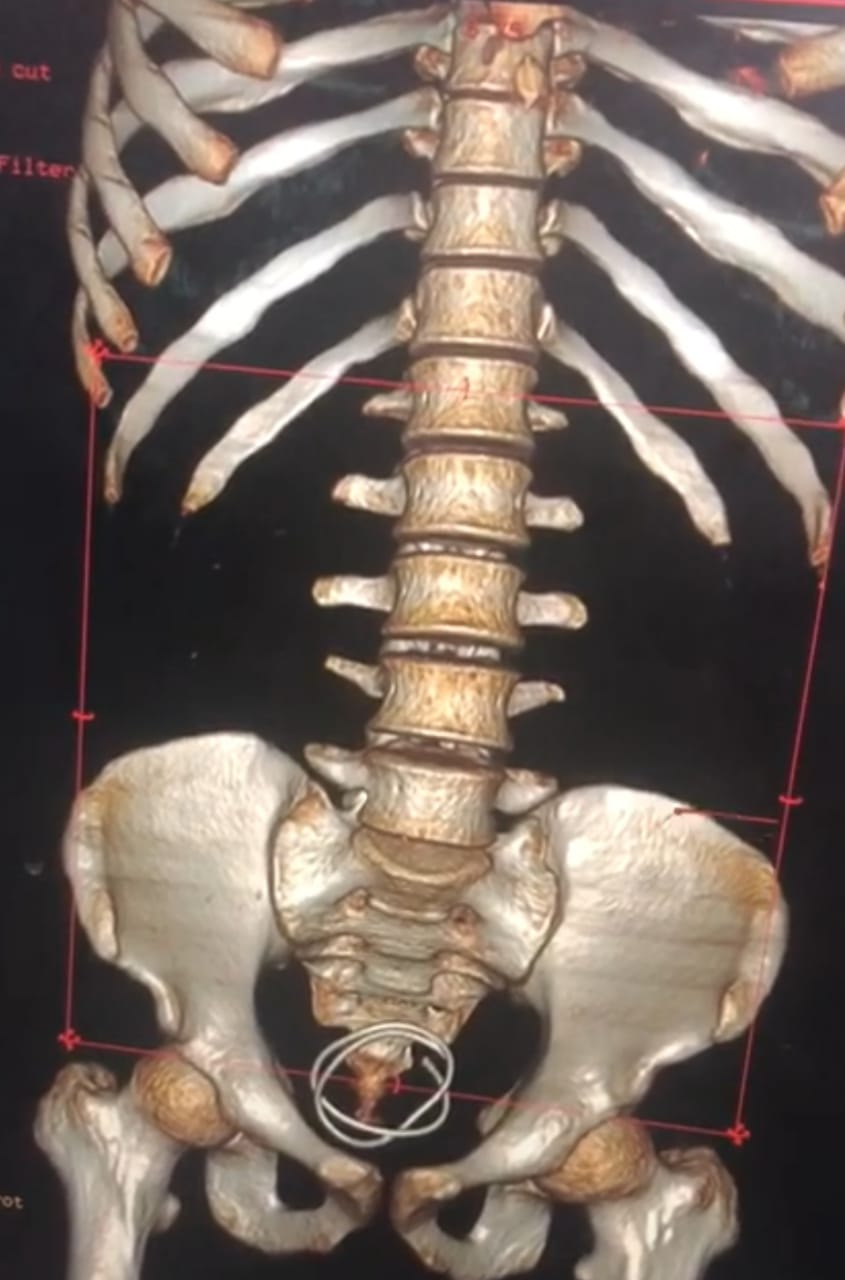
Urine culture subsequently showed a growth of E.Coli at a concentration of 107 colony counts/mm3. Due to the duration of the complaints, an ultrasound scan KUB was also ordered. The ultrasound study showed a long hyperechoic entity within the bladder which was reported to possibly be a displaced DJ stent. An x-ray KUB was subsequently done which showed a radiopaque foreign body in the bladder. Since the patient gave no history of any previous medical procedures, a CT KUB study was planned. CT KUB shows a coiled wire in the urinary bladder on 3D reconstruction [Fig1]. The need for endoscopic retrieval was explained to the patient including the possibility of open surgery and he was subjected to a urethrocystoscopy under spinal anesthesia. A coiled wire with encrustation was seen in the bladder.

Figure 1: CT KUB showed a coiled wire in the urinary bladder on 3D reconstruction.
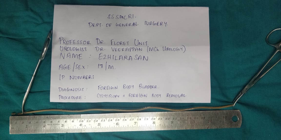
On manipulation, it was rigid and not pliable. A moray micro forceps was used to grasp and break the encrustation at the tip which fragmented and exposed an electrical wire with insulation. The tip of the wire was held with biopsy forceps and carefully negotiated transurethrally with difficulty. The entire wire was successfully removed transurethrally itself and the patient was catheterized with 14 Fr Foley’s catheter after revisualisation of the bladder and ensuring there were no iatrogenic injuries [fig2]. The patient did not disclose any history regarding the insertion of the foreign body per urethra. A psychiatric consult was obtained but the patient’s confidence could not be gained. He was treated with antibiotics and was discharged after the removal of the foleys in three days.

Figure 2: Coiled insulated electric wire after removal transurethrally ( 30cm in length).
Discussion:
While foreign bodies in the male urinary bladder and urethra are a relatively uncommon case to see in the OPD, a sizeable number of such cases have been reported in literature. The nature of these objects is quite interesting and some even outright scary. They include sharp and lacerating objects like needles and pencils, plants and vegetables like cucumber, glass ampules, batteries, fluids like glue, and even powders like cocaine. (1-4) The most common reason for the self-insertion of a foreign body into the male urethra is erotic or sexual in nature. While eroticism and sexual urges are common to the entire human race, one may argue that inserting objects into the urethra is anything but pleasurable and not normal. (5,6).
A few interesting psychiatric-psychoanalytic theories have been postulated. According to Kenney RD – the initiating event is the coincidentally discovered pleasurable stimulation of the urethra, followed by a repetition of this action with objects of unknown danger, driven by a particular psychological predisposition to sexual gratification. (7) Wise TN et al considered urethral manipulation as a paraphilia combining sadomasochistic and fetishist elements where the orgasm of the individual depends on the presence of the fetish. He believed it showed a regression to a urethral stage of erotism due to a traumatic event or a strong libidinal drive. Sexual experimentation and fetishes among sexual partners had been found to be a reason for foreign bodies in the urethra or bladder. (8) Alcohol or drug intoxication has also been found to be associated with the insertion of foreign bodies per urethra, probably due to sexual deviation or even sexual assault following incapacitation. Sexual assault is an important reason and often in the majority of cases, the patient does not seek medical help out of fear and embarrassment.
Other instances of migration of foreign bodies or iatrogenic causes have also been identified. In our case, the patient had no previous medical history and was an alcoholic. He did not agree to self-insertion or sexual experimentation with a partner and was unwilling to pursue any conversation in that direction. From a clinical standpoint, these patients must undergo a psychiatric evaluation, based on theories that this act may be an indication of a potentially self-punishing impulsive behavior, which may even lead to suicide.
On the other hand, the psychiatric evaluation of these patients is often controversial as many of these patients may have no psychological abnormality. In our case, a psychiatric evaluation was performed, however, the patient’s confidence could not be gained and he did not review for follow-up.
A common pattern of clinical presentation is with lower urinary tract symptoms mimicking urinary tract infections. Some patients present asymptomatically only to incidentally find the foreign body on radiological evaluation for some other disease. Others may present with dysuria, hematuria, pyuria, poor urinary stream or urinary retention, urethritis, bloody or purulent urethral discharge, and ascending urinary tract infections. While a similar case hasbeen described earlier, open surgery is usually the method of choice as endoscopic retrieval is often difficult and tricky. Removal of the foreign body may be quite challenging requiring imagination, spontaneous innovation, and a high level of surgical skills.
It is not possible to describe the standard operation to be done for different types of foreign bodies. Objects may be stuck in different parts of the lower urinary tract and the technique of retrieval should be individually decided on a case-to-case basis depending on the merits of the case and the best possible outcome. While endoscopic retrieval should ideally be given preference, other more invasive options such as open surgery for a foreign body in the bladder or meatotomy for a urethral foreign body should not be looked down upon. The authors disagree with the general idea that what was pushed in through the meatus can also come out through the meatus. While the act of inserting it in is often done in a sexually delicate fashion using one's fingers, retrieval of the same endoscopically requires the use of a grasper which should be able to hold onto the foreign body without breaking it or tearing it and causing more harm. In cases where endoscopic procedures are unsuccessful, then open surgery is usually the only way forward.
Follow up:
The patient was admitted for 3 days after the foreign body removal and watched for bleeding or any other complications. And discharged on the 4th day, followed up after 10 days, and found no significant problems.
Conclusion:
A prompt evaluation with the right history, examination, and imaging is advised to ascertain the item's size and location and to improve efficient extraction. Even the distantly situated FB can be successfully removed transurethrally with endoscopic retrieval combined with fluoroscopy (CT - KUB). A more comprehensive management strategy is needed, one that addresses infection control procedures in addition to reducing the risk of more urethral injuries, identifying and recording more serious underlying injuries, and keeping an eye on postponed problems.
Source of funding: Self.
Declaration of patient consent:
There are no Ethical issues, declared by the author.
The patient willingly gave his consent, along with the guardian for the usage of all relevant clinical information and photos given academic importance.
He was aware the names and initials would remain anonymous and that every effort would be taken to hide the patient’s identity.