Saeed Asgary
Iranian Centre for Endodontic Research, Research Institute of Dental Sciences, Shahid Beheshti University of Medical Sciences, Evin, Tehran, Iran.
*Corresponding author: Saeed Asgary, Iranian Centre for Endodontic Research, Research Institute of Dental Sciences, Shahid Beheshti University of Medical Sciences, Evin, Tehran, Iran.
Received: December 22, 2023
Accepted: January 10, 2023
Published: January 18, 2023
Citation: Asgary S. (2024) “Successful Management of a Molar with Vertical Root Fracture through Root Amputation: A Case Report.”. International Surgery Case Reports, 6(1); DOI: http;//doi.org/07.2024/1.1068.
Copyright: © 2024 Saeed Asgary. This is an open access article distributed under the Creative Commons Attribution License, which permits unrestricted use, distribution, and reproduction in any medium, provided the original work is properly cited.
This case report showcases the effective utilization of a less commonly employed treatment approach to salvage a diseased multi-rooted tooth as a preferable alternative to extraction and implantation. Root amputation provides a means to preserve multi-rooted teeth afflicted with furcation involvement and/or defective roots. The case involved a 38-year-old female patient with a failed mesiobuccal root in her maxillary left first molar, which had previously undergone endodontic surgery and received a well-fitted crown; however, it later failed due to a vertical root fracture. The procedure included a mini mucoperiosteal flap and amputation of the affected root, followed by postoperative instructions for the patient. Subsequent one-year follow-up, involving clinical examinations, periapical radiographs, and cone-beam computed tomography, revealed the tooth's integrity and functionality, with no reported signs or symptoms except for minor radiolucency. This case illustrates that root amputation can effectively manage seemingly hopeless teeth.
root amputation; vertical root fracture; endodontic surgery
Introduction
Vertical root fractures in molars often pose a significant clinical challenge, typically leading to a poor prognosis and the need for extraction [1]. These fractures can result from various factors, including endodontic procedures and the normal wear and tear of daily oral functions. When a molar exhibits signs of vertical root fracture, the treatment choices may seem limited, with extraction and subsequent implantation frequently viewed as the primary solution.
However, in this case report, we present an alternative approach that has been infrequently employed but holds promise in the management of multi-rooted molars with vertical root fractures. Root amputation, a surgical procedure that involves the surgical removal of an affected root while retaining the remaining healthy roots [2], offers a means to salvage such teeth with a poor prognosis. This procedure not only preserves the natural dentition but also avoids the complexities and costs associated with implant-based rehabilitation.
Our case centers on a 38-year-old female patient who presented with a failed mesiobuccal root in her maxillary left first molar. This tooth had previously undergone successful endodontic surgery and received a well-fitted crown but faced an uncertain fate due to a vertical root fracture.
Case Report
A 38-year-old female patient was referred to our dental clinic with a chief complaint of persistent discomfort and pain associated with her maxillary left first molar. On clinical examination, it was evident that the patient's molar had undergone previous surgical endodontic retreatment and was restored with a well-fitted crown. The patient reported a history of recurrent discomfort and episodes of mild pain, particularly when chewing and applying pressure on the tooth. The OPG of patient which was taken 1 year ago revealed a successful root-end surgery (Figure 1A); however, radiographic evaluation using periapical radiographs revealed a concerning finding – a wraparound lesion in the mesiobuccal root of the first molar demonstrating a vertical root fracture (Figure 1B). The diagnosis of a vertical root fracture was further supported by the clinical examination and presence of a localized deep pocket alongside the fracture line. Upon further inquiry, the patient disclosed that she had experienced a notable increase in discomfort and sensitivity in the tooth in recent weeks. The patient's dental and medical histories were reviewed, and no relevant systemic conditions or contributory medications were identified. The patient had maintained good oral hygiene practices.
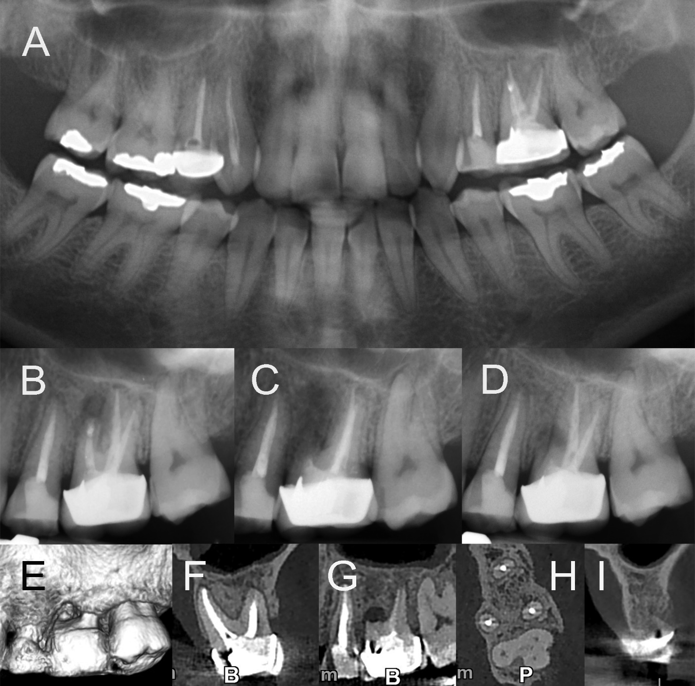
Figure 1: Preoperative, Diagnostic, Postoperative, and follow-up Radiographs of the case. A) Preoperative radiograph of the patient taken one year ago, illustrating a successful root-end surgery on the maxillary left first molar. B) Diagnostic periapical radiograph demonstrating a large lesion in the mesiobuccal root of the maxillary left first molar. C) An image depicting the surgical procedure of root amputation, with the mesiobuccal root being surgically removed while preserving the integrity of the remaining healthy roots. D) Periapical radiograph taken at the one-year follow-up, showing the healing process and stability of the treated tooth. E-I) A series of Cone-Beam Computed Tomography images providing detailed three-dimensional views of the surgical site, illustrating the postoperative condition and the integrity of the surrounding structures.
Given the clinical presentation and radiographic evidence, the diagnosis of a vertical root fracture in the mesiobuccal root of the maxillary left first molar was confirmed. The challenging nature of vertical root fractures, their poor prognosis, and the potential for further complications, such as infection, led to a comprehensive discussion of treatment options and prognosis with the patient. The range of available treatments, including extraction followed by implantation, and root amputation, was presented to the patient. Ultimately, after careful consideration and weighing the risks and benefits, the patient and the dental team decided to proceed with root amputation as the chosen treatment approach for salvaging the affected tooth. The rationale for this choice was based on the desire to retain natural dentition, avoid the complexities and costs associated with implant-based rehabilitation, and provide the patient with a suitable long-term solution for her dental health. The patient provided informed consent, and the root amputation procedure was scheduled.
The root amputation procedure was conducted in accordance with established surgical protocols. The patient was informed about the surgical process and provided with preoperative instructions to ensure optimal conditions for the procedure. On the day of the root amputation, local anesthesia was administered. A mini mucoperiosteal flap was carefully raised to access the affected root and surrounding area. The mesiobuccal root was then amputated surgically, removing the fractured portion while preserving the integrity of the remaining healthy roots (Figure 1C). The site was thoroughly irrigated, and meticulous care was taken to ensure hemostasis. Suturing was performed to close the surgical site, and the patient was given postoperative instructions and medications for pain management and infection control.
A critical aspect of the treatment plan included a comprehensive follow-up strategy to monitor the healing and overall condition of the treated tooth. Clinical examinations, including assessment of occlusion and periodontal health, were conducted regularly to ensure that the tooth remained functional and free from discomfort. Radiographic evaluations, including periapical radiographs (Figure 1D) and cone-beam computed tomography (Figure 1E-I), were performed at the one-year follow-up to assess the integrity of the tooth and its surrounding structures. The follow-up revealed a remarkable outcome. The tooth, following root amputation, maintained its integrity and functionality, with no reported signs or symptoms of discomfort. The radiographic evaluations showed favorable healing and stability, with only a minor radiolucency observed in the area of the surgical site, indicating the expected healing process.
Discussion
This case report demonstrates that root amputation can be a successful alternative for the management of multi-rooted molars with vertical root fractures. The patient's decision to opt for this approach was driven by the desire to preserve her natural dentition and avoid the complexities and costs associated with implant-based rehabilitation.
The successful outcome achieved in this case underscores the importance of meticulous treatment planning, accurate diagnosis, and case selection. It also emphasizes the role of maintaining excellent oral hygiene and vigilant follow-up in achieving long-term success. Root amputation provides a viable option for preserving seemingly hopeless teeth and offers an alternative to extraction and implantation in appropriate clinical scenarios.
Conclusion
The case presented here highlights the potential benefits of root amputation in managing multi-rooted molars with vertical root fractures. It demonstrates that, with careful consideration and a patient-centered approach, this less commonly employed technique can be a valuable addition to the armamentarium of dental practitioners, offering patients the opportunity to retain their natural dentition and achieve successful long-term outcomes.
Funding Statement: No source of funding
Disclosures: The authors have nothing to disclose

Open Access By Aditum Open Access Journals id licensed under Creative Commons Attribution 4.0 International License. Based On a Work at aditum.org