International Surgery Case Reports
OPEN ACCESS | Volume 7 - Issue 1 - 2025
ISSN No: 2836-2845 | Journal DOI: 10.61148/2836-2845/ISCR


Loraine Entringer Falqueto 1, Lissa Tiemi Araki 2, Jaline de Souza Bonetti 3, Lucas Koji Matsuzaki 4, Kellen Regina Ferri 5, Fernando Antônio Bersani Amado 6*
1 Pediatric Surgery of “Hospital Pequeno Príncipe”, Parana-Brazil.
2 Medicine of Pontifical Catholic University of Parana, Parana-Brazil.
3 Medicine of Pontifical Catholic University of Paraná, Parana-Brazil.
4 Pediatric Surgery of “Hospital Pequeno Príncipe”, Parana-Brazil.
5 Neonatology of “Hospital Pequeno Príncipe”, Parana-Brazil.
6 Pediatric Surgery of “Hospital Pequeno Príncipe”, Parana-Brazil.
*Corresponding Author: Fernando Antônio Bersani Amado, Address: Rua Desembargador Motta, nº 1070, Água Verde, Curitiba, Paraná, Brasil.
Received date: December 01, 2021
Accepted date: December 06, 2021
published date: December 10, 2021
Citation: Loraine E Falqueto, Lissa T Araki, Souza Bonetti J D, Lucas K Matsuzaki, Kellen R Ferri, (2021) “Surprise in The Delivery Room: Scrotoschisis, Case Report.”. International Surgery Case Reports, 3(3); DOI: http;//doi.org/11.2021/1.1042.
Copyright: © 2021 Fernando Antônio Bersani Amado. This is an open access article distributed under the Creative Commons Attribution License, which permits unrestricted use, distribution, and reproduction in any medium, provided the original work is properly cited
The congenital defect in the scrotal wall through which a testicle is prolapsed and exposed is known as scrotoschisis. This birth abnormality is very rare, with less than 25 cases reported in the international literature to date.(1) It normally affects healthy infants with no family recurrency or ethnical preference. Its etiology is not yet well understood, but there are various theories to explain its physiopathology. Currently the most accepted hypothesis points to the outcome of meconial periorchitis and its subsequent inflammation.(2) We report a case of scrotoschisis in neonate of 34 weeks of gestation, premature due to mother’s development of uterine rupture. He was surgically treated with an orchidopexy associated with inguinal herniorrhaphy and closure of the scrotal sac. His recovery was uneventful.
Introduction
Scrotoschisis is a rare congenital defect that affects the scrotal wall normally leading to a unilateral testicular extrusion of otherwise healthy infants. There are less than 25 cases reported in the international literature today [1]. Its etiology is not yet well understood, but its primary hypothesis suggests that it a result of meconial perorchitis.(2) The present report describes a case of scrotoschisis in a premature newborn delivered by emergency cesarian after mother’s uterine rupture.
Case Report
Male neonate, premature of 34 weeks of gestation, was born by emergency cesarean delivery on account of uterine rupture. Even though the pregnancy was achieved by in vitro fertilization due to mother’s endometriosis, and her being 35 years of age at the time of insemination, there were no complications during pregnancy. There were also no fetal malformations detected nor were other maternal illnesses.
During delivery, patient developed bradycardia and bradypnea, with satisfactory response to one cycle of positive-pressure ventilation, APGAR 4 and 8, birth weight of 2320g. On physical examination at the delivery room, it was detected an extrusion of the vaginal sac containing the right scrotum (Figure 1).
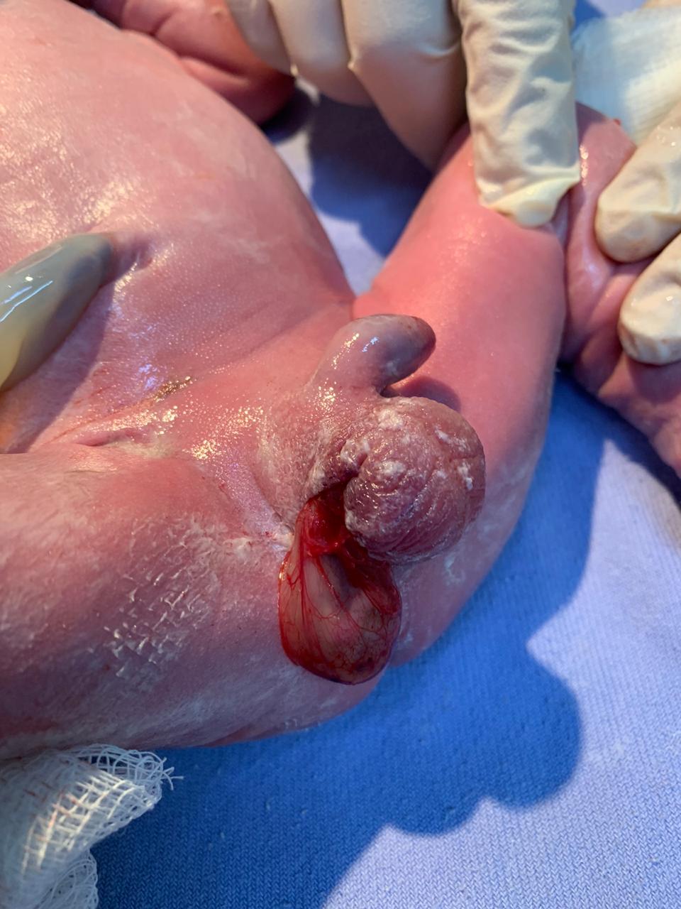
Figure 1: Scrotoschisis. Diagnosis in the delivery room. Note the tunica vaginalis preserved with extrusion due to scrotal open.
Facing the diagnosis of scrotoschisis the patient was transferred to a pediatric specialized hospital unit and operated on day of life one. Other malformations were excluded with complementary investigation.
Scrotoschisis surgical treatment was done by transscrotum approach with patient under general anesthesia. It consisted of right orchidopexy associated with inguinal herniorrhaphy and closure of the scrotal sac (Figure 2).
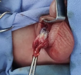
Figure 2: Surgery treatment, high ligature of persistence of the peritoneum-vaginal conduit and orchidopexy.
During the surgical evaluation there were no signs of recent trauma, infection, inflammation, or meconium residue on the defect. After extubation in the operating room the patient returned to the neonatal intensive care unit (NICU). Due to prematurity and need of weight gain, he remained in the NICU for 18 days, and was discharged on the 19th day of hospitalization.
Post-operative ultrasound showed no morphological alterations, with testis in adequate to age dimensions and absence of hydrocele, varicocele, or other collections. During ambulatory follow-up the complete surgical wound healing (Figures 3), and adequate testicular development were achieved.
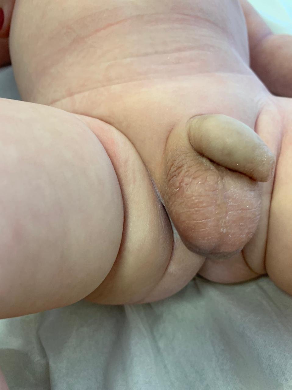
Figure 3: Final aspect, 3 months later.
There were no signs of testicular atrophy and size was similar to the contralateral organ. Child has remained well at 12 months of follow-up.
Discussion
Scrotoschisis is a rare congenital anomaly with idiopathic etiology, normally occurring unilaterally. The first scrotoschisis report was published in 1963 by Von der Leyern et al. Since than less than 25 cases were reported in international literature [3].
This anomaly doesn’t appear to have relation with any other congenital alterations, being found only 2 cases associated with Beckwith-Wiedmann syndrome. There was no literary evidence of recurrence in family members or ethnical preference. It occurs most often in the right hemiscrotum but can also happen on the left side or even bilaterally in a few cases of middle line lesions. The scrotum wall defect through which the testis exits is more frequently found in anterior and cranial regions, ipsilateraly to the defect and immediately lateral to the median raphe of the scrotum – as was our case. The prolapsed testicles are mostly viable (as long as there are no associated testicular torsions). They are surrounded by their anatomical layers, excluding the skin, and are suspended only by the spermatic cord [2].
A variety of hypothesis as to the physiopathology of scrotoschisis were brought up. Among them is the result of an inadequate development of the gubernacular mesenchyme that could lead to the rupture of the scrotum wall by avascular necrosis, which was brought up by Gongaware et al. in 1991. This theory has little acceptance due to the scrotum’s rich vasculature. Another theory says that it is caused by iatrogenesis during cesarian delivery, but this too have little acceptance for most of the reported cases deliveries were vaginal, and for the absence of signs of recent trauma in the lesions. A third hypothesis affirms that the defect is a consequence of embryologic errors on the fusion of the labioscrotal folds or nondevelopment of the cremasteric muscle. However, the development of this structures take place in the first semester of gestation, while the descent of the testicles happens at least 2 months before term. Besides, in vitro tests in rats have shown that defects on the scrotal wall made on fetal life have a tendency to regenerate before birth [2].
Currently the most accepted hypothesis is that scrotoschisis is a consequence of a meconial periorchitis. Its physiopathology is based on the theory that an intestine rupture late in pregnancy leads to the overflow of meconium to the peritoneal cavity. This meconium would than pass through the patent peritoniovaginal conduct, resulting in a meconial periorchitis that would cause local inflammation. The final outcome would be the rupture of the scrotum wall and extrusion of the testis [4]. There were 5 reported cases in which meconial residue was present at birth mostly in the hemiscrotum wall ipsilateral to the lesion. This hypothesis also explains the constant location in which the anomaly is found as it is related to the positioning of the peritoniovaginal conduct [1, 2,4,5,6].
Diagnosis is confirmed by physical examination. Initial management includes prophylactic antibiotics and physical protection of the organ covering the scrotoschisis with a moist and sterile dressings. It is fundamental to exclude the possibility of associated testicular torsion by direct visualization of the testis and spermatic cord’s coloration, considering that the anomaly raises the chances of torsion. If present, emergency surgery is needed [7].
Utter treatment is based on orchidopexy and primary closure of scrotal wall. High suture of the peritoniovaginal conduct, even if made by scrotal approach, will prevent future development of inguinal hernia [8].
Early evaluation and intervention of scrotoschisis are extremely important for ensuring testicular viability as well as for preventing local infection. After correction, the prognosis is good. Long term follow-ups that show testicular atrophy after surgical intervention, in absence of torsion, have not yet been reported. Data about fertility rate have also not been reported to date. Considering it is a very rare malformation, multicentric studies with multiple retroactive cases would be necessary to evaluate long term results, however limited due to the low incidence of cases [7,9,10].
Conclusion
Scrotoschisis is a rare congenital anomaly which normally takes place in a unilateral form and affects male neonates with non-eventful gestations. It doesn’t appear to have any association with other congenital abnormalities or any genetic aspect. It has good short-term prognosis as long as it doesn’t occur associated with testicular torsion. Long-term follow-ups have not yet been reported, however, as its defect happens at the end of the fetal life, it is expected to have a favorable outcome [2,7].
Funding Sources: No funding was received for the development of this research.
Acknowledgement: None.
Conflict of Interests: None.


Open Access By Aditum Open Access Journals id licensed under Creative Commons Attribution 4.0 International License. Based On a Work at aditum.org