International Surgery Case Reports
OPEN ACCESS | Volume 8 - Issue 1 - 2026
ISSN No: 2836-2845 | Journal DOI: 10.61148/2836-2845/ISCR


Sabatino D'Archi *, Gianluca Franceschini, Beatrice Carnassale, Lorenzo Scardina, Alejandro Martin Sanchez, Riccardo Masetti,
Dipartimento Scienze della Salute della donna e del Bambino, Multidisciplinary Breast Center, Fondazione Policlinico Universitario Agostino Gemelli IRCCS, Rome, Italy
*Corresponding Author: Sabatino D’Archi, Multidisciplinary Breast Center, Fondazione Policlinico Universitario Agostino Gemelli IRCCS, Largo Agostino Gemelli 8, 00168 Rome, Italy.
Received date: May 21, 2021
Accepted date: May 29, 2021
published date: June 11, 2021
Citation: Sabatino D'Archi *, Franceschini G, Carnassale B, Scardina L, Alejandro M Sanchez, Masetti R, (2021) “Giant Recurrent Sarcoma of the Breast in a Young Patient: A Case Report.”. International Surgery Case Reports, 2(4); DOI: http;//doi.org/03.2021/1.1024.
Copyright: © 2021 Prachi. This is an open access article distributed under the Creative Commons Attribution License, which permits unrestricted use, distribution, and reproduction in any medium, provided the original work is properly cited
Primary breast sarcomas are rare, locally aggressive tumors with a high rate of local recurrence. We present the case of a young patient affected by a recurrent giant sarcoma of the breast, who underwent a wide lumpectomy with reconstruction by latissimus dorsi flap.
Introduction
Breast sarcoma is a very rare mesenchymal tumor and accounts for about 0.2-1% of the total breast malignancies and less than 5% of all soft tissue sarcomas 1.
The most common classification of breast sarcoma is based on the heterogeneity of cells present in mammary gland mesenchymal tissue such as endothelial cells, fat cells, and muscle cells, explaining the different histologic types described in the literature (angiosarcoma, liposarcoma, leiomyosarcoma, and the rarer chondrosarcoma and osteosarcoma) 2.
Surgery is the primary modality of treatment for all breast sarcomas with adjuvant chemotherapy and radiotherapy indicated in limited-stage disease.
Case Report
We present the case of a 26-year-old nulliparous woman with a 4-year history of a recurrent mass of the left breast. In May 2017, she reported a self-examination of a wide nodular mass of the upper quadrants of the left breast, which corresponded to a 6 cm hypoechoic lesion at the preoperative US scan. She underwent lumpectomy for the first time and the histological examination presented a 6 cm fibroadenoma. Four months after surgery, during a follow-up visit, a new palpable lump was discovered in the surgical site. The ultrasound scan performed in November 2017 showed a well-defined hypoechoic lesion measuring 7 cm located at the surgical bed. She underwent the second lumpectomy in December 2017 and the histological reports showed medium-grade phyllodes tumor.
In December 2018 a third tumor recurrence was diagnosed, clinically described as a palpable lump of the upper quadrants of the left breast, and correspondent at the preoperative mammography and US to a 4x2,7 cm lesion. A third lumpectomy was performed. The histopathological examination reported a malignant spindle cell tumor.
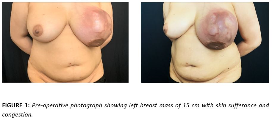
The patient came to our breast center’s attention in July 2020, presenting a subcutaneous lesion of the left breast that had grown rapidly over the past 6 months. Physical examination showed a wide lumpy 15 cm tumor fully occupying the upper part of the left breast, with areas of suffering and congested skin. On palpation, the mass appeared to have a very hard consistency, quite fixed to the deep layers and which distorted the normal breast profile. The nipple did not seem to be involved by the mass and there were no clinical signs of axillary involvement (Figure 1).
In our breast center, before surgery, she underwent a computer tomography and MRI, which showed a 13x11 cm breast mass with a multinodular confluent aspect and necrotic-colliquative components in the most caudal portion (Figure 2). The mass affected the soft tissues of the breast full-thickness, without signs of infiltration of large and small ipsilateral pectoral muscles or the skin above. It revealed no axillary pathological lymph nodes and nor distant metastasis.

She underwent a wide upper left lumpectomy in combination with a latissimus dorsi muscle flap (Figure 3).

The intraoperative histological examination did not show nipple involvement. The tumor did not appear macroscopically to be invading the chest wall. The mass was characterized by important angiogenesis. The resected tumor was 15x12x10 cm and weighted 1360 gr and appeared like a fleshy, multinodular confluent neo-formation with large necrotic areas (Figure 4).

The patient had a good post-surgical recovery and was discharged on the fourth postoperative day in good conditions with no wound complications. One month after surgery the result was good and the patient was satisfied, and she resumed all activities.
The histological examinations reported a malignancy spindle cell tumor with high mitotic activity (20 mitoses for 10HPF). The Ki67 index was 15%. The cancer elements were found to be positive for Vimentin and SMA (widespread positivity) and negative for immunohistochemical colorings for AE1/AE3, CAM5.2, 34BE12, CK5/6, CK7, CK14, CK17, p63, EMA, CD31, ERG, CD34, BCL2, CD99, Desmina, Melan-A, HMB45, S100, MDM2, STAT-6. There was a focal expression of CD68PGM1. The absence of perilesional in situ cancer component, as well as the negativity for epithelial markers and p63, was suggestive for high-grade fused cell sarcoma with morphological and immunophenotypic aspects of myofibroblastic differentiation.
The most appropriate oncological treatment has been decided in a multidisciplinary meeting, considering the overall conditions and the clinical history of the patient, the final pathological examination, and the TNM stage. The indication that emerged, in this case, was adjuvant chemotherapy with anthracycline and ifosfamide and radiotherapy on the chest wall.
Discussion
Because of their rarity, the treatment of breast sarcoma is still controversial. Mastectomy was considered as the gold standard from the 1950s and onward 3,4 but this option has been challenged over the years. Several studies have shown no differences in oncological outcomes between patients treated with breast conservative surgery compared with mastectomy5 .
The newest NCCN Guidelines 6 recommend surgical excisions with appropriately negative margins as the standard primary approach for most patients with soft tissue sarcoma, with post-operative radiotherapy indicated in selected cases, when margin status is uncertain or close (<1cm). Complete tumor removal is a primary prognostic factor for local recurrence. In the case of positive surgical margins on final pathological examination, re-resection to obtain negative margins should be strongly considered.
The treatment of giant sarcoma of the breast is always a challenge for the surgeon, who has to perform extensive removal respecting the natural shape of the breast, in particular in young patients.
Funding: None
Conflicts of interest: None declared
Ethical approval: Not required


Open Access By Aditum Open Access Journals id licensed under Creative Commons Attribution 4.0 International License. Based On a Work at aditum.org