International Surgery Case Reports
OPEN ACCESS | Volume 7 - Issue 1 - 2025
ISSN No: 2836-2845 | Journal DOI: 10.61148/2836-2845/ISCR


Chukwubuike Kevin Emeka
Pediatric Surgery Unit, Department of Surgery, Enugu State University Teaching Hospital, Enugu, Nigeria.
*Corresponding author: Chukwubuike Kevin Emeka, Department of Surgery, Enugu State University Teaching Hospital, Enugu, Nigeria.
*Corresponding author: Chukwubuike Kevin Emeka, Department of Surgery, Enugu State University Teaching Hospital, Enugu, Nigeria.
Received date: February 18, 2021
Accepted date: February 21, 2021
published date: February 26, 2021
Citation: Chukwubuike K Emeka. “Meckel’s Diverticulum Causing Intussusception In a 2-year Old Nigerian Child.”. International Journal of Surgery Case Reports and Images, 2(1); DOI: http;//doi.org/03.2021/1.1007.
Copyright: © 2021 Chukwubuike Kevin Emeka. This is an open access article distributed under the Creative Commons Attribution License, which permits unrestricted use, distribution, and reproduction in any medium, provided the original work is properly cited.
Meckel’s diverticulum is a congenital malformation of the gastrointestinal tract. Few cases of Meckel’s diverticulum causing intussusception have been reported. We report a case of Meckel’s diverticulum causing intussusception in a 2-year old Nigerian girl. Meckel’s diverticulum may be the pathologic lead point causing the intussusception.
1. Introduction
Meckel’s diverticulum is a true diverticulum and is a common congenital malformation of the gastrointestinal tract. Meckel’s diverticulum is quoted to occur in 2% of the population [1]. Embryologically, Meckel’s diverticulum arises due to persistence of the omphalomesenteric (vitelline) duct during fetal development. Meckel’s diverticulum typically present as a 2-inch (5 centimeters) out-pouching of the intestine at approximately 2 feet (60 centimeters) proximal to the ileocecal valve. Ectopic gastric or pancreatic tissue may be found in the Meckel’s diverticulum [2]. A good number of children who have Meckel’s diverticulum are without symptoms. Symptomatic Meckel’s diverticulum presents in the forms of melana stool, rectal bleeding, intussusception, volvulus and intestinal obstruction. It’s been estimated that the life time risk complication rate of untreated Meckel’s diverticulum is 4% [3].
Intussusception is the invagination of one bowel segment into another. Intussusception is a surgical emergency and one of the most common causes of intestinal obstruction in early childhood [4, 5]. Most cases of intussusception in children are idiopathic and are located in the ileocolic area. Occasionally, pathologic lead points may be a cause of intussusception in children at any point in the small or large bowel [6].
Merkel’s diverticulum causing intussusception is commonly reported in the adult population [7]. Intussusception resulting from Meckel’s diverticulum is a rare occurrence in children. We report a rare case of intussusception resulting from a Meckel’s diverticulum in a 2-year old female.
2. Case presentation
A 2-year old female was referred from a peripheral hospital on account of a 5-day history of abdominal pain, vomiting, and passage of red currant jelly stool. Abdominal pain was colicky, intermittent and located in the mid region of the abdomen. Vomiting was non projectile and vomitus was bilious. There was no associated fever and no abdominal distension. Physical examination revealed a well-appearing female whose abdomen was soft, non-tender and a palpable mass in the epigastric region. Examination of the other systems found no abnormality. On digital rectal examination, no mass was palpable per rectum. The diaper and examining finger were stained with red currant stool (Figure 1).
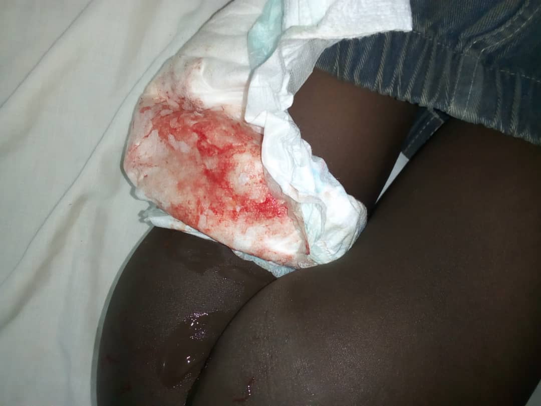
Figure 1: Red currant jelly stool.
An ultrasound of the abdomen revealed showed the pseudokidney and target signs (Figure 2). Plain abdominal radiograph was not supportive (Figure 3). Computed tomography (CT) scan was not done. The working diagnosis was intestinal obstruction secondary to intussusception.
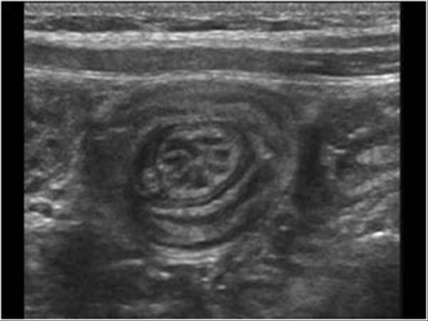
Figure 2: Ultrasound image
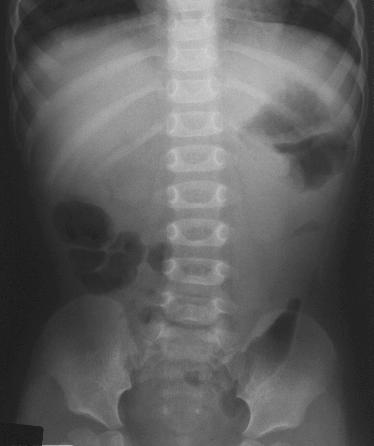
Figure 3: Plain abdominal x ray of the index patient
The patient was optimized and taken to theatre for laparotomy. At surgery, the intussusception was manually reduced and a residual mass could be palpated. This residual mass was located at the antimesenteric border of the ileum, 45 centimeter from the ileocaecal valve (Figure 4).
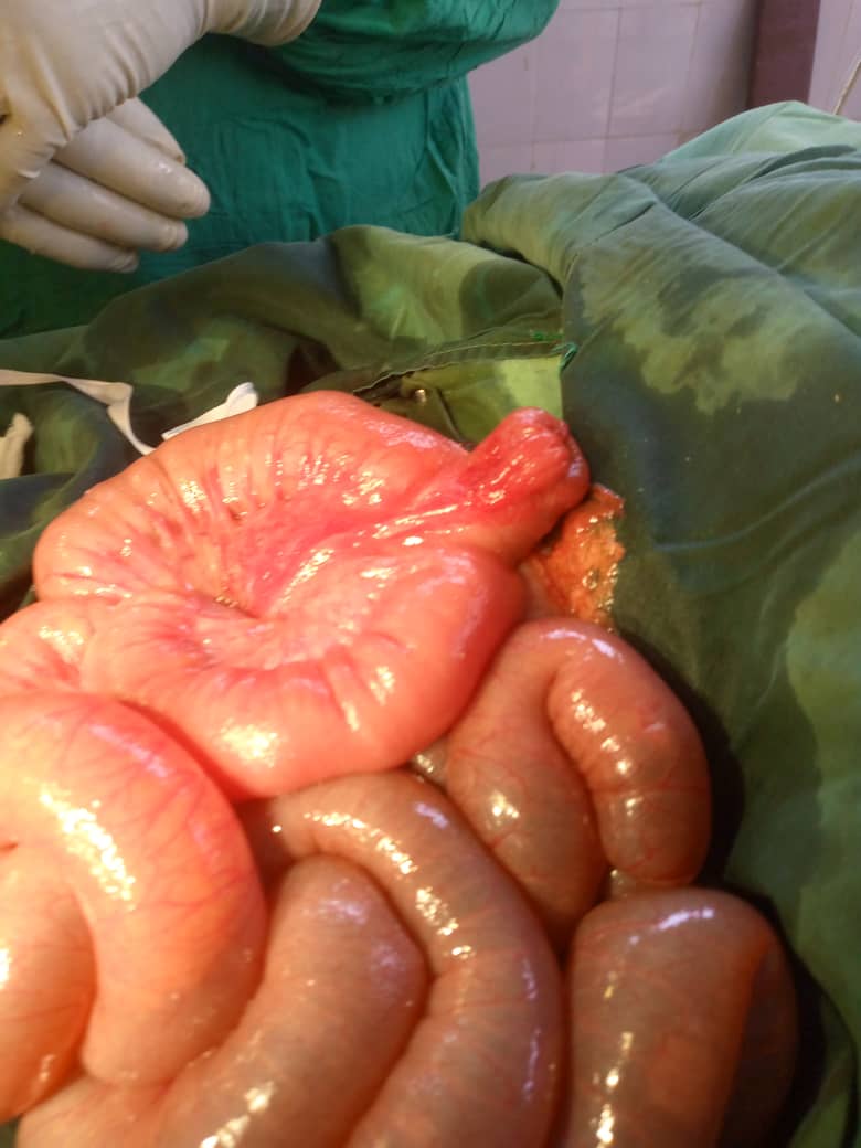
Figure 4: Meckel’s diverticulum in antimesenteric border of the ileum (arrow).
Segmental resection of the ileum containing Meckel’s diverticulum was performed and the continuity of the bowel restored by end to end ileal anastomosis. The specimen (Meckel’s diverticulum) was sent for histology. Figure 5 shows the resected specimen.
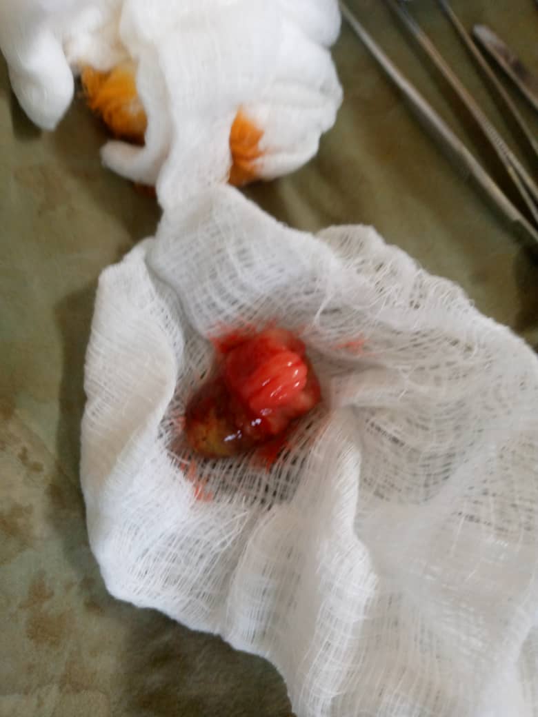
Figure 5: Resected Meckel’s diverticulum
Post operatively, the patient’s recovery was uneventful and was discharged home on the 6th day post op. She is currently being followed up in the clinic and has no complaint.
Histology of the resected specimen confirmed Meckel diverticulum without ectopic pancreatic or gastric tissues.
3. Discussion
Failure of obliteration of the omphalomesenteric (viteline) duct during fetal development is the etiopathogenesis of Meckel’s diverticulum. The presence of the Meckel’s diverticulum may lead to bleeding, intestinal obstruction or vomiting. Meckel’s diverticulum may be a lead point for intussusception if the diverticulum becomes inverted. What leads to the inversion of the Meckel’s diverticulum is not known. Some authors have suggested that the presence of ectopic tissues (gastric, pancreatic or both) may be stimulus for the inversion process and subsequent occurrence of intussusception [8].
The patient in the current case report is a female. Two reports also documented Meckel’s diverticulum causing intussusception in a female [8, 9]. However, Lima et al reported a case of intussusception resulting from the diverticulum in a boy [10]. The reason for the gender disparity in the incidence of Meckel’s diverticulum causing intussusception is not clear.
Meckel’s diverticulum causing intussusception occurs at about 2 years of age. This is consistent with the report of other similar series on intussusception [8, 10]. Our patient was also 2 years of age. The reason why symptoms of Merkel’s diverticulum occur at about the age of 2 years is still being a subject of research. Howbeit, it is worthy to note that the manifestation of Meckel’s diverticulum, including intussusception, can occur at any age [1].


Open Access By Aditum Open Access Journals id licensed under Creative Commons Attribution 4.0 International License. Based On a Work at aditum.org