
Yi-Xue Zhou1, 2, 3, Qiao-Yun Tong1, 2, 3, Wei Liu1, 2, 3*
1The First College of Clinical Medical Science, China Three Gorges University, Yichang, China
2Institute of Digestive Disease, China Three Gorges University, Yichang, China.
3Department of Gastroenterology, Yichang Central People’s Hospital, Yichang, China.
*Corresponding author: Wei Liu, Institute of Digestive Disease, China Three Gorges University, 8 Daxue Road, Yichang 443000, China.
Received date: March 08, 2023
Accepted date: June 16, 2023
Published date: June 28, 2023
Citation: Yi-Xue Zhou, Qiao-Yun Tong, Wei Liu. (2023) “A Rare Endoscopic Finding: Gastric Diverticulum”, J of Gastroenterology and Hepatology Research, 4(1); DOI: http;//doi.org/06.2023/2.10138.
Copyright: © 2023 Wei Liu. This is an open access article distributed under the Creative Commons Attribution License, which permits unrestricted use, distribution, and reproduction in any medium, provided the original work is properly cited.
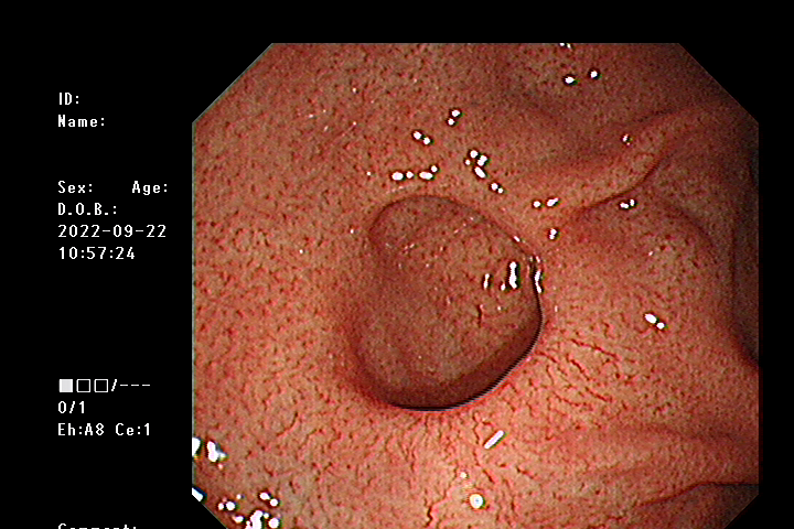
A 40-year-old man received upper gastrointestinal endoscopic scan during a health check-up. There was no history suggestive of Helicobacter pylori eradication, reflux esophagitis, peptic ulcer disease and upper abdominal surgery
Introduction:
A 40-year-old man received upper gastrointestinal endoscopic scan during a health check-up. There was no history suggestive of Helicobacter pylori eradication, reflux esophagitis, peptic ulcer disease and upper abdominal surgery. Endoscopy revealed a wide-mouthed diverticulum of the size of 1.5×2 cm between the fundus and greater curvature of the stomach (Figure 1A). Gastric diverticulum is the least common type of diverticular disease of the gastrointestinal tract which is usually incidentally diagnosed[1]. As a form of diverticular disease, gastric diverticulum is an outpouching of the gastric wall that is equally distributed between men and women. It is usually single, varying in size from 1 to 3 cm. Gastric diverticulum may be classified into congenital (true diverticula), involving all the layers on the posterior wall in the area near the gastric fundus, and acquired (false diverticula) that lack the muscularis in the prepyloric region[2]. Clinical feature may include dyspepsia, upper abdominal pain, reflux or even hemorrhage and perforation[3]. However, it remains asymptomatic in most cases. The appropriate management depends on the presentation and complications of the diverticulum. When the diverticulum is large or the symptoms do not respond to medical management, surgical intervention is considered. Intraoperative endoscopy is helpful because the diverticulum is easily missed during surgical exploration.
Footnote
Conflicts of Interest: The authors have no conflicts of interest to declare.
Ethical Statement:
The authors are accountable for all aspects of the work in ensuring that questions related to the accuracy or integrity of any part of the work are appropriately investigated and resolved. Written informed consent was obtained from the patient for publication of this “GI Image”.

Open Access By Aditum Open Access Journals id licensed under Creative Commons Attribution 4.0 International License. Based On a Work at aditum.org