Alisa Farokhian*, David Schwartzberg and Bo Shen
Center for Inflammatory Bowel Diseases and Division of Colorectal Surgery, Columbia University Irving Medical Center/NewYork Presbyterian Hospital, New York, NY, USA.
*Corresponding author: Alisa Farokhian, Center for Inflammatory Bowel Diseases and Division of Colorectal Surgery, Columbia University Irving Medical Center/NewYork Presbyterian Hospital, New York, NY, USA.
Received date: February 05, 2024
Accepted date: February 12, 2024
Published date: February 15, 2024
Citation: Alisa Farokhian, David Schwartzberg and Bo Shen. (2024) “Endoscopic Septectomy for the Augmentation of Volume of “Rabbit Ear” Pouch.”, J of Gastroenterology and Hepatology Research, 5(1); DOI: 10.61148/2836-2888/GHR/046.
Copyright: ©22024 Alisa Farokhian. This is an open access article distributed under the Creative Commons Attribution License, which permits unrestricted use, distribution, and reproduction in any medium, provided the original work is properly cited.
Restorative proctocolectomy with Ileal pouch anastomosis remains the surgical treatment for both medically refractory ulcerative colitis and familial adenomatous polyposis. Surgical complications include a residual septum that is often referred to as apical pouch bridge syndrome. This configuration, although traditionally has been treated surgically, can now be treated endoscopically using septectomy.
restorative proctocolectomy
Introduction:
Ileal pouch anal-anastomosis (IPAA) remains the standard surgical treatment for medically refractory ulcerative colitis. [1] IPAA improves the quality of life for most patients, however it does not remain without complications. The most frequent complications that are seen include abscess, anastomotic leaks, pouchitis, and pouch strictures. [2] These complications can hinder both quality of life and pouch function. In rare cases a residual septum can lead to evacuation difficulties that is often known as the apical pouch bridge syndrome [3]. A pouch septum may also result in bleeding, tenesmus, anal pain, or evacuation difficulty. In this case report we describe a case of apical pouch bridge syndrome caused by a rabbit ear septum configuration that was treated with septectomy endoscopically.
Case report:
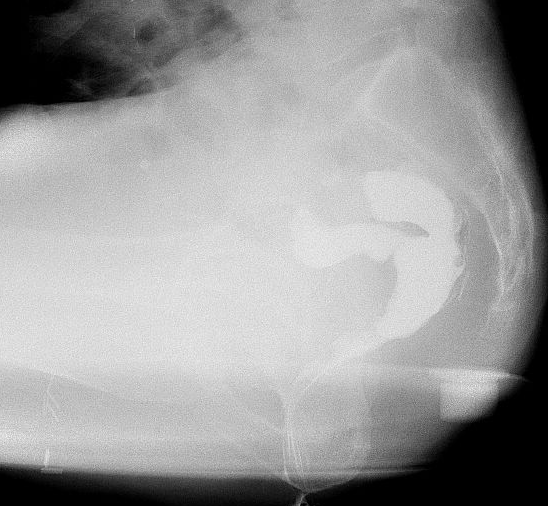
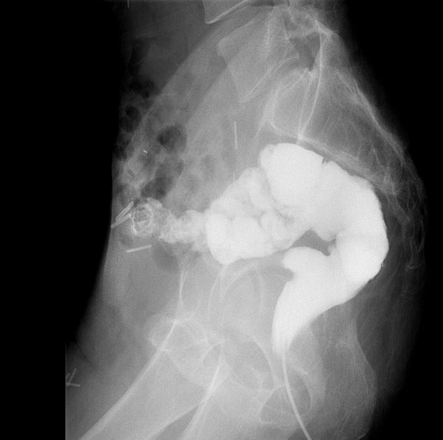
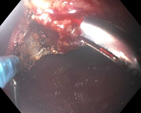
A 53-year-old male who was diagnosed with ulcerative colitis (UC) and underwent a 2-stage restorative proctocolectomy IPAA in 2001 for medically refractory colitis. His course after surgery was complicated by anastomotic leak, chronic pouchitis, and later on diagnosis of Crohn’s disease of the pouch. The decision was made to begin treatment with infliximab and 6-mercaptopurine however after worsening symptoms he was ultimately switched to vedolizumab. Despite medical therapy he never felt complete symptomatic improvement and In 2020 he presented to our center with dyschezia and tenesmus. A pouchoscopy was performed which showed relatively mild inflammation however was significant for a rabbit ear configuration of 10 cm. The decision was made to obtain a Barium defecography and anorectal manometry. (Figure 1) There was concern of pelvis floor dysfunction and the patient was sent for 10 sessions of biofeedback. Despite the biofeedback sessions he experienced only 10% of resolution of symptoms. The decision was made to proceed to treat the apical pouch bridge or rabbit ear configuration endoscopically. During the first septectomy for pouch augmentation 3 cm was treated. Soon after the patient returned underwent a second septectomy for the remaining 7 cm along (figure 3) with another 10 sessions of biofeedback. After follow up in the clinic, he endorsed roughly 80% of improvement in symptoms. A repeat barium defecography was done showing a resolution in the rabbit ear configuration confirming that treatment of the residual apical bridge was not only the cause of the symptoms but that endoscopic therapy was a viable option to treat our patient. (Figure 2).

Figure 1: Barium defecography showing septal bridge.

Figure 2: Barium defecography after septectomy showing rounded contour.

Figure 3: Pouchoscopy with needle knife and endoclip placement.
Discussion:
Roughly 20% of IPAA patients experience structural complications after surgery [4]. Structural complications often hinder the quality of life of these patients. Although surgery has been the traditional therapy for these complications, endoscopic therapy has been seen to emerge as an alternative option. [5] Recently it has been shown that endoscopic therapy has proven to manage closure of fistula of J pouch [6], obstructing pouch twists, and strictures. [7] This “rabbit ear” configuration that was seen in our patient, can be caused by septum formation leading to dyschezia and ultimately an obstruction or outlet issue of the pouch. Endoscopic therapy includes septectomy by advancing the colonoscope through the ileoanal anastomosis via the anus and advancing to the ileoanal pouch and into the neo-terminal ileum. Using needle knife therapy, the septum can be cut and 10 endoclips were placed to allow healing in the correct configuration (figure 4). For our patient, this process was repeated three months after using the same technique to allow for not only healing of the initial therapy but to see if there was any symptomatic improvement. Barium defecography that was done after complete treatment of the apical bridge via endoscopic septectomy showed improvement of the rabbit ear pouch with good transit into the afferent small bowel. A similar approach was done at Mayo clinic, in which the septum was divided using a laparoscopic stapler [8]. As laparoscopic staplers and robotic surgical interventions increase in the surgical world, it is important to know they may come with complications that may not have been traditionally noted as hand sewed anastomosis. Learning not only the complications but how to treat these complications endoscopically provides both enhancement in patient care but also in quality of life. Ultimately using endoscopic septectomy provides patients and providers and alternative rather than surgery and avoidance of loop ileostomy.