Boubacar Siddi Diallo 1*, Boubacar Alpha Diallo 1, Abdoulaye Sylla 3, Ibrahima Conte 2, Diallo Yaya 2, A Diakite 3, Ibrahima Sory Balde 2, Moussa Koulibaky 3, Telly Sy2, Yolande Hyjazi 1, Namory Keita 1
1University Department of Gynecology Obstetrics , Donka national Hospital , Conakry Guinea
2 University Department of Gynecology Obstetrics , Ignace Deen national Hospital , Conakry Guinea
3 University Department of Anatomy Pathology , Donka national Hospital , Conakry Guinea
*Correspondence: Diallo Boubacar Siddi, Gynecologist- Obstetrician. Lecturer at Conakry University Teaching Hospital.
Received date: January 28, 2021
Accepted date: February 01, 2021
Published date: February 11, 2021
Citation: Boubacar S Diallo, Boubacar A Diallo, Sylla A, Conte I, Yaya D,., (2021) Female Genitomammary Tuberculosis: Frequency and Anatomo-Histoclinical Aspects at Conakry University Teaching Hospital. Guinea. Women’s Health Care And Analysis, 2(1) ; DOI: http;//doi.org/03.2021/1.1007
Copyright: © 2021 Diallo Boubacar Siddi, This is an open access article distributed under the Creative Commons Attribution License, which permits unrestricted use, distribution, and reproduction in any medium, provided the original work is properly cited.
Objectives: Calculate the frequency of female genito-mammary tuberculosis, describe the epidemiological profile and describe the anatomo-histoclinical aspects of female genito-mammary tuberculosis at the Conakry University Hospital.
Methodology: this was a descriptive retrospective study lasting 10 years, from January 1, 2008 to December 31, 2018, all cases of histopathologically confirmed female tuberculous genital and breast lesions were excluded. All cases of female genital and breast lesions for which the diagnosis of tuberculosis has been ruled out. The limitations or constraints of the study were the absence of certain information on the pathological examination request forms and the absence of a molecular biology study. We carried out an exhaustive examination of the data available in the registers of the anatomo-pathologies service of Conakry University Teaching Hospital.
Results: The frequency of genito-mammary tuberculosis was 2.38% (n = 17) among benign genital and breast pathologies (n = 669) and 2.55% (n = 17) among those of genital and mammary pathologies. malignant (n = 623). The epidemiological profile was that of a woman of the age group of 20-29 years (32.96%), housewives (47.04%), nulligestes (28.56%), nulliparous (42.84%). The tumor mass was the main reason for consultation (76.47%). The main presumptive clinical diagnosis was pelvic tumor (41.16%). HIV serology was positive in 42.88% of cases. The samples represented by the operative parts constituted the bulk of the samples examined (74.44%). The topography of tuberculosis in the tubes was the highest (29.41%). Tuberculous lesions with a budding appearance made up the bulk of samples (58.80%). Cases of tuberculous lesions with heterologous elements represented (58.80%) followed by cases with heterologous necrotic elements (17.64 %). Granuloma was the main tuberculous type (82.32%). Lesions visualized under the microscope consisted of follicles of the same age (76.44%). Basic histological lesions were most observed on single follicles (29.40%). The cases of female genital tuberculosis without associated visualized lesions represented (41.16%) followed by cystic lesions (17.64%). The Zielh Nelsen histochemical staining technique was positive in 66.66%.
Conclusion: Genito - mammary tuberculosis constitutes organic lesions whose recrudescence increases with opportunistic infections (AIDS), more particularly affecting young women in full genital activity, which lead to complications with irreparable sequelae (primary and secondary sterility, ectopic pregnancies). Its diagnosis is complex due to the absence of pathognomonic signs and most often discovered during an infertility assessment. Anatomopathological examination is a valuable diagnostic aid. It makes it possible to rule out or confirm the diagnosis and to look for any associated lesions.
female genital tuberculosis; female breast tuberculosis; frequency
Introduction :
Genitomammary tuberculosis affects the genitals and mammary glands by the tuberculosis bacillus. It represents chronic mutilating organic diseases of latent evolution, which can cause irreparable damage [1]. It is a rare condition, but extremely serious, and revealed in 47-50% of cases by the couple's infertility. It represents a real public health problem in developing countries [2]. The genital and breast involvement can be primary or secondary or included in a table of tuberculous polyseritis [4]. In the majority of cases, the diagnosis of genital tuberculosis remains a surprise on histological study of the biopsy or the operative specimen after surgery for genital tumor [5]. Symptoms or signs of genital tuberculosis are most often latent. Symptomatic forms do not show symptoms or pathognomonic signs [6]. 50% of diagnoses are made during a sterility assessment. It is therefore probable that a systematic search in these patients undergoing sterility assessment would make it possible to obtain a more objective frequency of genital tuberculosis disease [8, 9].
The elevation of CA 125 levels in serum and body fluids in a woman with genital tuberculosis is not a specific indicator, but it does allow us to judge the effectiveness of anti-bacillary treatment. The pathological examination is the key to the diagnosis. Standard histology is used to assess the age of the follicles. ZielhNeelsen stain histochemistry can identify Mycobacterium, but its negativity does not immediately eliminate tuberculosis. PCR after culture could identify the species of Mycobacterium [10]. Female genital tuberculosis has seen its incidence decrease in Western countries thanks to improved living conditions and prevention strategies.
However, it remains a public health concern in developing countries where it is still a common cause of couple infertility [2]. Their overall frequency is difficult to estimate, as many forms are asymptomatic. The incidence of tuberculosis during infertility varies from country to country. It is around 1% in North America and 19% in India. In Tunisia, 10% of infertility is thought to be due to genital tuberculosis [3]. The objectives of this study were to calculate the frequency of female genito-mammary tuberculosis, describe the epidemiological profile and describe the anatomo-histoclinical aspects of female genito-mammary tuberculosis at Conakry University Teaching Hospital
Methodology:
This was a retrospective descriptive-type study lasting 10 years, from January 1, 2008 to December 31, 2018, all cases of histopathologically confirmed female tuberculous genital and breast lesions were included. Cases of female genital and breast lesions for which the diagnosis of tuberculosis have been ruled out were all excluded. The limitations or constraints of the study were the absence of certain information on the pathological examination request forms and the absence of a molecular biology study. We carried out an exhaustive examination of the data available in the registers of the anatomo-pathologies service of the Conakry University Teaching Hospital. The variables studied were epidemiological (frequency, age, gestity, parity, socio-professional categories), clinical (reasons for consultation, presumptive clinical diagnosis, serological status), anatomopathological: macroscopic (type of sample, topography, appearance, associated rearrangements) and histological (tuberculous type, age of follicles, basic histological lesions, lesions associated with genital tuberculosis, histochemical technique).
Results:
I - Frequency: The frequency of genito-mammary tuberculosis was 2.38% (n = 17) among benign genital and mammary pathologies (n = 669) and 2.55% (n = 17) among those of pathologies genital and malignant breasts (n = 623).
II-The epidemiological profile:
1-Age: the age group of 20-29 years was the most concerned (32.96%), followed by that of 30-39 years (31, 30%). The average age was 36.4 years with extremes of 26 and 65.
2-Gesture: The nulli gestures were the most represented (28.56%) followed by the primigestes (21.42%).
3-Parity: The nulliparas constituted the majority of the cases observed (42.84%).
4-Socio-professional categories: Housewives were the most concerned (47.04%).
III-Clinic:
a) Reasons for consultation: The tumor mass constituted the main reason for consultation (76.47%).
Table I (n = 17)
|
Motive |
NUMBER |
PERCENTAGE |
|
Tumor mass |
13 |
76,47 |
|
Hypermenorrhea Amenorrhea |
6 |
35,29 |
|
Infertility |
7 |
41,17 |
|
Abdomino-pelvic pain |
9 |
52,94 |
|
Metrorrhagia |
5 |
29,41 |
|
Hydrorrhea
|
4 |
23,52 |
|
Pyorrhea |
3 |
17,64 |
|
Weight loss |
6 |
35,29 |
|
Fever |
3 |
17,64 |
|
Total
|
56 |
100 |
b) Presumptive clinical diagnosis: The main presumptive clinical diagnosis was pelvic tumor (41.16%) followed by mammary tumor 17.64%.
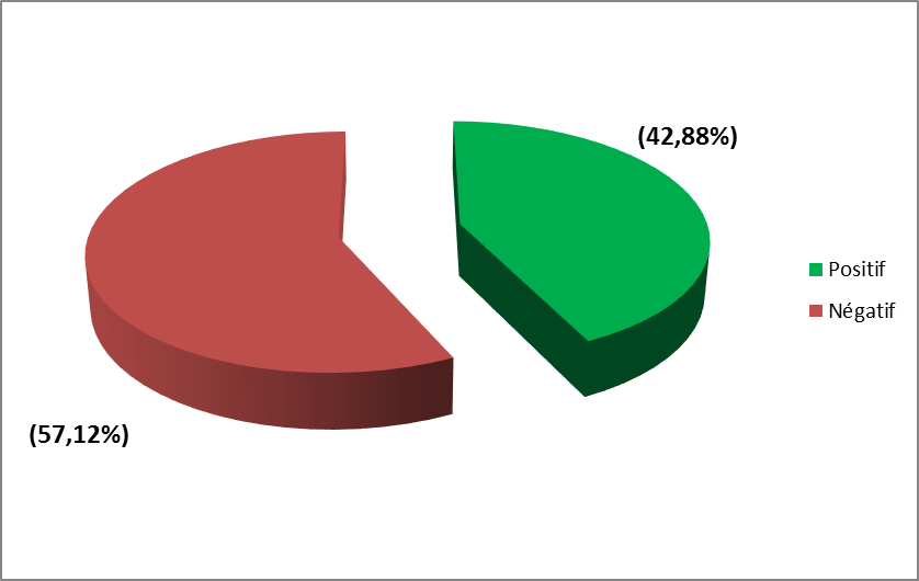
c) Serological status: HIV serology was positive in 42.88% of cases. Figure 1.

IV-Anatomo-pathology:
A-Macroscopy:
• Type of sample: The samples represented by the operative documents constituted the bulk of the samples examined (74.44%).
• Topography: The topography of tuberculosis in the tubes was the highest (29.41%). Table II.
|
TOPOGRAPHY
|
NUMBER |
PERCENTAGE |
|
Uterine body |
2 |
11,76 |
|
Cervical
|
3 |
17,64 |
|
Ovarian |
2 |
11,76 |
|
Tubal |
5 |
29,41 |
|
Vaginal
|
1 |
5,88 |
|
Vulva
|
1 |
5,88 |
|
Breast
|
3 |
17,64 |
|
Total |
17 |
100 |
• Appearance: Tuberculous lesions with a budding appearance made up most of the samples (58.80%) followed by lesions with an ulcerative budding appearance (35.76%).
• Associated changes: Cases of tuberculous lesions with heterologous elements represented (58.80%) followed by cases with heterologous necrotic elements (17.64%) then cases with heterologous calcium element 11.76%.
B-Histology:
• Tuberculous type: Granuloma was the main tuberculous type (82.32%).
• Age of follicles: Lesions seen under the microscope consisted of follicles of the same age (76.44%).
• Basic histological lesions: simple follicles were the most observed basic histological lesions (29.40%). Table III
|
BASIC LESIONS
|
NUMBER
|
PERCENTAGE |
|
|
Exudative
|
1 |
5,88 |
|
|
Cellular
|
Simple follicle |
5
|
29,40 |
|
Follicular caseo |
4
|
23,52 |
|
|
Caseo follicular cave |
2
|
11,76 |
|
|
Healing and fibrous organization |
Fibrous follicle |
2
|
11,76 |
|
Caseo-fibrous |
2
|
11,76 |
|
|
cavernofibrous |
1
|
5,88 |
|
|
|
Total |
17
|
100 |
• Lesions associated with genital tuberculosis: Cases of female genital tuberculosis without associated lesions visualized represented (41.16%) followed by ovary cystic lesions (17.64%).
|
ASSOCIATED LESIONS
|
NUMBER
|
PERCENTAGE |
|
Without associated lesions |
7 |
41,16 |
|
Adenomyosis |
2 |
11,76 |
|
Pregnancy |
1 |
5,88 |
|
Ovarian cyst |
3 |
17,64 |
|
Leiomyoma |
2 |
11,76 |
|
Adenomatoid tumor |
1 |
5,88 |
|
Polyp |
1 |
5,88 |
|
Total |
17 |
100 |
• Histochemical technique: The Ziehl Neelsen histochemical staining technique was positive in 66.66%.
Discussion :
I - Frequency: The frequency of genito-mammary tuberculosis was 2.38% (n = 17) among benign genital and mammary pathologies (n = 669) and 2.55% (n = 17) among those of genital pathologies and malignant breasts (n = 623). Hablani. N and Coll [19] reported in their study that female genital tuberculosis accounts for 6-10% of all tuberculosis sites.
II-The epidemiological profile:
1-Age: the age group of 20-29 years was the most concerned (32.96%), followed by that of 30-39 years (31, 30%). The average age was 36, 4 years with extremes of 26 and 65. This result is comparable to that found by A, ABOUL FALAH.Y. et al. [1] or 29.30% in the age group of 20 and 40 years with an average age of 35 years. Most authors [7,13, 20] are unanimous that the preferred period for the onset of genital and mammary tuberculosis is the period of genital activity. This is because the estrogen impregnation makes the mucosa more abundant and fertile for the development of tuberculous follicles.
2-Gestity: The nulli gestity were the most represented (28.56%) followed by the primigestes (21.42%). This result is lower than that found by E.RAVELOSOA and Coll. [13] in nulli gestures is 54.54% and could be explained by the fact that genital and mammary tuberculosis is a pathology causing irreversible organic lesions, especially of the tubes (destruction of the vibratile cilia), compromising fertility.
3-Parity: The nulliparas constituted the majority of the cases observed (42.84%). This result is similar to that found by A-HAMMAMI: et al. [24] or 45, 31%.
4-Socio-professional categories: Housewives were the most concerned (47.04%). This result corroborates those of the literature [13, 23, 24]. The low socio-economic level is found with most of the authors.
III-Clinic :
a) Reasons for consultation: The tumor mass was the main reason for consultation (76.47%) followed by abdomino-pelvic pain 52.94% then infertility 41.17%. This result is similar to several studies [1, 13,23]. This observation shows that genital and mammary tuberculosis is an organic pathology with polymorphic symptoms without any pathognomonic sign or symptom. This often makes diagnosis difficult or late. Its discovery is most often histopathological after removal of the lesion. The clinic cannot under any circumstances immediately confirm the diagnosis of genital tuberculosis without doing pathological examination.
b) Presumptive clinical diagnosis: The main presumptive clinical diagnosis was pelvic tumor (41.16%) followed by mammary tumor 17.64%. This result is similar in several studies [2,7,9]. This observation can be explained by the fact that genital and mammary tuberculosis are degenerative organic lesions mutilating the genitals and mammary organs, which can become chronic, ulcerated or budding giving macroscopically an abscess or tumor. It is a clinically latent lesion. Imaging tests will simply appreciate the heterogeneous nature but will not be able to confirm the diagnosis of tuberculosis. Hence the interest of the pathological examination on an excisional biopsy or an operative specimen for histopathological study of the lesion in order to confirm the diagnosis of tuberculosis is necessary.
c) Serological status: HIV serology was positive in 42.88% of cases.
IV-Anatomo-pathology:
A-Macroscopy:
• The type of sample: The samples represented by the surgical specimens constituted the bulk of the samples examined 74.44%. This result corroborates those in the literature [9,13,19].
• Topography: The topography of tuberculosis at the level of the tubes was the highest (29.41%). This result can be superimposed on that found by HABLANI N. et al. [19] or 33%. This observation seems to be classic. Tubal damage is the most described in the literature. Tubal involvement of tuberculosis would explain that the infertility and sterility that are often associated with this pathology.
• Appearance: Tuberculous lesions with a budding appearance constituted most of the samples (58.80%) followed by lesions with an ulcerative budding appearance (35.76%). Tuberculous lesions are polymorphic lesions. in their macroscopic aspect taking on several aspects, bringing them closer to cancerous lesions. These aspects rarely suggest the tuberculous lesion at the outset, unless it is associated with caseous necrosis.
• Associated changes: The cases of tuberculous lesions with heterologous elements represented (58.80%) followed by cases with heterologous necrotic elements (17.64%) then cases with heterologous calcium element 11.76%. This would explain the frequent association of tuberculous lesions with mostly associated heterologous elements.
B-Histology:
Conclusion:
Genito - mammary tuberculosis constitutes organic lesions whose recrudescence increases with opportunistic infections (AIDS), more particularly affecting young women in full genital activity, which lead to complications with irreparable sequelae (primary and secondary sterility, ectopic pregnancies). Its diagnosis is complex due to the absence of pathognomonic signs and most often discovered during an infertility assessment. Anatomopathological examination is a valuable diagnostic aid. It makes it possible to rule out or confirm the diagnosis and to look for any associated lesions.