Journal of Dermatology and Venereology
OPEN ACCESS | Volume 4 - Issue 1 - 2026
ISSN No: 3065-677X | Journal DOI: 10.61148/3065-677X/JDV


Jean Zevallos, Cecilia Vera Cornejo and Jhoann Aurich Rojas
Dermatology Service, Department of Medicine, Hospital Maria Auxiliadora, Lima, Peru.
*Corresponding author: Jean Zevallos, Dermatology Service, Department of Medicine, Hospital Maria Auxiliadora, Lima, Peru.
Received Date: November 15, 2023
Accepted Date: November 20, 2023
Published Date: November 25, 2023
Citation: Zevallos J, Cecilia V Cornejo and Jhoann A Rojas. (2023) “Tinea capitis mimicking cicatricial plaque on the scalp and acne keloidalis.” Journal of Dermatology and Venereology, 1(1); DOI: http;//doi.org/10.2023/11.1001.
Copyright: © 2023 Jean Zevallos. This is an open access article distributed under the Creative Commons Attribution License, which permits unrestricted use, distribution, and reproduction in any medium, provided the original work is properly cited.
Tinea capitis is a common infection caused by dermatophyte fungi in children and can sometimes occur in adults. The clinical features are variable due to the diverse immune responses produced by fungal infection1. Atypical presentations have been reported within which scarring alopecia due to tinea capitis has also been described2,3. We present a case of tinea capitis which manifests as cicatricial alopecia and acne keloidalis.
Introduction
Tinea capitis is a common infection caused by dermatophyte fungi in children and can sometimes occur in adults. The clinical features are variable due to the diverse immune responses produced by fungal infection1. Atypical presentations have been reported within which scarring alopecia due to tinea capitis has also been described2,3. We present a case of tinea capitis which manifests as cicatricial alopecia and acne keloidalis.
Case report
A 32-year-old woman with a medical history of systemic lupus erythematous presented for evaluation with a twelve-year history of itching and hair loss. Her medications included prednisone, hydroxychloroquine and enalapril. Physical examination revealed an erythematous cicatricial plaque with associated alopecia and difusse dry scales on the scalp and firm confluent papules on the posterior neck (Figure 1).
Figure 1:
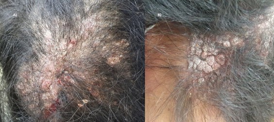
Figure 1. Cutaneous finding of our patient. (A) Erythematous plaque with cicatricial aspect with associated alopecia and diffuse dry scales on the scalp. (B) Confluent papules on the posterior neck.
Trichoscopy showed clobbestoned cicatricial like papules with single hairs arising in some areas. Tufted hair were not observed. A scalp biopsy was performed. Histopathology revealed a mixed inflammatory infiltrate composed of lymphoplasma cells and fibrosis surrounded the hair shafts. Clusters of spores and hyphaes within some hair shafts were found. Tissue culture isolated Trichophyton tonsurans. Therapy was started with oral terbinafine at a dose of 250mg per day for 2 months with complete resolution of lesions (Figure 2). After 6 months of follow-up, no recurrence was observed.
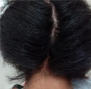
Figure 2. Complete resolution of lesions after 2 months of treatment with terbinafine.
Discussion
Tinea capitis is a frequent dermatomycosis most commonly seen in children. Despite its frequency, TC can represent a diagnostic challenge due to the variation in clinical presentation. Some cases of TC due to Trichophyton tonsurans mimicking folliculitis, chronic furuncles, canalizing pyoderma and tufted hair folliculitis have been reported2,3. Likewise, a few cases of TC mimicking cicatricial alopecia have also been described. A case of a child with tinea capitis with apparent loss of follicular markings treated successfully with oral terbinafine was reported by Mirmirani4. In addition, Ferenczi described a woman with acne keloidalis caused by fungal infection with near-complete resolution after 2 months of treatment with griseofulvin5. In both cases, patients were diagnosed with long-standing tinea capitis due to Trichophyton tonsurans and showed good response to antifungal therapy.
Clinical presentation of TC depends on the causative fungus and the inmune status of the patient4.A immunohistochemical study found evidence that supports the involvement of both cell-mediated and humoral mechanisms in the chronic and non-inflammatory TC6. In a proposed model which could explain the long-standing non-inflammatory scalp infections in some patients, an immun complex formation between IgE and antigen produced by T. tonsurans would bind mast cells at the site of infection realising histamine. Histamine would activate H2 receptors of T-suppressor cells which could prevent T-helper cell proliferation and inhibit MIF expression resulting in immune anergy7. We believe patient treatment could additionally contribute to the immune anergy.
The role of mast cells and hystamine have been studied in the pathogenesis of primary cicatricial alopecia8. Furthermore, a study by Hoda found IL-1β and MIF cytokine network induce MMP-1 and contribute to the loss of interstitial dermal collagen9. We hypothesize histamine could also play a role in scarring alopecia due to tinea capitis and MIF expression could induce MMP which would lead collagen degradation explaining the excellent response to antifungal treatment, albeit further studies are needed to determine the exact pathway.
This case shows tinea capitis can mimic scarring alopecia and should be considered in the differential diagnosis of apparent cases of primary cicatricial alopecia.


Open Access By Aditum Open Access Journals id licensed under Creative Commons Attribution 4.0 International License. Based On a Work at aditum.org