International Journal of Medical Case Reports and Medical Research
OPEN ACCESS | Volume 5 - Issue 1 - 2026
ISSN No: 2994-6905 | Journal DOI: 10.61148/2994-6905/IJMCRMR


Abdulwahab F. Alahmari
Radiology Specialist, Radiology Department, Al-Namas General Hospital, Ministry of Health, Al-Namas City,Saudi Arabia.
*Corresponding Author: Abdulwahab F. Alahmari, Radiology Specialist, Radiology Department, Al-Namas General Hospital, Ministry of Health, Al-Namas City, Saudi Arabia.
Received Date: January 26, 2024
Accepted Date: February 07, 2024
Published Date: February 10, 2024
Citation: Abdulwahab F. Alahmari. (2024) “Acute on chronic subdural hemorrhage on CT: Images in radiology.”, International Journal of Medical Case Reports and Medical Research, 2(1); DOI: 10.61148/2994-6905/IJMCRMR/026.
Copyright: © 2024. Abdulwahab F. Alahmari. This is an open access article distributed under the Creative Commons Attribution License, which permits unrestricted use, distribution, and reproduction in any medium, provided the original work is properly cited.
This is a case of a 72-year-old male patient with history of a subdural hemorrhage before 5 years (Figure 1). The subdural hemorrhage caused a mass effect. The patient suffers from hypertension which causes this subdural hemorrhage. The subdural hemorrhage turned into a hygroma. After 5 years, the patient came with a new subdural and inter-parenchymal hemorrhage. A CT scan after a week showed a unique hematocrit effect. The acute on chronic subdural hemorrhage appear like the chronic hemorrhage below and the acute hemorrhage above. In this case, both hemorrhages are side by side. The new subdural hemorrhage was pushed to the midline which appears as an inter-parenchymal hemorrhage. This effect appears in patients with coagulopathy [1].
Acute on chronic subdural hemorrhage on CT:
This is a case of a 72-year-old male patient with history of a subdural hemorrhage before 5 years (Figure 1). The subdural hemorrhage caused a mass effect. The patient suffers from hypertension which causes this subdural hemorrhage. The subdural hemorrhage turned into a hygroma. After 5 years, the patient came with a new subdural and inter-parenchymal hemorrhage. A CT scan after a week showed a unique hematocrit effect. The acute on chronic subdural hemorrhage appear like the chronic hemorrhage below and the acute hemorrhage above. In this case, both hemorrhages are side by side. The new subdural hemorrhage was pushed to the midline which appears as an inter-parenchymal hemorrhage. This effect appears in patients with coagulopathy [1].
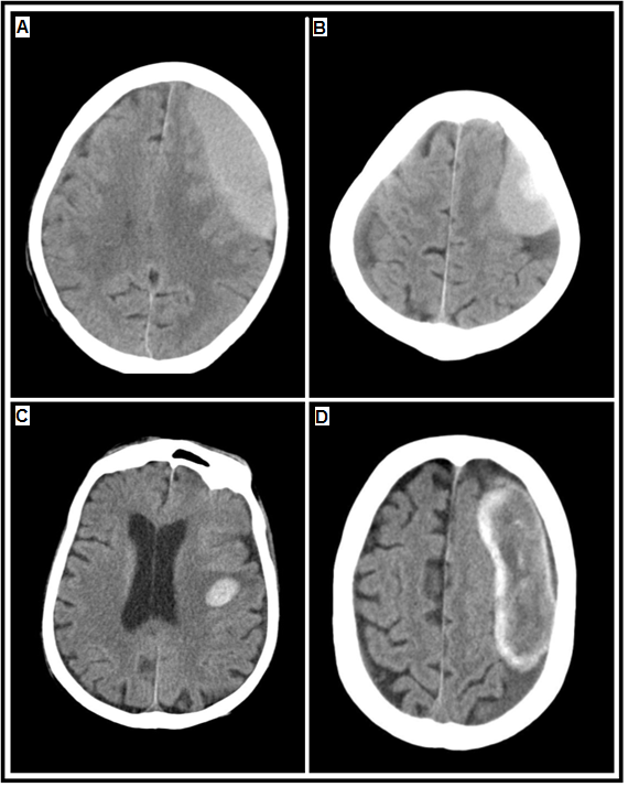
Figure 1: A brain axial CT of a 72-year-old male patient. A: shows a subdural hemorrhage on the left side. This CT scan was done 5 years before the following CT scans. B: The 2nd scan shows a new hemorrhage formed in the same location. C: The 2nd scan shows an inter-parenchymal hemorrhage, causing the brain to shift. D: The 3rd scan shows a hematocrit effect (i.e. sedimentation of blood) after 1 week.
CT: Computed Tomography.
Funding:This paper received no external funding.
Conflicts of interest:The author declares that they have no conflicts of interest.
Ethical disclosures:
Protection of Individuals: This study was conducted in compliance with the Declaration of Helsinki (1964) and its subsequent amendments.
Data Confidentiality: The authors declare they followed their center’s protocol for sharing patient data.
Right to privacy and informed consent: Informed consent was not required to analyze and publish routinely acquired clinical and imaging data.


Open Access By Aditum Open Access Journals id licensed under Creative Commons Attribution 4.0 International License. Based On a Work at aditum.org