
Sanjaya Walawe Nayaka
Post graduate institute of Medicine, University of Colombo, Sri Lanka.
*Corresponding Author: Sanjaya Walawe Nayaka, Post graduate institute of Medicine, University of Colombo, Sri Lanka.
Received date: December 22, 2023
Accepted date: January 05, 2024
Published date: January 12, 2024
Citation: Sanjaya Walawe Nayaka. (2024). “Case series of broad ligament lesions due to four different pathologies”. International J of Clinical Gynaecology and Obstetrics, 4(1); DOI: 10.61148/2836-0737/IJCGO/030.
Copyright: © 2024 Sanjaya Walawe Nayaka. This is an open access article distributed under the Creative Commons Attribution License, which permits unrestricted use, distribution, and reproduction in any medium, provided the original work is properly cited.
Broad ligament is a double layered peritoneal fold which consist of various anatomical structures and cell types which gives rise to different types of lesions. Symptomatology and the ultrasonic appearance of a broad ligament lesions may be difficult to distinguish from an ovarian tumor. This is a case series of broad ligament lesions with four different pathologies presented in four different backgrounds.
broad ligament; cystadenoma; hematoma; dyspareunia; fibroids; benign metastasizing leiomyoma
Introduction:
Broad ligament is a double layered peritoneal fold consist of mesothelial cells which drapes over the uterus and fallopian tubes. It connects uterus, fallopian tubes and ovaries with the lateral pelvic wall. The broad ligament contains smooth muscle fibers, blood vessels (uterine and ovarian vessels), uterosacral ligaments posteriorly, cardinal ligaments laterally, ovarian ligament medially and suspensory ligament of the ovary laterally, a portion of the ureters, round ligaments anteriorly and embryonic rests (such as mesonephric duct remnants, paramesonephric duct remnants, adrenocortical cell remnants and heterotopic hilar cell clusters) [1]. This can be divided into 3 parts (mesosalpinx, mesovarium and mesometrium commonly known as broad ligament).
Lesions of the broad ligament can be broadly classified as follows [2].
Fibroids are the commonest neoplasm in the broad ligament [3]. They may have originated from uterus and extended into the broad ligament or they may have originated from broad ligament without having any connection to the uterus. Very rarely this may be a part of Benign Metastasizing Leiomyoma [4]. Some times these fibroids may undergo cystic degeneration as well.
Most of the broad ligament cysts are from paramesonephric origin [5]. There may be cysts from mesonephric origin as well2. 10% of all adenexial cysts are broad ligament cysts [6]. These cysts may be benign, border line or malignant just like in epithelial ovarian tumors. Pain during and after the sexual intercourse is called dyspareunia [7]. The causes can be broadly classified as
Common causes for secondary dyspareunia in reproductive age women are endometriosis, Pelvic inflammatory disease, pelvic tumors, surgical adhesions and vulvar-vaginal lesions.
Broad ligament hematomas are more common during pregnancy and post-partum. But some time it may occur following pelvic surgeries. Most of these hematomas may present immediately after the procedure.
Case 1:
Previously healthy, 38-year-old, mother of three children (delivered vaginally) presented with gradually developing dyspareunia during last six months. She didn’t have secondary dysmenorrhea and chronic pelvic pain which suggestive of endometriosis.
Her abdominal examination revealed a non-tender abdomino-pelvic mass in left lower abdomen which compatible with 16 weeks size gravid uterus. Ultra sound scan (USS) of the abdomen and pelvis showed bilocular cystic lesion measuring 15x10cm which located in left adenexia. There were no solid areas and ipsilateral ovary was not seen separately. Uterus was displaced towards right and otherwise appeared normal. Right ovary was normal and there was no free fluid in the peritoneal cavity. Her CA 125 level was 8 IU/ml. At the end of history, examination and investigation working diagnosis of “left ovarian benign tumor with possible adhesions to uterus” was made.
After careful counselling and consent taking she agreed for mini-laparotomy and cystectomy. Supra-pubic transverse incision was made to enter into the peritoneal cavity which showed a large cystic left broad ligament cyst which displaced the uterus towards right. Her both tubes, ovaries and then Pouch of Douglas appear normal with out any adhesions or ascites (figure 1).
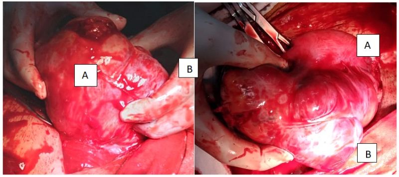 Figure 1: intra operative view of the broad ligament cyst (B) with the displaced uterus (A).
Stretched left round ligament was identified and dissected to enter into the space between two leaves of left broad ligament. Surgeons dominant hand’s fingers were inserted between broad ligament leaf and the cyst wall to enucleate the cyst. The cyst had been extended up to the deeper level of the pelvis. Careful digital enucleation of the cyst was done to prevent the damage of adjacent ureter. After the enucleation of the cyst (figure 2), oozing was noted in between the two broad ligament leaves which was managed with application of local compression for few minutes. She had an uneventful recovery. Her histology report came as serous cystadenoma of the left broad ligament.

Figure 2: left broad ligament cystadenoma after excision.
Case 2:
Previously healthy, 30-year old, multi-parous lady presented with gradually developing dyspareunia and chronic pelvic pain during last 10 months. Her abdominal examination revealed a non-tender abdomino-pelvic mass in right lower abdomen which compatible with 20 weeks size gravid uterus Ultra sound scan (USS) of the abdomen and pelvis showed solid lesion with cystic areas measuring 15x15cm which located in right adenexia. The ipsilateral ovary was not seen separately. Uterus was displaced towards left and otherwise appeared normal. Left ovary was normal and there was no free fluid in the peritoneal cavity. Her CA 125 level was 10 IU/ml. At the end of history, examination and investigation working diagnosis of “fibroid uterus with cystic degeneration” was made.
After careful counselling and consent taking she agreed for mini-laparotomy and myomectomy. Supra-pubic transverse incision was made to enter into the peritoneal cavity which showed a large right broad ligament fibroid which displaced the uterus towards left. Her both tubes, ovaries and then Pouch of Douglas appeared normal without any adhesions or ascites.
Stretched right round ligament was identified and dissected to enter into the space between two leaves of right broad ligament. Enucleation of the myoma was done with the help of a myoma screw. The fibroid had been extended up to the deeper level of the pelvis. Special attention was paid to prevent the damage of adjacent ureter. Her histology report came as multiple leiomyomas with cystic degeneration.
Case 3:
A 36-year-old unmarried nulliparous lady presented with 34 weeks size fibroid uterus for the myomectomy. Multiples leiomyomas (more than 30) removed through 3 uterine incisions during the open myomectomy and 10 out of them were located within the broad ligament and retroperitoneal space without any connection with the uterus. Histology report confirmed the presence of multiple leiomyomas.
She was investigated for lower abdominal pain and right lower limb pain four months after the surgery and found to have soft tissue mass measuring 12cm deep in the pelvis, below the level of cervix which displace the uterus towards the left.
MRI scan of the pelvis showed multi lobulated deep pelvic mass with extension into the retroperitoneum and right S1 neural foramen without typical MRI features of uterine fibroids. Plexiform neurofibroma, paraganglioma and leiomyosarcoma were the differential diagnoses suggested by radiologist.
Multi-disciplinary team meeting was arranged with the participation of a general surgeon and the patient and decide to proceed with exploratory laparotomy. She had multiple leiomyomas deep in the pelvis below the level of cervix, with in the bilateral broad ligaments and pelvic peritoneum which were extended towards the sacrum and retroperitoneal space. Surgery abounded due to the risk of uterine ischemia and hysterectomy following the removal of pelvic mass. Patient was thoroughly explained about the condition and available treatment options. She selected the total abdominal hysterectomy and bilateral salphingo oophorectomy (TAH+BSO) as she didn’t want to suffer anymore and not having fertility wishes. Patient agreed to accept all possible complications of selected treatment modality. Her abdomen was re-opened two days after the initial surgery and TAH+BSO done with the removal of all deep pelvic, broad ligaments and retroperitoneal fibroids. She was started on estrogen only Hormone Replacement Therapy (HRT).
Case 4:
67-year-old multiparous, menopaused woman underwent vaginal hysterectomy and repair (VH & R) for a second-degree utero-vaginal prolapse. She complained of right side sever lower abdominal pain during the latter part of the surgery day. By next day she become restless due to the pain which not responded to opioids analgesics. USS showed mixed echogenic mass (10x10cm) in the right side of the pelvis. Considering her hemoglobin drop after the surgery, decision was taken to perform an emergency laparotomy. It revealed right broad ligament hematoma due to damaged infundibulo-pelvic ligament and bleeding from ovarian vessels into the broad ligament. Right side oophorectomy and right ovarian vessels ligation were done. She had uneventful recovery and hematoma was gradually reabsorbed with the time.
Discussion:
These patients in case 1 and two were previously healthy ladies who didn’t have dyspareunia. So, there is a high probability of having intra abdomino-pelvic cause for the dyspareunia. Other than dyspareunia, as her history was not suggestive of having pelvic inflammatory disease or endometriosis, presence of pelvic mass should be born in physician’s mind. The mass detected during the abdominal examination was less mobile. This could be due to adhesions or due to mass in the broad ligament. Slightly deviated cervix away from the mass give a clue as well.
Best investigation to assess an adenexial cyst is ultra sound scan (USS) due to its diagnostic accuracy, availability and low cost. Both trans abdominal and trans vaginal route should be used in this patient as this lesion had come out from the pelvis into the abdomen. Due to non-visualization of the left ovary, diagnosis of left broad ligament cyst had been missed even though it had not changed the management and patient outcome. Their risk of malignancy indexes (RMI) were very low. Absence of ascites, low CA 125 level, low RMI and pre-menopausal age were directed the physician towards a benign cause. If there was a suspicious of non-epithelial ovarian tumor, another three tumor markers should be done as she is less than 40-year-old [8].
If there were any inconclusion in diagnosis, contrast enhanced computerized tomographic (CT) scan could have been done. But in these particular cases there were no such requirements as there were no any high suspicion about a malignancy.
Surgical management is the best option for this kind of large lesions. The two routes of entry are laparoscopy and laparotomy. Both these methods have their own merits and demerits. Even though these are relatively larger lesions, laparoscopic cystectomy could have been done with an expert hand in a well-developed laparoscopy center. Care should be taken to prevent the spillage of the lesion content even though they appeared benign. This can be achieved by using an endo-bag for the specimen retrieval procedure. Laparoscopy route will avoid a laparotomy which gives more post-operative pain. But laparotomy is still a good option in a low resource setting which gives a better idea of the lesion to the surgeon with both visual and tactile sensation [9]. Same time it will reduce the possibility of spilling of the cyst content into the peritoneal cavity. Entry into the broad ligament to enucleate the cyst can be best achieved by dividing the round ligament. Sweeping movements of the surgeon’s fingers will easily enucleate the cyst if it goes in the correct tissue plane. As this method is a blind procedure, care should be taken to prevent the damage to ureter. The bleeding from the cyst bed can be easily managed with the combination of diathermy and applying local pressure. As this is a benign cyst she doesn’t need any further treatment and follow up. But if it was a papillary cystadenoma, possibility of von Hippel–Lindau disease should be excluded [10,11].
Case 3 is a little different and rare presentation of benign metastasizing leiomyoma (BML)4. Even though the commonest sites of BML are lungs, lymph nodes, retro-peritoneal space and deep pelvic spaces, BML can be seen in broad ligaments as well. MRI is the best investigation to assess the deep pelvic soft tissue tumors even though preliminary assessment can be done with USS. Management is difficult in her case due to nulliparity, younger age and rapid re-growing of fibroids in the retro-peritoneal space and broad ligaments. But in this case, she opted total pelvic clearance including bilateral oophorectomy which facilitated the management in physician’s side. But she had to get HRT after the surgery.
Case 4 is an acute condition which was an immediate and rare complication of VH & R. Unnecessary traction during surgery or weak ligaments due to menopause may have led to avulsion of ovarian vessels during the surgery. The loosing blood easily drained to broad ligament space which can expand easily. This case shows the importance of knowing the anatomy of the broad ligament. In a well-equipped laparoscopy center, this second surgery could have been easily done with a laparoscopy.
The following graph shows a comparison between four cases.
|
|
Case 1 |
Case 2 |
Case 3 |
Case 4 |
|
presentation |
Long term history |
Long term history |
Long term history |
Acute presentation |
|
Diagnosis |
cystadenoma |
Fibroid with cystic degeneration |
BML |
Broad ligament hematoma. |
|
Main symptoms |
dyspareunia |
dyspareunia |
Abdominal mass |
Severe post-operative pain |
|
Investigations |
USS |
USS |
USS and MRI |
USS |
|
CA125 |
Normal |
Normal |
Not done |
Not done. |
|
Route of surgical management |
Laparotomy |
laparotomy |
Laparotomy |
Laparotomy |
|
Entry into the broad ligament |
By dividing the round ligament |
By dividing the round ligament |
By dividing the round ligament. |
By dividing and ligation of infundibulo-pelvic ligament. |
Table 1: comparison of the four cases.
Conclusion:
Lesions of the broad ligament can be due to various reasons which need careful history, examination and investigations to come to a correct diagnosis and proper management in order to reduce the patient’s morbidity and mortality. USS is the best first line investigation to assess the broad ligament lesions. Care should be taken to prevent ureteric damage during the surgeries for broad ligament lesions.
Declarations
Acknowledgements: Not applicable.
Competing interest: None.
Sponsorship: None.
Funding source: None.
Ethical approval: As this is a case report which does not contain any patient identification details, ethical approval is not required.
Informed consent: Informed written consent was obtained from the patient.
Author contribution: The author confirms sole responsibility for study conception and design, data collection, analysis and interpretation of results, and manuscript preparation.

Open Access By Aditum Open Access Journals id licensed under Creative Commons Attribution 4.0 International License. Based On a Work at aditum.org