
Nagaveni NB
Consultant Pediatric Dentist ‘Garike Dental Care’ Davangere, Karnataka, India.
*Corresponding author: Nagaveni NB, Consultant Pediatric Dentist ‘Garike Dental Care’ Davangere, Karnataka, India.
Received Date: February 19, 2024
Accepted Date: March 01, 2024
Published Date: March 05, 2024
Citation: Nagaveni NB. (2024) “Radiculomegaly (Rhizomegaly/Root Gigantism) in permanent teeth of a non-syndromic patient – A rare case report”. Dental Science and Innovative Research, 3(1); DOI: 10.61148/DSIR/005.
Copyright: © 2024 Nagaveni NB. This is an open access article distributed under the Creative Commons Attribution License, which permits unrestricted use, distribution, and reproduction in any medium, provided the original work is properly cited.
The purpose of this article is to present a case of non-syndromic Indian female patient diagnosed with an occurrence of Radiculomegaly (Root Gigantism or Rhizomegaly or extremely long root) affecting the permanent maxillary right lateral incisor and all canines except mandibular right canine. Root dilaceration was also observed in mandibular left canine, first premolar and right first premolar.
permanent canine; root anomaly; radiculomegaly
Introduction
A 45-year-old female patient reported to a private dental clinic complaining of fractured tooth in the upper front region. On physical examination, patient was apparently normal, with no signs and symptoms of any associated systemic, metabolic, or syndromic disorders observed. On intraoral examination, all permanent teeth were erupted in the oral cavity. In maxillary left region, the permanent left central incisor had Ellis class III fracture which was treated long back in some other dental clinic. No other peculiar abnormality in other teeth was observed. To know the status of fractured tooth, patient was subjected to radiographic evaluation. Orthopantomograph (OPG) radiograph exhibited some radiopaque filling material within the root canal of maxillary left central incisor suggesting attempted root canal treatment. On further examination of the radiograph, it was evident that root of permanent maxillary right lateral incisor appeared extremely long (measured 30 mm in length) compared to left lateral incisor. Even the maxillary canines (right canine - 30 and left canine - 29 mm) and mandibular left canine (29 mm) appeared very long and bulky (Figure 1). Root dilaceration was also observed in mandibular left canine, first premolar and right first premolar. The OPG was taken in normal angulation and no elongation in radiographic exposure was evident. No other dental malformations were observed. Based on literature search, the case was diagnosed as Radiculomegaly involving lateral incisor. Regarding chief complaint of the patient, a treatment consisting of retreatment was advised for the maxillary left central incisor.
|
Patient Age |
Gender |
Ethnicity |
Tooth affected with Radiculomegaly |
Lenth of the Root |
Associated Anomalies |
|
47 years |
Female |
Indian |
Maxilla Right Lateral Incisor Right Canine Left Canine
Mandible Left Canine |
30 mm
30 mm 29 mm
29 mm |
Root Dilaceration in Mandibular Left Canine + Right First Premolar |
Table 1: Details of the present case diagnosed with Radiculomegaly involving permanent maxillary right lateral incisor.
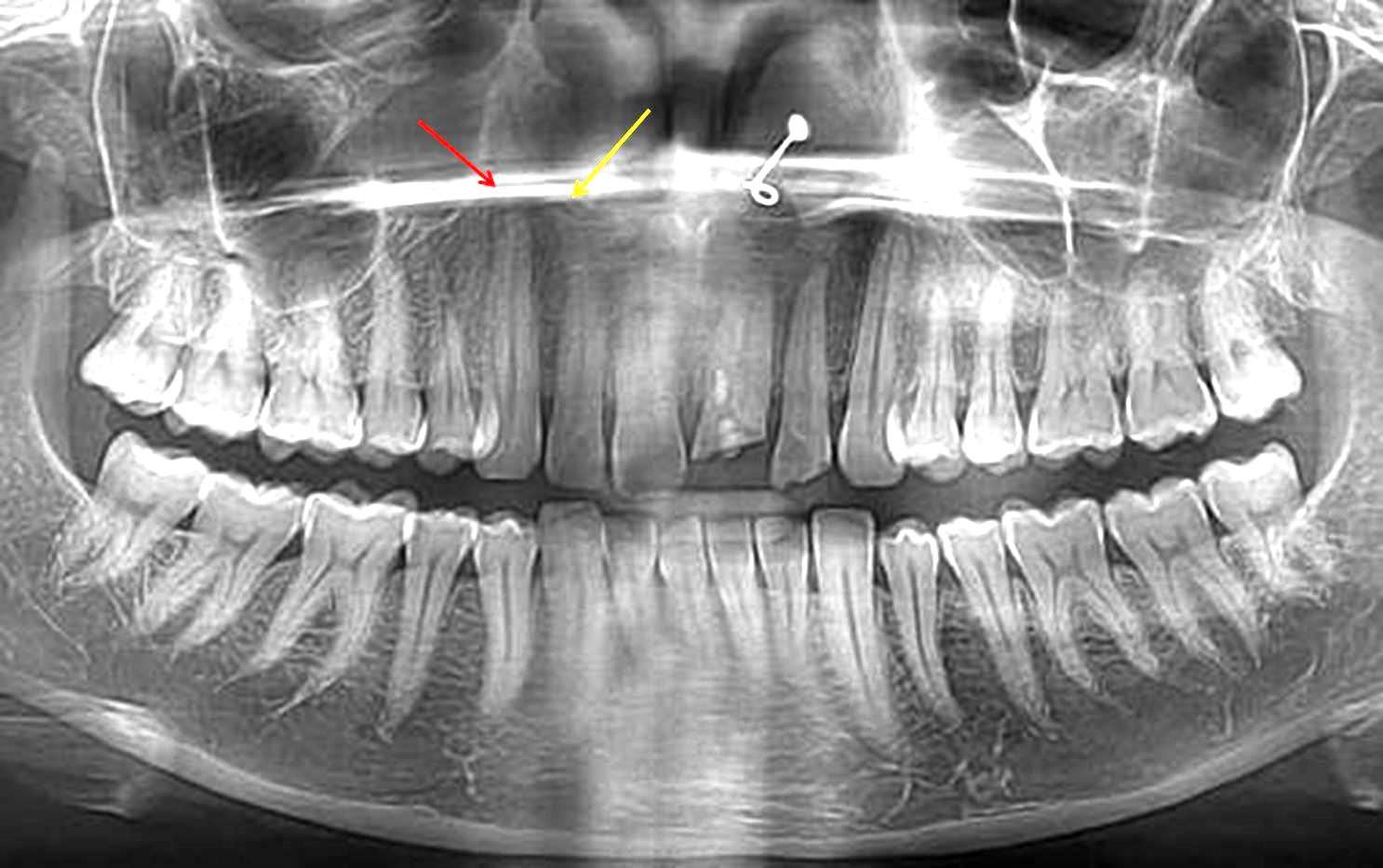
Figure 1: Orthopantomograph showing extremely long root (30 mm) in permanent maxillary right lateral incisor (apex is marked – yellow arrow) and almost equal to root of right canine (apex is marked – red arrow). Permanent maxillary both right (30 mm) and left canines (29 mm) and mandibular left canine’s root (29 mm) also appeared long. Root dilaceration was also observed in mandibular left canine and first premolar and right first premolar.
Dental anomalies involving tooth crown are very common in children [1]. Anomalies involving root structure are quire uncommon as these are identified only on radiographic examination and hence, they remain unnoticed most of the time during clinical examination. “Radiculomegaly” also referred by various synonyms such as ‘Root Gigantism’, ‘Long root’ ‘Rhyzomegaly’ or ‘Extremely long roots’ as suggested by Hayward [2] is rarely been reported in the literature. The exact etiology behind the lengthening of roots is not known. Literature suggests as it may develop from either developmental disorder of the root alone or disorders of radicular development as part of general tooth dysplasia. Clinically the coronal structure of the tooth associated with long roots appear apparently normal with respect to size and shape and it can occur in any gender and can occur single or generalized.
The first case of radiculomegaly was described by Hayward in 1980 [2]. Later two more cases were reported related to nonsyndromic type. Radiographically is characterized by pronounced elongation in dental roots. This root anomaly most frequently affects roots of permanent canines and incisors. Publications of radiculomegaly involving primary tooth roots is not reported till date. In case of canine, radiculomegaly is seen reaching the cortical plate of the orbit in the maxillary arch and inferior border of the mandible in the mandible. It is associated with a clinical significance as it is closely related to oculofaciocardiodental syndrome (OFCD) [3]. Therefore, in differential diagnosis this syndrome should be ruled out. However, Literature search revealed very few reports on occurrence of this anomaly in nonsyndromic patients or non-familial radiculomegaly. The first publication on this root anomaly is related to a characteristic feature of the X-linked rare congenital syndrome, oculofaciocardio-dental syndrome (OFCD). Apart from this syndrome, it is also reported with other syndromes like Cockayne syndrome and Klinefelter syndrome [4,5].
Normally the length of the permanent canines in adults varies from 24.6 to 27.4 mm with an average of 26 mm. The root length in maxillary lateral incisor is about 13 mm. Hence, to consider it as long root, the length of the canine and lateral incisor should be more than this. Moreover, the apices of the canines do not close until adulthood and closely approaching the inferior border of the mandible. The highlighting feature of this report is that radiculomegaly was noticed in permanent maxillary lateral incisor along with canines which is not reported so far according to best of best author knowledge following extensive review of the literature except for those reports showing generalized occurrence of rhizomegaly. Therefore, the present case is the first report showcasing the occurrence of radiculomegaly in the permanent maxillary lateral incisor along with canines. The length of the root in right lateral incisor appeared was almost to the length of the right canine reaching the maxillary sinus compared to left lateral incisor (Table 1). In the present case the length was about 30 mm. Hence this case was diagnosed as radiculomegaly (Table 1).
Recently in 2018, Smith et al [4] in his review of literature reported 67 cases of radiculomegaly in association with OFCD syndrome. In this investigation, he found only one case of non-syndromic occurrence of radiculomegaly. He also stated that, as this radicular anomaly is the primary feature of OFCD syndrome, dentist should be aware of the clinical and radiographic features of this anomaly. Therefore, this condition poses a great challenge to dentists and there are sparse reports showing cases where dental therapy is provided in this condition. Therefore, early diagnosis of the syndrome helps in prevention of dental challenges and improve in prognosis.

Open Access By Aditum Open Access Journals id licensed under Creative Commons Attribution 4.0 International License. Based On a Work at aditum.org