
Walid Abid, Abdessalem Hentati, Hèla Ben Jmaa, Ahmed Ben Ayed, Ghassen Ben Hlima, Aiman Dammak, Zied Chaari, Imed Frikha.
Thoracic and cardiovascular surgery department. Habib Bourguiba Hospital. University of Sfax, Tunisia.
*Corresponding authors: Hèla Ben Jmaà, department of cardiovascular and thoracic surgery Habib Bourguiba Hospital Sfax Tunisia.
Received Date: January 15, 2023
Accepted Date: February 20, 2023
Published Date: February 27, 2023
Citation: Abid W, Hentati A, Hèla B Jmaa, Ahmed B Ayed, Ghassen B Hlima (2023). “Thoracoscopic Approach In Hybrid Technique For Treatment Of Post Traumatic Subclavian Artery Injury”. Clinical Research and Clinical Case Reports, 4(1); DOI: http;//doi.org/02.2023/1.1061.
Copyright: © 2023 Hèla Ben Jmaà. This is an open access article distributed under the Creative Commons Attribution License, which permits unrestricted use, distribution, and reproduction in any medium, provided the original work is properly cited.
Subclavian artery SCA traumatism are uncommon, and are often associated with severe traumatism. They represent an emergency regarding the risk of bleeding and limb ischemia. Classically, surgical repair requires large approaches that may increase complication rates. Less invasive techniques had been reported including mainly endovascular solutions, that are not always available in different center, mainly in poor and developing countries.
We report a case of thoracoscopic ligation of SCA followed by a surgical bypass in a patient who was suffering from a violent traumatism with occlusion of the artery and signs of ischemia.
Introduction:
Traumatism of subclavian artery (SCA) is a sign of severity of the trauma. It is often associated with clavicle fracture, and it represents a serious emergency regarding the risk of bleeding by its rupture, and the risk of upper limb ischemia by its occlusion.
These lesions repair is challenging, because of associated injuries.
We report in this case a hybrid approach for repairing a left subclavian artery contusion, including a thoracoscopic ligation of the artery by a double port approach, and a carotid-subclavian bypass by a prosthetic graft.
Case presentation:
A 24-year-old man, without any medical history, was violently crashed to the wall by a speedy car. The patient was admitted to the intensive care unit. Hemodynamic parameters were conserved. A body scanner showed multiple rib fractures, with a total right pneumothorax, a mild left hemothorax with a contusion of the proximal part of the subclavian artery, and signs of hemorrhagic suffusion of mediastinal fat (figure 1). There was no clavicle fracture.
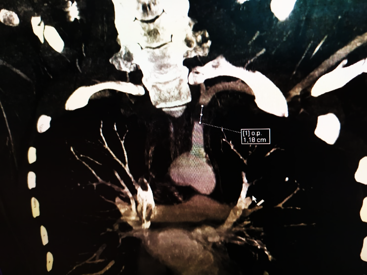
Figure 1 : CT scan showing an occlusion of the subclavian artery and a mediastinal effusion.
Left pulses of the superior left limb were absent with clinical signs of the beginning of ischemia.
Emergent surgical repair was indicated because endovascular graft was not available.
After drainage of the pneumothorax, the patient was intubated by a double lumen tube and putted in a supine position with a slight rotation to the right. The head was turned to the right. A first trocar was inserted at the 3rd intercostal space on the mid clavicular line from which a 30° optic was introduced. A 3 cm incision was done at the 4th intercostal space between anterior and middle axillary line for introducing instruments.
There was an important chest wall contusion, with infiltration of mediastinal pleura at the level of supra-aortic vessels.
Dissection was started from the beginning of the descending aorta to the aortic arch, using an energetic device. Subclavian artery was then dissected and encircled after identification of its roots. It was complicated to use vascular stapler because of the risk of rupture of the vessel which was fragile; so, we ligated it by surgical knots (video) (figure2).
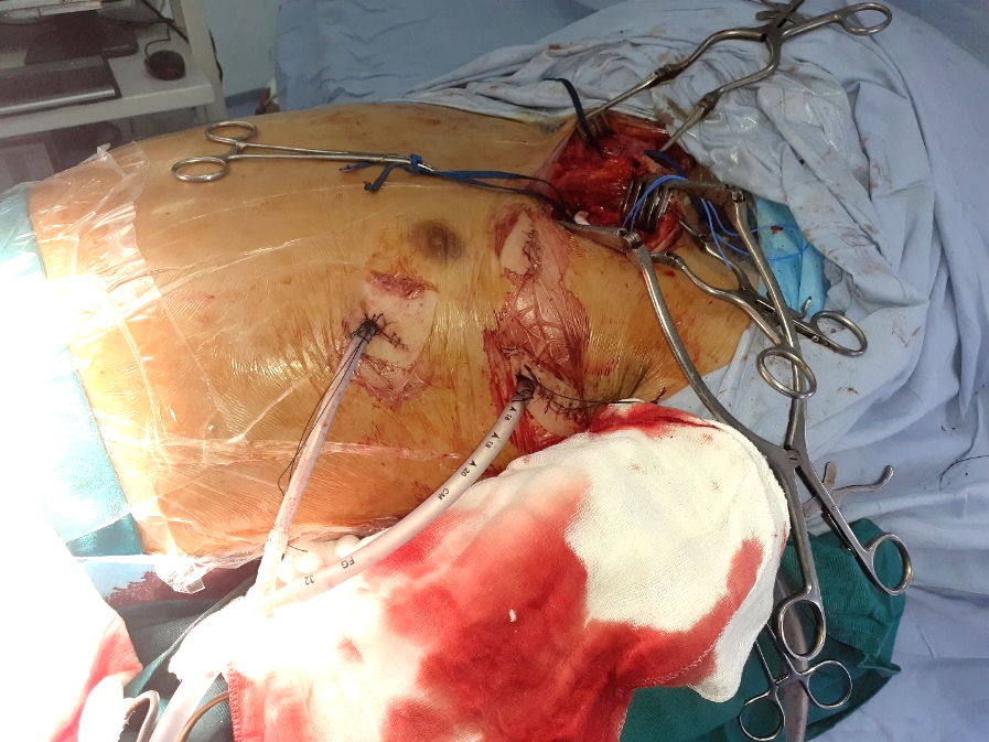
Figure 2: Per operative view showing surgical approaches.
The second time was the vascular bypass. Incision was done on the anterior edge of the sternocleidomastoid muscle and prolonged to the inferior edge of the clavicle (like skin incision in Cormier Dartevelle Grunenwald approach). Left common carotid artery and distal portion of subclavian left artery were controlled. The distal part of the SCA was ligated. Bypass was done with a prosthetic armed graft of 6 mm of diameter and was passed under the sternoclavicular articulation. At the end of intervention, all pulses were recovered. Chest drainage was done by two chest tubes.
Post-operative course was complicated with a left hemothorax. A thoracoscopic revision was performed at the 3rd post-operative day. No active bleeding was found. Parietal contusion was very important because of ribs’ fractures and the adjunction of heparin. The patient was discharged at the 8th post-operative day, with a normal chest X ray, present radial and cubital pulses, and no signs of ischemia. There was no signs of carotid steal syndrome or vertebral symptoms.
Discussion:
Subclavian Artery (SCA) trauma are rare and represent 2% of vascular trauma [1]. It represents a serious and life-threating condition that may lead to death in 30% of cases [2]. it is often seen in violent traffic accidents. Other trauma are frequently associated and increase the mortality rate.
Trauma of SCA must be suspected in traumatism involving the upper part of the chest wall, and particularly shoulders. Clinical signs may include signs of hemorrhagic shock, parietal hematoma, and signs of limb ischemia with loss of pulses [3].
As computed tomography became the standard in the exploration of trauma, lesion of the SCA is more frequently diagnosed on CT angiography compared to conventional angiography. According to angiographic findings, injuries of SCA can be classified into low grade and severe depending on the type of lesion and the diameter of injuries.
Management of SCA traumatic lesions is usually emergent, especially when there is signs of active bleeding, hemodynamic shock or signs of limb ischemia. The choice of therapeutic strategy depends mainly on the hemodynamic state of the patient and the severity of the injury. Thus, for low grade injuries, an active watching is sufficient with angiographic control. This situation include intimal disruption, arterial occlusion without signs of ischemia and small pseudo-aneurysms [2]. For severe injuries, like arterial transection for laceration of more than 50% of circumference, surgical repair is frequently needed. The aim is to prevent or to control an active bleeding and then to restore vascularization of the limb.
It is important to mention that surgical approach of the SCA is complicated, especially for the left side because of anatomical configuration. Indeed, the root of the left SCA is intra thoracic, and so, for surgical control, a dilapidating approach is needed like median sternotomy, anterior thoracotomy or event hemi clamshell approach [4-6]. Less invasive procedures are possible, mainly endovascular solution which can be exclusive or in association with surgery [2, 7]. Several studies reported the use endovascular balloon for the control of an active bleeding, followed by stent placement or a surgical bypass for revascularization [2, 3, 8]. These endovascular procedures require a hybrid operating room, offering the possibility of surgical conversion when necessary. It is undoubtable that these techniques offer less morbidity and mortality in patients suffering from associated injuries [9]: for open surgery through median sternotomy or anterolateral thoracotomy, morbidity and mortality rate are about 40% et 5% respectively in this context [8]. It is important to mention that there are some limiting conditions for endovascular approach like the hemodynamic state of the patient, the availability of a technical platform, and the experience of the surgeon [2]. Like in our case, endovascular treatment was impossible firstly because endovascular grafts were unavailable, and secondly because injury of the SCA was considered as severe.
Thoracoscopic approach was another less invasive approach for the control of the artery’s root, avoiding an open surgical approach mainly in this patient who was suffering from severe chest injuries. To the best of our knowledge, there is only three cases reported in the literature for the thoracoscopic approach of SCA. The first one is a case of direct repair of SCA injury after placement of a subclavian catheter [9]; and the two other cases were reported for a temporary endothoracic clamping of the SCA followed by a vascular bridge [1]. No case of ligation of SCA through a thoracoscopic approach has been reported before. The main complication in our case was related to the associated rib fracture and the adjunction of heparin.
It was also reported in several studies that pain is lower in thoracoscopic approach compared to the open surgical approachs, with a direct and indirect effects on the respiratory exchanges [9].
Conclusion:
SCA trauma are rare but serious. Their management depends on the severity of the injury, hemodynamic state of the patient, and accessibility of theapeutic tools. Even though open surgery and endovascular approach remain the most frequent way to manage SCA injuries, thoracoscopic control of this artery is an interesting option in some situations, offering a reduced rate of mortality and morbidity.

Open Access By Aditum Open Access Journals id licensed under Creative Commons Attribution 4.0 International License. Based On a Work at aditum.org