Clinical Research and Clinical Case Reports
OPEN ACCESS | Volume 7 - Issue 1 - 2026
ISSN No: 2836-2667 | Journal DOI: 10.61148/2836-2667/CRCCR


Hèla Ben Jmaà*, Aiman Dammak, Arwa Ziadi, Fatma Mhiri, Majdi Gueldich, Mohamed Seddik, Imed Frikha.
Department of cardiovascular and thoracic surgery Habib Bourguiba Hospital Sfax Tunisia.
*Corresponding author: Hèla Ben Jmaà, Department of cardiovascular and thoracic surgery Habib Bourguiba Hospital Sfax Tunisia.
Received: April 21, 2022
Accepted: May 04, 2022
Published: May 06, 2022
Citation: Hèla Ben Jmaà, Aiman Dammak, Arwa Ziadi, Fatma Mhiri, Majdi Gueldich et al (2022) “ Aortic Aneurysms in Patients with Behçet Diseases : Report of Two Cases and Literature Review”. Clinical Research and Clinical Case Reports, 3(2); DOI: http;//doi.org/05.2022/1.1051.
Copyright: © 2022 Hèla Ben Jmaà. This is an open access article distributed under the Creative Commons Attribution License, which permits unrestricted use, distribution, and reproduction in any medium, provided the original work is properly cited.
Behçet disease is a multisystemic vasculitis of unknown origin. In this disase, arterial aneurysms are often multiple and are characterized by a saccular configuration with increased risk of unexpected rupture, thrombosis, and aneurysm recurrence.
We report the case of 2 patients with vasculo-Behçet disease with aortic false aneurysms, treated surgically by resection of aneurysms, and replacement by prosthesis.
Introduction :
Behçet disease is a multisystemic vasculitis of unknowen etioogy, with an undulating course of exacerbations and remissions [1].
The primary lesion in Behçet disease is a type of systemic vasculitis that primarily affects the eyes, skin, joints, blood vessels, and nervous system.
Vascular involvement is characterized by occlusion of the large vessels and aneurysm formation, that can cause fatal complications [2].
We report 2 unusual presentations of Behçet disease with abdominal aortic pseudo-aneurysms treated surgically.
Case report 1 :
A 42-year-old-man, with a Behçet disease, was admitted to the emergency department with complaints of abdominal pain and gangrena of the right foot.
At physical examination his abdomen was tender, and femoral artery pulses were present.
Abdominal CT scan revealed 2 false aneurysms of the aorta, and a false aneurysm of the right iliac artery (figure 1).
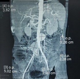
Figure 1 : abdominal CT scan showing 2 false aneurysms of the aorta, and a false aneurysm of the right iliac artery.
The patient received corticosteroids, and after stability of his inflammatory syndrome, he underwent surgery.
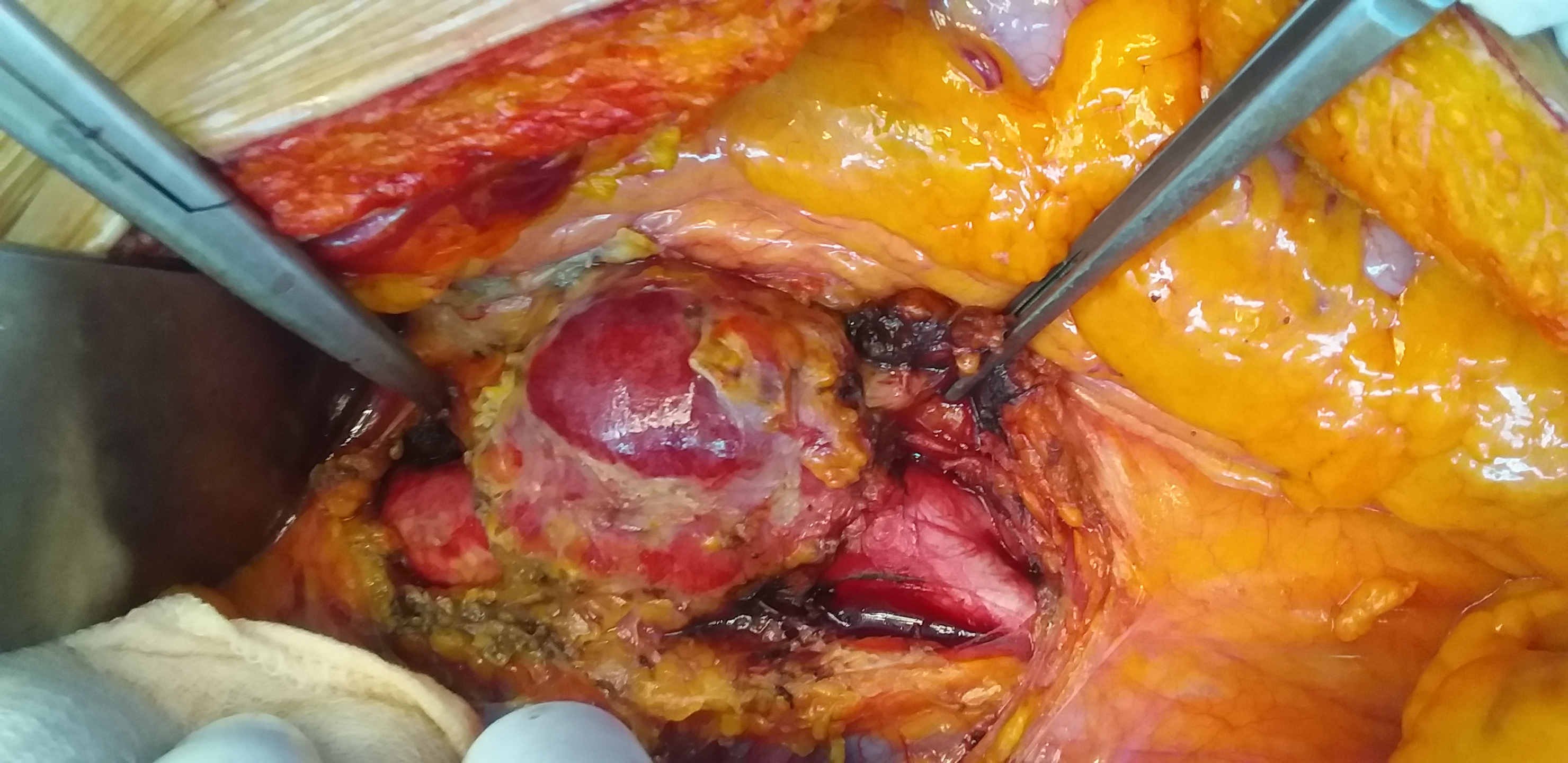
After median laparotomy, the aorta was dissected.
There was three saccular false aneurysms of the distal abdominal aorta, and the right iliac artery.
The proximal aorta below the renal arteries was clamped, and the aneurysm sacs was opened (figures 2 and 3).
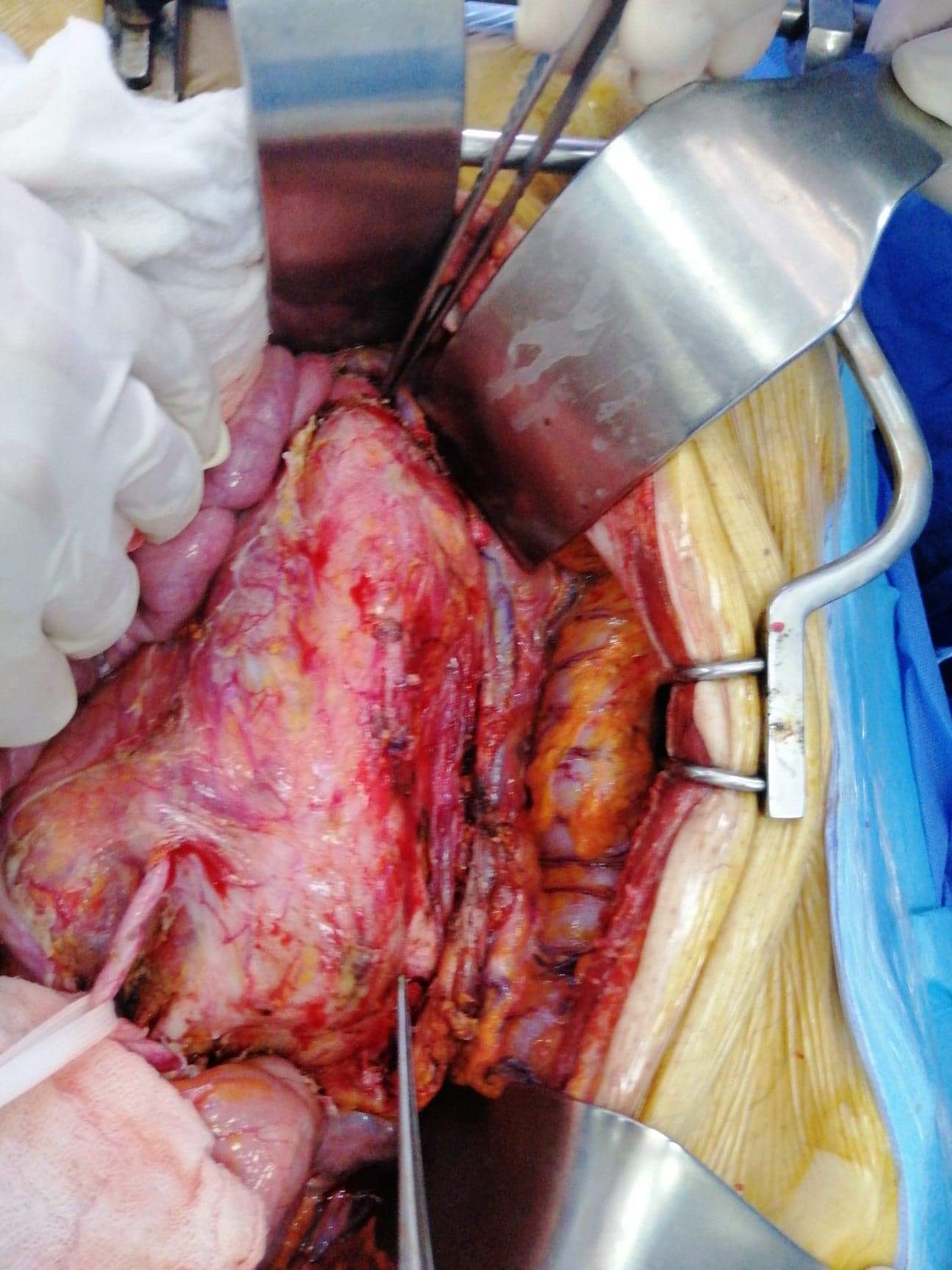
Figures 2 and 3 : Intra-operative views revealing the aorta and iliac arteries after resection of the aneurysms.
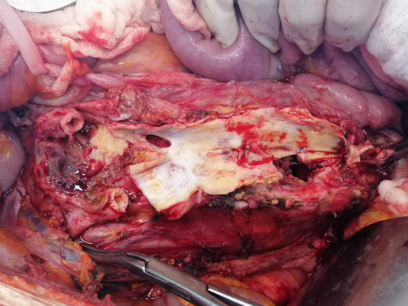
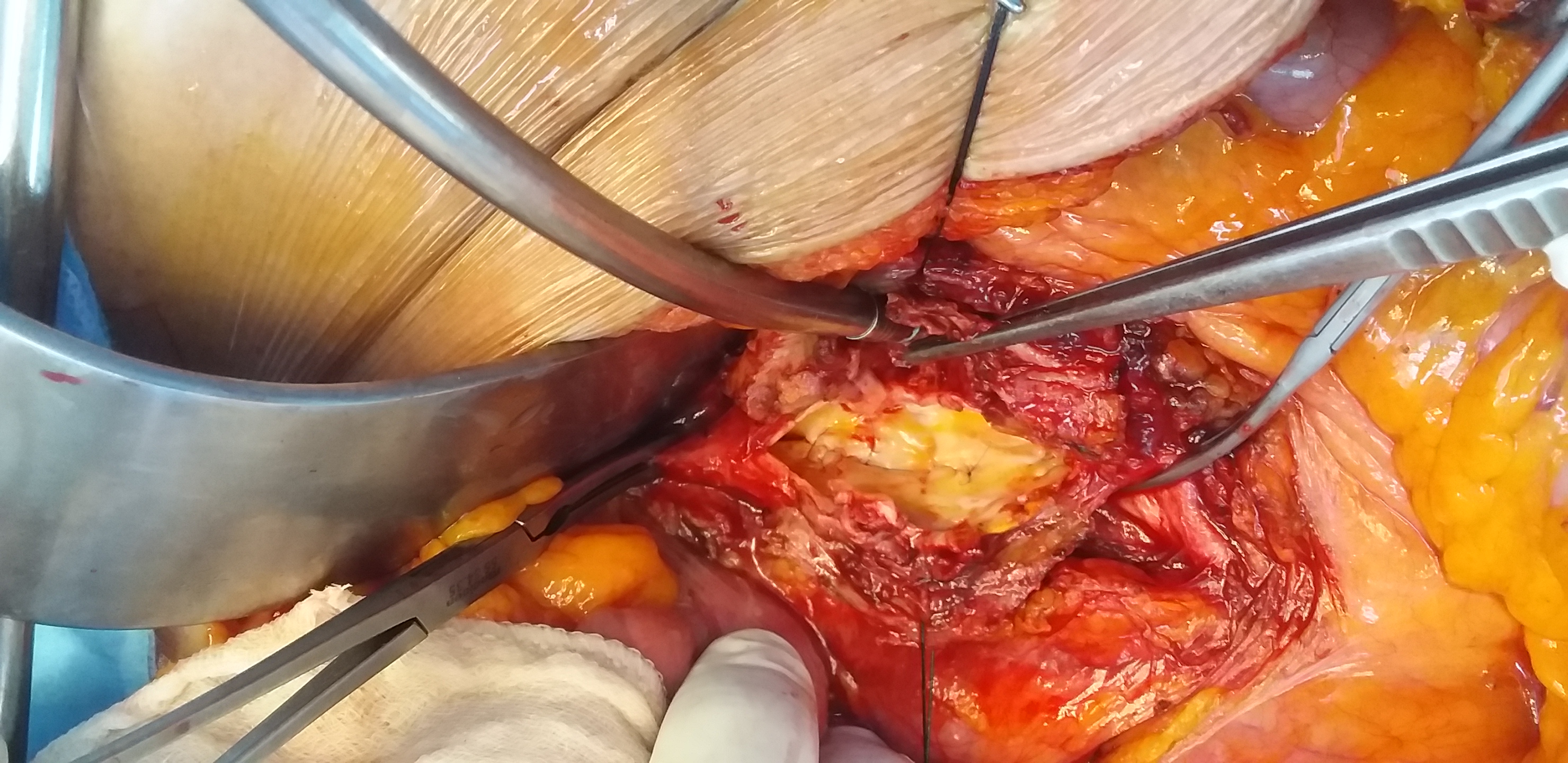
Abdominal aorta reconstruction was performed with a bifurcated Dacron prosthesis, below the renal arteries to the orifice of the two common iliac arteries (figues 4).
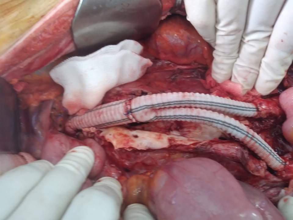
Figure 4 : Intra-operative view of the bifurcated posthesis.
After intensive care therapy for 5 days, rapid healing was observed, and the patient was transferred to the rheumatology clinic for diagnosis.
Case report 2 :
A 58-year-old man, with a Behçet disease, complainted of acute abdominal pain.
Physical examination was without anomalies.
Abdominal ultrasound showed an aortic mass. So, a CT scan was performed.
He revealed a saccular aneurysm of the infra-renal aorta (figure 5).
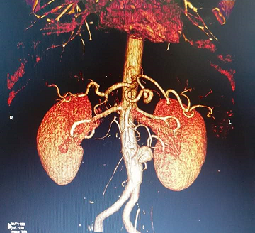
Figure 5 : Aortic CT scan showing a saccular aneurysm of the abdominal aorta (arrow).
The patient underwent conventional surgery under median laparotomy. The surgery consisted in a resection of the aneurysm and interposition of a tubular graft (figures 6, 7 and 8).
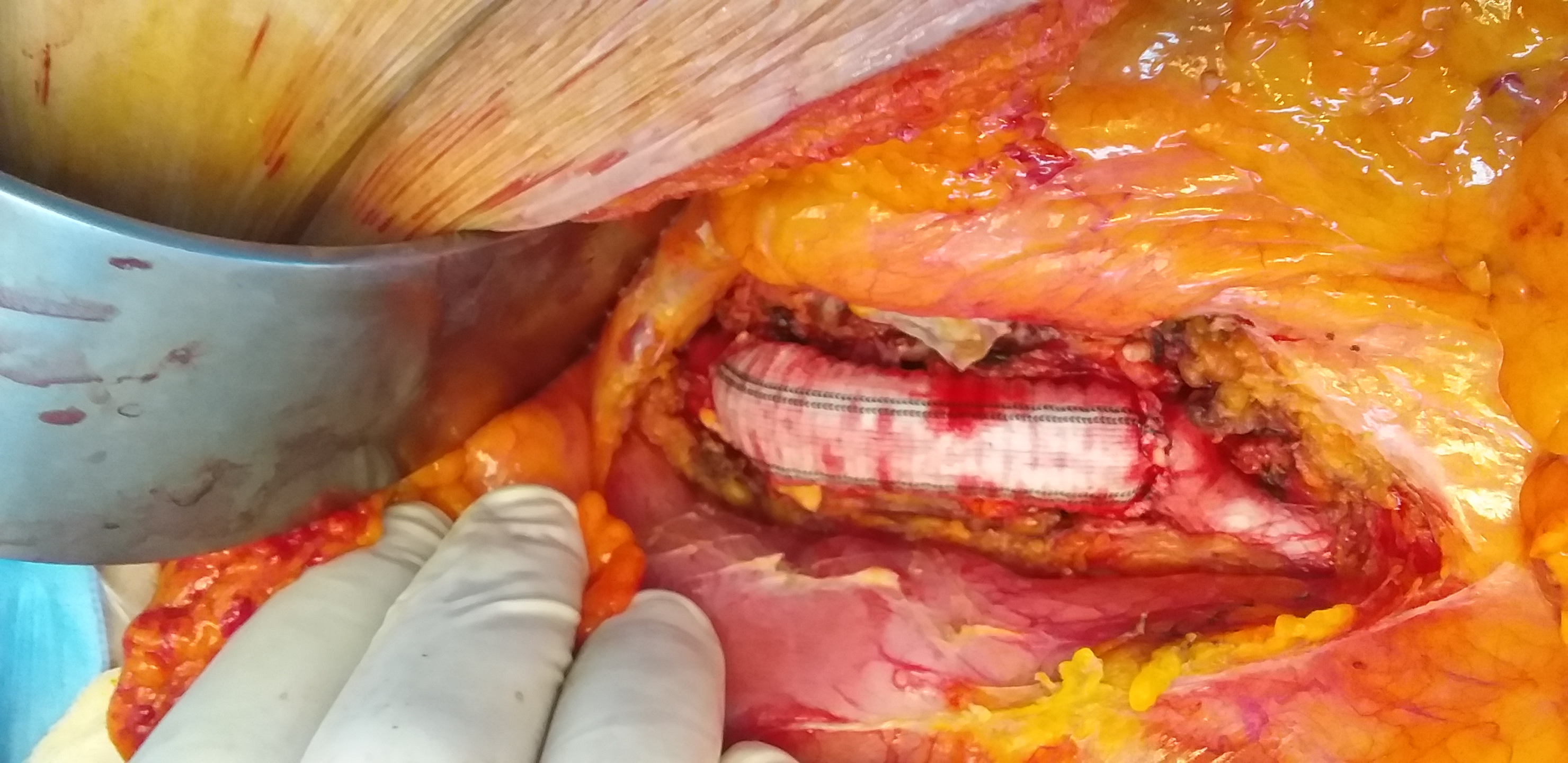
Figures 6, 7, and 8 : Intra-operative views showing the saccular aneurysm dissected, opened, and replaced by a tubular prosthesis.
The postoperative course was uneventful, and the patient was discharged from hospital 4 days after surgery.
Discussion :
Inflammatory abdominal aortic aneurysms are characterized by marked thickening of the aneurysm wall [3]. The diagnosis of vasculitis requires histologic confirmation in most cases, and the classification is based on the size of the predominant involved vessels.
The pathogenesis of aneurysms in Behçet disease is multifactorial and quite different from that of atherosclerotic aneurysms. Abnormalities of neutrophils, enhanced adherence to endothelial cells, and increased adhesion molecules have been suggested as responsible in the pathophysiology of vascular involvement [3].
Large arterial aneurysms, which are more common than occlusions, are a major cause of death, because of the risk of rupture [4].
The most common location for the formation of the aneurysm is the abdominal aorta [5].
Spontaneous rupture of these aneurysms is the most common cause of death [6].
Because of potentially fatal complications, early diagnosis is important. Open surgery and endovascular procedures are the available treatments in patients with arterial aneurysms.
Immunosuppressive therapy should be started in patients with arterial disease before the surgical intervention and should be continued after surgery to avoid postoperative complications [7].
Conclusion :
Early diagnosis of vascular involvement is helpful for planning effective management and improving the prognosis, and long-term follow-up is essential because of the relapsing nature of the disease.


Open Access By Aditum Open Access Journals id licensed under Creative Commons Attribution 4.0 International License. Based On a Work at aditum.org